. 2023 Sep 18:11:1261048.
doi: 10.3389/fcell.2023.1261048. eCollection 2023.

Ubiquitination of the protocadherin-γA3 variable cytoplasmic domain modulates cell-cell interaction

Affiliations

Ubiquitination of the protocadherin-γA3 variable cytoplasmic domain modulates cell-cell interaction

Albert Ptashnik et al. Front Cell Dev Biol. .

Abstract

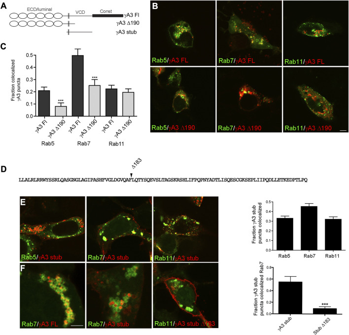
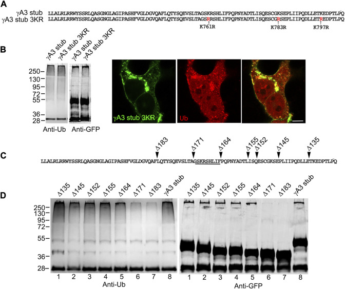
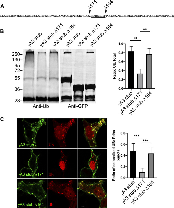
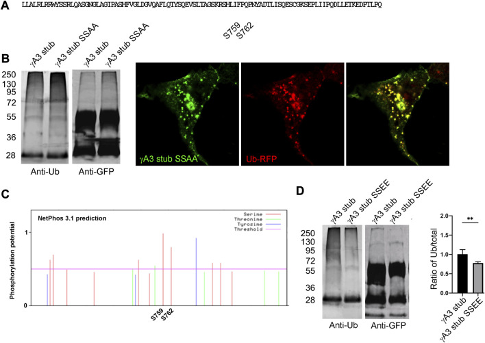
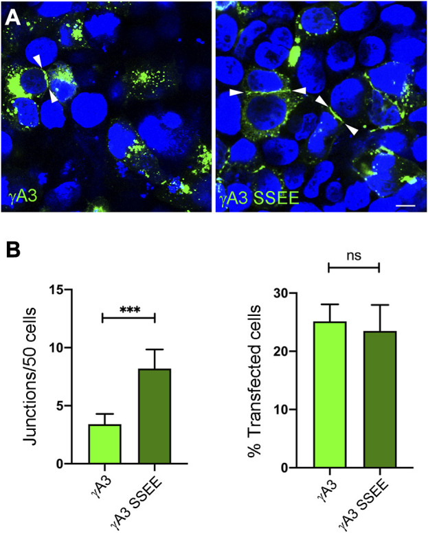
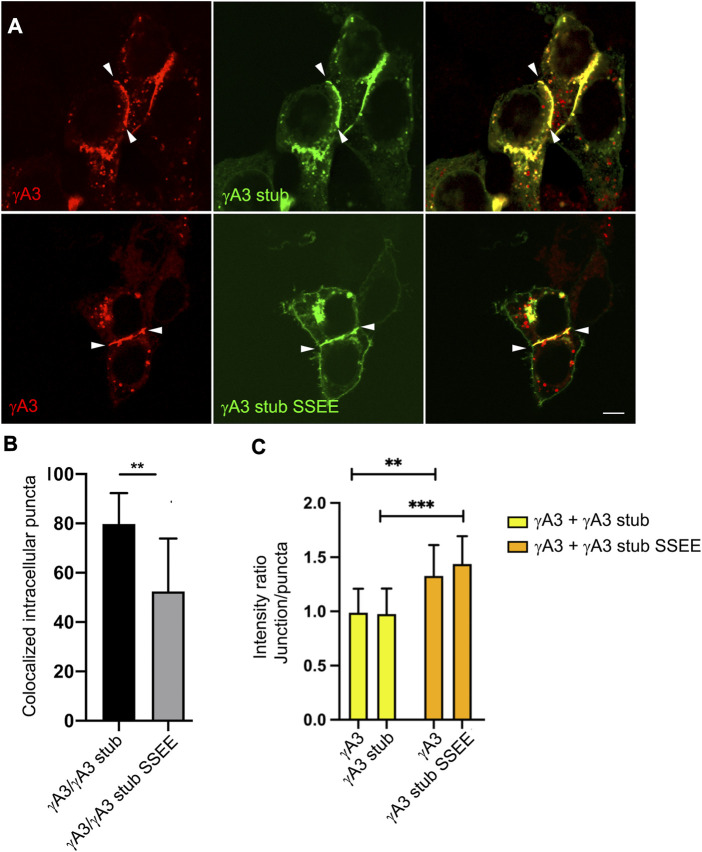
The family of ∼60 clustered protocadherins (Pcdhs) are cell adhesion molecules encoded by a genomic locus that regulates expression of distinct combinations of isoforms in individual neurons resulting in what is thought to be a neural surface "barcode" which mediates same-cell interactions of dendrites, as well as interactions with other cells in the environment. Pcdh mediated same-cell dendrite interactions were shown to result in avoidance while interactions between different cells through Pcdhs, such as between neurons and astrocytes, appear to be stable. The cell biological mechanism of the consequences of Pcdh based adhesion is not well understood although various signaling pathways have been recently uncovered. A still unidentified cytoplasmic regulatory mechanism might contribute to a "switch" between avoidance and adhesion. We have proposed that endocytosis and intracellular trafficking could be part of such a switch. Here we use "stub" constructs consisting of the proximal cytoplasmic domain (lacking the constant carboxy-terminal domain spliced to all Pcdh-γs) of one Pcdh, Pcdh-γA3, to study trafficking. We found that the stub construct traffics primarily to Rab7 positive endosomes very similarly to the full length molecule and deletion of a substantial portion of the carboxy-terminus of the stub eliminates this trafficking. The intact stub was found to be ubiquitinated while the deletion was not and this ubiquitination was found to be at non-lysine sites. Further deletion mapping of the residues required for ubiquitination identified potential serine phosphorylation sites, conserved among Pcdh-γAs, that can reduce ubiquitination when pseudophosphorylated and increase surface expression. These results suggest Pcdh-γA ubiquitination can influence surface expression which may modulate adhesive activity during neural development.

Keywords: cell adhesion; endocytosis; endosome; pseudophosphorylation; self-avoidance.


Conflict of interest statement

The authors declare that the research was conducted in the absence of any commercial or financial relationships that could be construed as a potential conflict of interest. The authors declared that they were an editorial board member of Frontiers, at the time of submission. This had no impact on the peer review process and the final decision

Figures

FIGURE 1
FIGURE 1
The Pcdh-γA3 VCD is sufficient to promote trafficking to the late endosome. (A) Diagram of Pcdh-γA3 constructs. (B) Pcdh-γA3 full length (γA3 FL) or the cytoplasmic deletion (γA3 Δ190), fused to RFP, were cotransfected with different Rab-GFP markers. (C) Quantification of colocalization. γA3 FL preferentially colocalized with Rab7, a marker for the late endosome and this colocalization was significantly diminished for γA3 Δ190. Colocalization with Rab5, a marker for late endosomes, was also significantly diminished upon cytoplasmic deletion. Colocalization with Rab11, a marker for recycling endosomes, was unaffected by cytoplasmic deletion. (D) Sequence of the Pcdh-γA3 VCD which comprises the intracellular segment of the γA3 stub construct. Location of the Δ183 deletion in the γA3 stub is indicated. (E) The γA3 stub preferentially colocalizes with Rab7, similar to the full length molecule. (F) Full length Pcdh-γA3 and the γA3 stub are trafficked to the interior of Rab7 positive vesicles (left and middle panels). In contrast, γA3 stub Δ183 does not colocalize significantly with Rab7 positive vesicles. Bar = 5 μm in (B,E) and (F) right panel, 300 nm in (F) left and middle panels. ***p <0.001.
FIGURE 2
FIGURE 2
Ubiquitination of the γA3 stub is associated with intracellular trafficking. (A) γA3-GFP stub puncta colocalize with ubiquitin-RFP (top, arrowheads) while γA3 stub Δ183 (bottom) exhibits less colocalization with ubiquitin-RFP as quantified in (B) (C) γA3 stub GFP immunoprecipitates probed with anti-ubiquitin exhibit a high molecular weight smear that was not present in immunoprecipitates of γA3 stub Δ183 (left). Probing with anti-GFP reveals the equivalent expression levels of γA3 stub and γA3 stub Δ183 (right). ***p <0.001.
FIGURE 3
FIGURE 3
A segment of the Pcdh-γA3 VCD regulates ubiquitination. (A) Sequence of the γA3 VCD. The indicated lysines were mutated to arginine in γA3 stub 3KR. (B) Both γA3 stub and γA3 stub 3KR exhibit a similar ubiquitin smear when probed with anti-ubiquitin antibodies at similar expression levels (left panels). γA3 stub 3KR shows colocalization with ubiquitin (right panels). (C) Deletions of the γA3 stub are indicated. (D) GFP precipitates probed with anti-ubiquitin show similar ubiquitin smears for the γA3 stub Δ135, Δ145, Δ152, Δ155 and Δ164 deletions, but reduced smears for the Δ171 and Δ183 deletions indicating the presence of a critical segment in the VCD [underlined in (C)] that regulates ubiquitination.
FIGURE 4
FIGURE 4
Quantification of γA3 stub, γA3 stub Δ164 and γA3 stub Δ171 ubiquitination. (A) Pcdh-γA3 VCD sequence with indicated deletions analyzed in the stub backbone. The segment required for ubiquitination is underlined. (B) γA3 stub, γA3 stub Δ164 and γA3 stub Δ171 immunoprecipitates were probed for anti-ubiquitin and GFP. Quantification of ubiquitin smear relative to GFP is shown at right. **p <0.005 (C) Cotransfection of γA3 stub, γA3 stub Δ164 and γA3 stub Δ171 with ubiquitin-RFP. Quantification of colocalization is shown at right. ***p <0.001.
FIGURE 5
FIGURE 5
Serines 759 and 762 are not substrates for ubiquitination but may be phosphorylation sites that affect ubiquitination. (A) The indicated serines were mutated to alanine to generate γA3 stub SSAA. (B) γA3 stub and γA3 stub SSAA immunoprecipitates were probed for anti-ubiquitin and cotransfected with ubiquitin-RFP. Both exhibited similar ubiquitin smears and colocalization with ubiquitin. (C) Analysis of the VCD region of Pcdh-γA3 by NetPhos 3.1. Both serine 759 and 762 were the most likely within the VCD to be substrates for phosphorylation. (D) γA3 stub and pseudophosphorylated γA3 stub (γA3 stub SSEE) immunoprecipitates were probed with anti-ubiquitin and anti-GFP. γA3 stub SSEE exhibited a reduced ubiquitin smear as compared to γA3 stub as quantified on the right. **p <0.005.
FIGURE 6
FIGURE 6
Pseudophosphorylation at S759 and S762 affects trafficking of the full length molecule. (A) Cells were transfected with full length Pcdh-γA3 or Pcdh-γA3 SSEE. Junctions between two cells (arrowheads) were identified. (B) Quantification of cell junctions per 50 transfected cells. The number of junctions formed by Pcdh-γA3 SSEE was significantly increased relative to wild-type Pcdh-γA3 (left) while the number of transfected cells was equivalent between the 2 constructs (right). Bar = 5 μm ***p <0.001.
FIGURE 7
FIGURE 7
Pseudophosphorylated γA3 stub increases recruitment of wild-type full length Pcdh-γA3 to cell junctions. (A) Cells were cotransfected with full length Pcdh-γA3 and either γA3 stub (top) or γA3 stub SSEE (bottom). (B) Intracellular puncta colocalization of γA3 stub or γA3 stub SSEE. (C) Pcdh-γA3 and cotransfected γA3 stub had a roughly equivalent intensity ratio between junctions and puncta (yellow bars) whereas when Pcdh-γA3 when cotransfected with γA3 stub SSEE both were more intense at the junctions than the intracellular puncta. **p <0.005, ***p <0.001.

References

    1. Brasch J., Goodman K. M., Noble A. J., Rapp M., Mannepalli S., Bahna F., et al. (2019). Visualization of clustered protocadherin neuronal self-recognition complexes. Nature 569, 280–283. 10.1038/s41586-019-1089-3 - DOI - PMC - PubMed
    1. Buchanan S. M., Schalm S. S., Maniatis T. (2010). Proteolytic processing of protocadherin proteins requires endocytosis. Proc. Natl. Acad. Sci. U. S. A. 107, 17774–17779. 10.1073/pnas.1013105107 - DOI - PMC - PubMed
    1. Canzio D., Maniatis T. (2019). The generation of a protocadherin cell-surface recognition code for neural circuit assembly. Curr. Opin. Neurobiol. 59, 213–220. 10.1016/j.conb.2019.10.001 - DOI - PMC - PubMed
    1. Chen J., Lu Y., Meng S., Han M.-H., Lin C., Wang X. (2009). alpha- and gamma-Protocadherins negatively regulate PYK2. J. Biol. Chem. 284, 2880–2890. 10.1074/jbc.M807417200 - DOI - PMC - PubMed
    1. Flaherty E., Maniatis T. (2020). The role of clustered protocadherins in neurodevelopment and neuropsychiatric diseases. Curr. Opin. Genet. Dev. 65, 144–150. 10.1016/j.gde.2020.05.041 - DOI - PMC - PubMed

LinkOut - more resources