. 2023 Aug 9;8(9):1081-1097.
doi: 10.1016/j.jacbts.2023.05.007. eCollection 2023 Sep.

Empagliflozin Suppresses the Differentiation/Maturation of Human Epicardial Preadipocytes and Improves Paracrine Secretome Profile

Affiliations

Empagliflozin Suppresses the Differentiation/Maturation of Human Epicardial Preadipocytes and Improves Paracrine Secretome Profile

Masayuki Takano et al. JACC Basic Transl Sci. .

Abstract

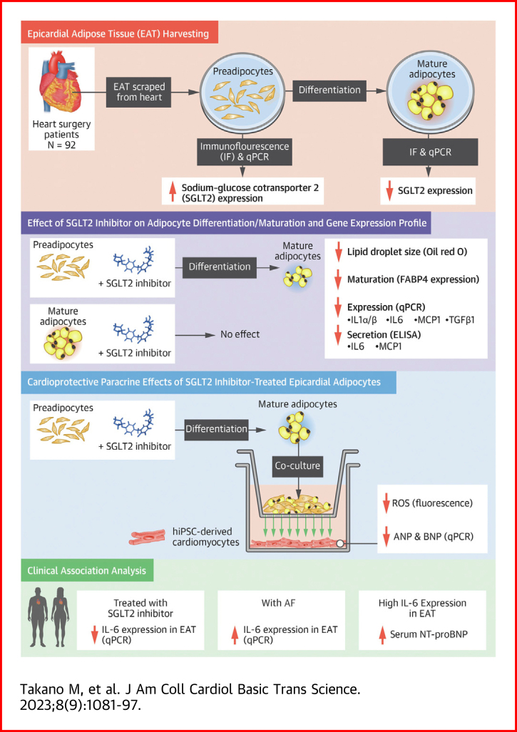
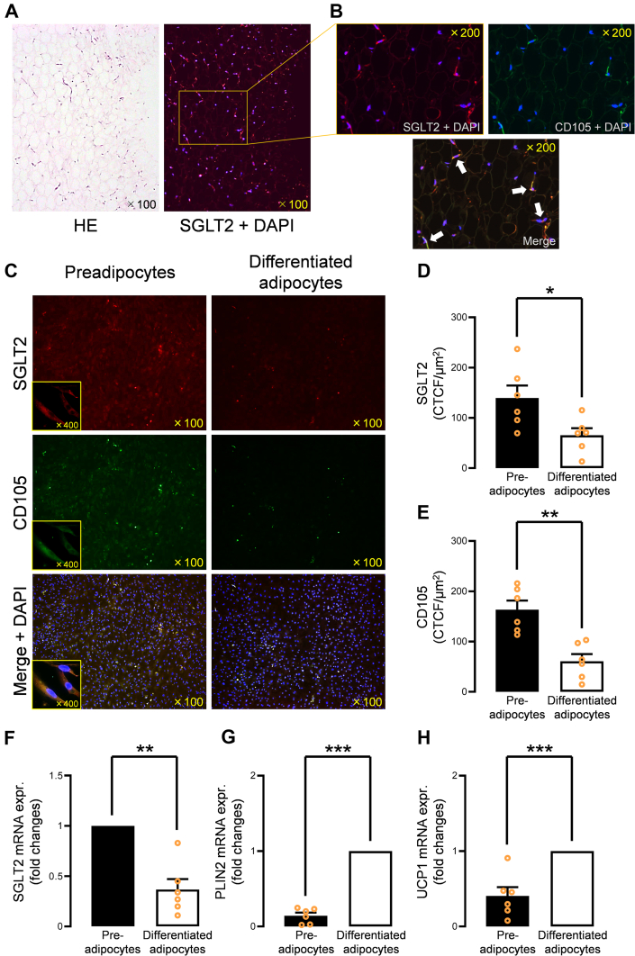
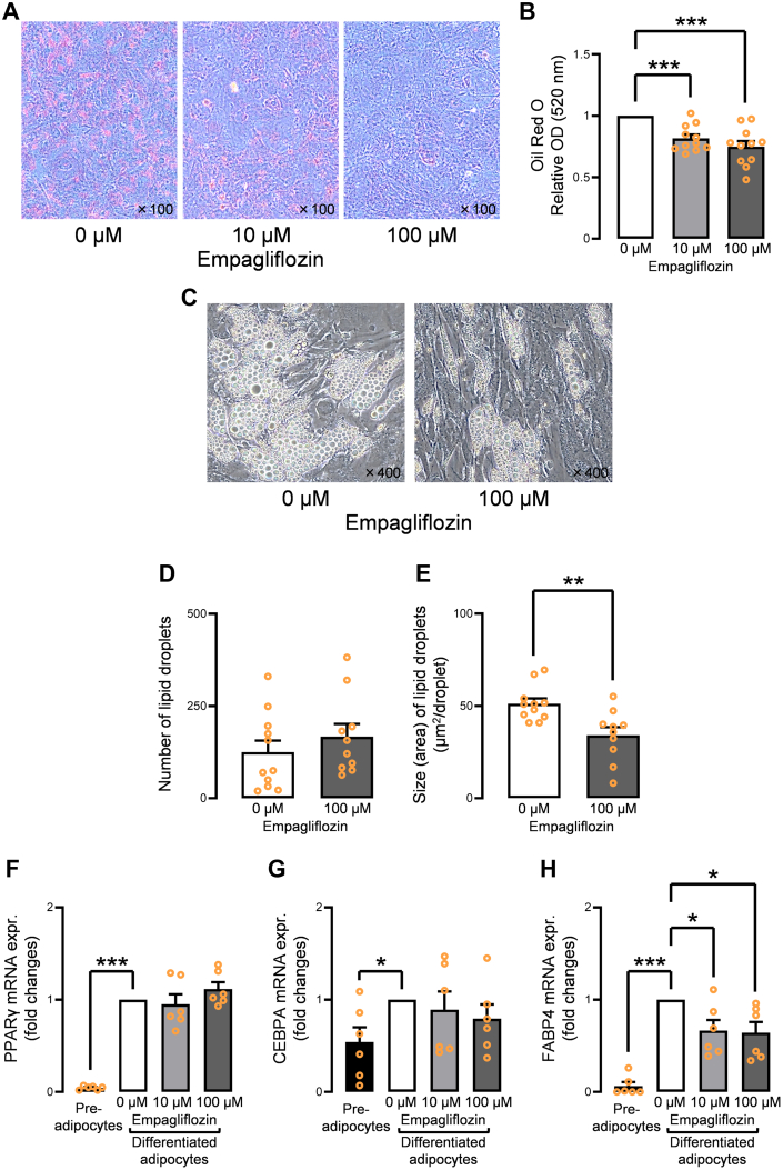
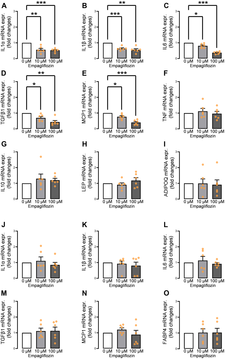
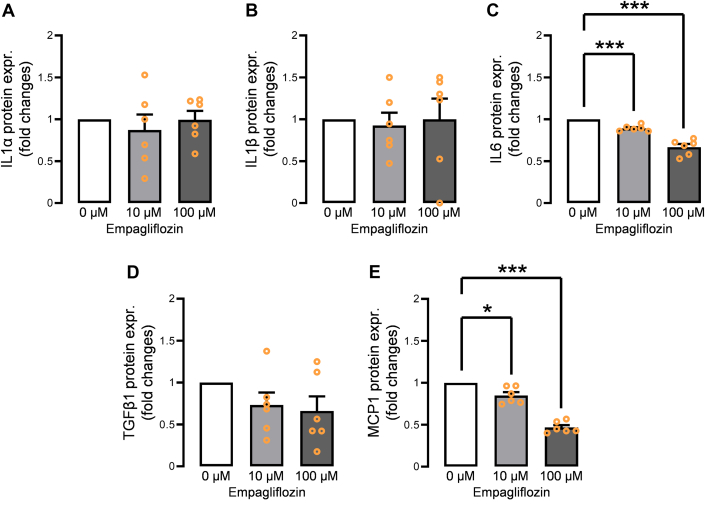
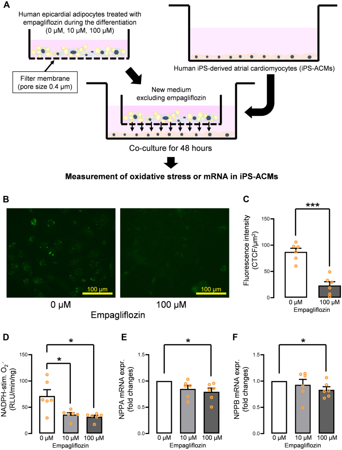
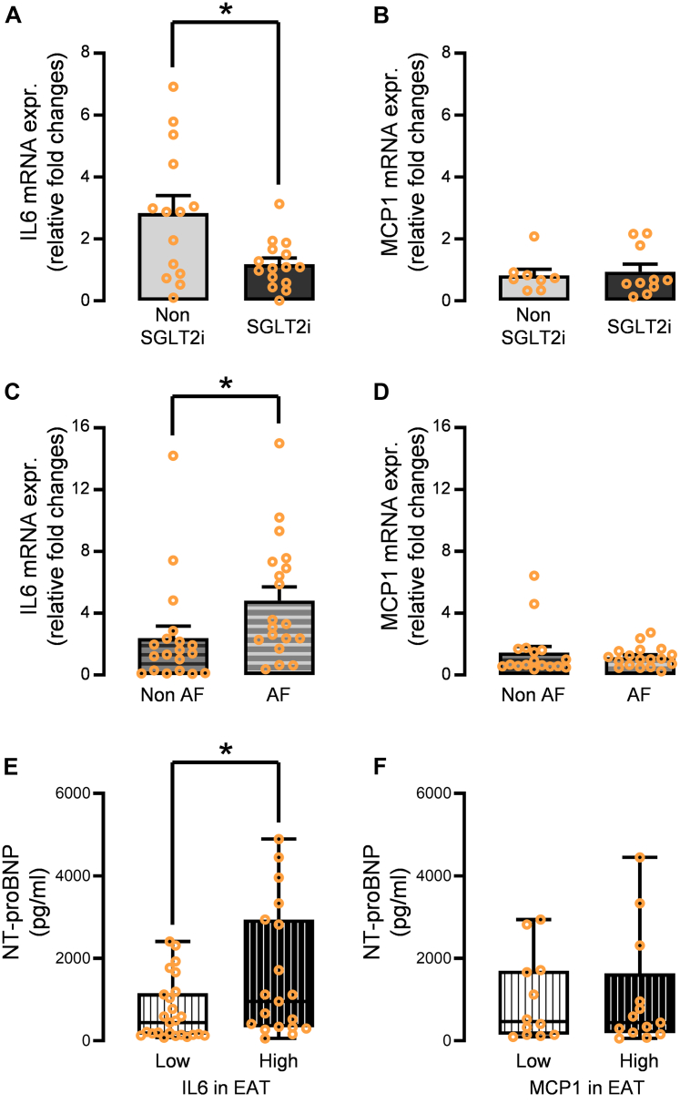
Sodium-glucose cotransporter 2 (SGLT2) inhibitors reduce epicardial adipose tissue (EAT) in humans, enhancing cardioprotective effects on heart failure and atrial fibrillation. We investigated the direct effect of the SGLT2 inhibitor empagliflozin on human primary epicardial adipocytes and preadipocytes. SGLT2 is primarily expressed in human preadipocytes in the EAT. The expression levels of SGLT2 significantly diminished when the preadipocytes were terminally differentiated. Adipogenesis of preadipocytes was attenuated by empagliflozin treatment without affecting cell proliferation. The messenger RNA levels and secreted protein levels of interleukin 6 and monocyte chemoattractant protein 1 were significantly decreased in empagliflozin-treated adipocytes. Coculture of human induced pluripotent stem cell-derived atrial cardiomyocytes and adipocytes pretreated with or without empagliflozin revealed that empagliflozin significantly suppressed reactive oxygen species. IL6 messenger RNA expression in human EAT showed significant clinically relevant associations. Empagliflozin suppresses human epicardial preadipocyte differentiation/maturation, likely inhibiting epicardial adipogenesis and improving the paracrine secretome profile of EAT, particularly by regulating IL6 expression.

Keywords: epicardial adipose tissue; interleukin 6; paracrine effect; preadipocyte; sodium-glucose cotransporter 2 inhibitor.


Conflict of interest statement

The authors have reported that they have no relationships relevant to the contents of this paper to disclose.

Figures

None
Graphical abstract
Figure 1
Figure 1
Flowchart of 92 Consecutive Patients Who Underwent Open Heart Surgery and Were Enrolled in the Multicenter Cohort SGLT2 expression levels in human EAT and preadipocytes/adipocytes were evaluated in 25 nondiabetic patients. In vitro incubation studies or coculture experiments using human adipocytes/preadipocytes and human induced-pluripotent stem cell–derived cardiomyocytes included the same nondiabetic 25 patients to confirm the pure effect of SGLT2 inhibitors on preadipocytes/adipocytes (study 1). A cohort-wide association study to clinically validate the effect of SGLT2 inhibitors on EAT included 29 diabetic patients treated with SGLT2 inhibitors or other antidiabetic drugs (study 2). In addition, the gene expression levels of inflammatory adipocytokines were statistically compared using samples from 19 patients with AF and 19 patients without AF (study 3). EAT = epicardial adipose tissue; SGLT2 = sodium-glucose cotransporter.
Figure 2
Figure 2
SGLT2 Expression in Human EAT (A) SGLT2 was abundantly expressed in the human EAT. (B)SGLT2 was primarily expressed in CD105-positive preadipocytes in human EAT. (C) SGLT2 expression was predominantly expressed in human epicardial preadipocytes, and SGLT2 expression was greatly diminished in the differentiated and mature adipocytes. (D) Fluorescence intensity of SGLT2 was significantly decreased in differentiated adipocytes. (E) Fluorescence intensity of CD105 was significantly decreased in differentiated adipocytes. (F) mRNA SGLT2 expression was significantly down-regulated in the differentiated adipocytes. (G)PLIN2 and (H)UCP1 mRNA expression levels were up-regulated in the differentiated adipocytes compared to the preadipocytes. Data are presented as mean ± SD and compared using the Student’s t-test or Mann-Whitney U test. ∗P < 0.05, ∗∗P < 0.01, and ∗∗∗P < 0.001. CTCF = corrected total cell fluorescence; DAPI = 4’,6-diamidino-2-phenylindole (DAPI); expr. = expression; HE = hematoxylin and eosin; mRNA = messenger RNA; other abbreviations as in Figure 1.
Figure 3
Figure 3
Effect of Empagliflozin on the Differentiation/Maturation of Human Epicardial Preadipocytes (A) Incubation with empagliflozin (10 μmol/L, 100 μmol/L) attenuated the Oil Red O–stained area. (B) Oil Red O dye measured using spectrophotometer with an absorbance of 520 nm was significantly decreased by incubation with empagliflozin (10 μmol/L, 100 μmol/L). (C) Representative images show that the size of lipid droplets incubated with 100 μmol/L empagliflozin were apparently smaller than in the control. (D) The number of lipid droplets incubated with 100 μmol/L empagliflozin was not significantly decreased, whereas (E) the size of lipid droplets was significantly smaller than in the control. (F) Gene expression levels of PPARγ were not significantly suppressed by empagliflozin incubation. (G)CEBPA gene expression was not significantly suppressed by empagliflozin incubation. (H)FABP4 gene expression was significantly reduced by empagliflozin incubation. Data are presented as mean ± SD and compared using the Student’s t-test, Mann-Whitney U test, or analysis of variance. ∗P < 0.05, ∗∗P < 0.01, and ∗∗∗P < 0.001. M = mol/L; OD = optical density.
Figure 4
Figure 4
Effect of Empagliflozin on Adipokine Gene Expression Profiles of Human Epicardial Adipocyte Incubation of human preadipocytes with empagliflozin (10 μmol/L, 100 μmol/L) from the beginning of the differentiation significantly suppressed (A)IL1α, (B)IL1β, (C)IL6, (D)TGFβ1, and (E)MCP1; however, (F)TNF, (G)IL10, (H)LEP, and (I)ADIPOQ gene expression levels were not affected. In contrast, mature human epicardial adipocytes incubated with empagliflozin (10 μmol/L, 100 μmol/L) showed no significant down-regulation of (J)IL1α, (K)IL1β, (L)IL6, (M)TGFβ1, (N)MCP1, and (O)FABP4. Data are presented as mean ± SD and compared using Student’s t-test. ∗P < 0.05, ∗∗P < 0.01, and ∗∗∗P < 0.001. IL = interleukin; other abbreviations as in Figures 2 and 3.
Figure 5
Figure 5
Secreted Protein Levels From the Empagliflozin-Treated Adipocytes Adipocyte-secreted protein levels of (A) IL-6 and (B) MCP-1 significantly decreased in a dose-dependent manner upon incubation with empagliflozin, whereas those of (C) IL-1α, (D) IL-1β, and (E) TGF-β1 did not decrease. Data are presented as mean ± SD and compared using Student’s t-test. ∗P < 0.05 and ∗∗∗P < 0.001. Abbreviations as in Figures 2 and 3.
Figure 6
Figure 6
Confirmation of Improved Paracrine Effect Using Coculture of Human Epicardial Adipocytes Treated With Empagliflozin and Human iPS-ACM (A) Schematic of the coculture experiment. (B) Coincubation of iPS-ACM with empagliflozin (10 μmol/L, 100 μmol/L)–treated adipocytes suppressed the whole reactive oxygen species evaluated by the CellROX system. (C) Fluorescence intensity of reactive oxygen species was significantly decreased in the iPS-ACM coincubated with the adipocytes treated with 100 μmol/L empagliflozin. (D) NADPH-stimulated superoxide generation was significantly suppressed in the iPS-ACM coincubated with the adipocytes treated with 10 μmol/L and 100 μmol/L empagliflozin. (E)NPPA and (F)NPPB gene expression levels were suppressed in a dose-dependent manner. Data are presented as mean ± SD and compared using the Student’s t-test or Mann-Whitney U test. ∗P < 0.05 and ∗∗∗P < 0.001. iPS-ACM = induced pluripotent stem cell–derived atrial cardiomyocytes; RLU = relative light unit; stim. = stimulated; other abbreviations as in Figures 2 and 3.
Figure 7
Figure 7
Clinical Validation of the Impact of IL-6 and MCP-1 Using Data of the Cohort Study (A) Gene expression levels of IL6 in the EAT were significantly attenuated in the diabetic patients treated with SGLT2 inhibitor compared to those treated with other antidiabetic drugs. (B) Those of MCP1 was not significantly affected. (C)IL6 mRNA levels in the EAT were significantly increased in the patients with atrial fibrillation compared to those without atrial fibrillation. (D) No significant difference was observed in MCP1 between the 2 groups. (E) Patients with high IL6 expression in EAT have higher plasma NT-proBNP levels. (F) No significant difference was observed in NT-proBNP levels between the patients with high MCP1 expression in EAT and low MCP1 expression in EAT. Data are presented as mean ± SD and compared using the Mann-Whitney U test. ∗P < 0.05. NT-proBNP = N-terminal pro–B-type natriuretic peptide; other abbreviations as in Figures 1, 2, 3, and 4.

References

    1. Zinman B., Wanner C., Lachin J.M., et al. EMPA-REG OUTCOME Investigators Empagliflozin, cardiovascular outcomes, and mortality in type 2 diabetes. N Engl J Med. 2015;373:2117–2128. - PubMed
    1. Neal B., Perkovic V., Mahaffey K.W., et al. CANVAS Program Collaborative Group Canagliflozin and cardiovascular and renal events in type 2 diabetes. N Engl J Med. 2017;377:644–657. - PubMed
    1. Wiviott S.D., Raz I., Bonaca M.P., et al. DECLARE–TIMI 58 Investigators Dapagliflozin and cardiovascular outcomes in type 2 diabetes. N Engl J Med. 2019;380:347–357. - PubMed
    1. McMurray J.J.V., Solomon S.D., Inzucchi S.E., et al. DAPA-HF Trial Committees and Investigators Dapagliflozin in patients with heart failure and reduced ejection fraction. N Engl J Med. 2019;381:1995–2008. - PubMed
    1. Packer M., Anker S.D., Butler J., et al. EMPEROR-Reduced Trial Investigators Cardiovascular and renal outcomes with empagliflozin in heart failure. N Engl J Med. 2020;383:1413–1424. - PubMed