. 2024 Feb;20(2):819-836.
doi: 10.1002/alz.13436. Epub 2023 Oct 4.

APOE Christchurch-mimetic therapeutic antibody reduces APOE-mediated toxicity and tau phosphorylation

Affiliations

APOE Christchurch-mimetic therapeutic antibody reduces APOE-mediated toxicity and tau phosphorylation

Claudia Marino et al. Alzheimers Dement. 2024 Feb.

Abstract

Introduction: We discovered that the APOE3 Christchurch (APOE3Ch) variant may provide resistance to Alzheimer's disease (AD). This resistance may be due to reduced pathological interactions between ApoE3Ch and heparan sulfate proteoglycans (HSPGs).

Methods: We developed and characterized the binding, structure, and preclinical efficacy of novel antibodies targeting human ApoE-HSPG interactions.

Results: We found that one of these antibodies, called 7C11, preferentially bound ApoE4, a major risk factor for sporadic AD, and disrupts heparin-ApoE4 interactions. We also determined the crystal structure of a Fab fragment of 7C11 and used computer modeling to predict how it would bind to ApoE. When we tested 7C11 in mouse models, we found that it reduced recombinant ApoE-induced tau pathology in the retina of MAPT*P301S mice and curbed pTau S396 phosphorylation in brains of systemically treated APOE4 knock-in mice. Targeting ApoE-HSPG interactions using 7C11 antibody may be a promising approach to developing new therapies for AD.

Keywords: Alzheimer's disease; ApoE; ApoE Christchurch; Apolipoprotein E; HSPG; drug development; heparan sulfate proteoglycan; heparin; tau phosphorylation; tauopathy; therapeutic antibody.


Conflict of interest statement

Drs Y. Quiroz, F. Lopera and J. Arboleda‐Velasquez are inventors on a patent filed by Mass General Brigham to leverage therapeutics inspired by the APOE Christchurch findings. Dr Y. Quiroz received grants from the National Institute on Aging, the Alzheimer's Association and Massachusetts General Hospital ECOR. Dr Y. Quiroz is and Editorial Board Member of this journal but was not involved in the peer‐review process nor had access to any information regarding its peer‐review and serves as a consultant for Biogen. Drs. J. Arboleda‐Velasquez and L. A. Kim are co‐founders of Epoch Biotech, an LLC developing resilient case‐inspired therapeutics. F. Lopera received consulting fees from Biogen and Tecnoquimicas. All other authors have no conflicts to disclose. Author disclosures are available in the supporting information.

Figures
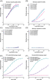
FIGURE 1
FIGURE 1
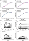
Anti‐ApoE3‐GAG antibody screening for top candidates. (A‐D) Screening of hybridoma cell supernatants via ELISA to test binding to peptides representing HSPG‐binding domains of ApoE3 (A), full‐length (FL) ApoE3 (B), ApoE3Ch peptide of ApoE3‐HSPG domain (C), and FL ApoE3Ch (D). Data show that all clones bind to HSPG region (ApoE peptide) with similar affinity (A) compared to binding between clones to the FL protein, in which clone 7C3.Ab (light purple) was the strongest, followed by 7C11.Ab (cyan), 1D6.Ab (green), 1H4.Ab (red), and 3A6.Ab (purple). Conversely, none of the antibodies tested showed binding for ApoE3Ch peptide (C) or FL ApoE3Ch (D), confirming the selectivity for the HSPG‐binding region of ApoE3. Binding expressed as averaged binding percentage normalized to the maximum intensity of absorbance as a function of logarithm of dilution factor ± SEM of three repeated measures. (E and F) Binding analysis via ELISA of purified monoclonal antibodies 7C11.mAb (E) and 1H4.mAb (F), using either FL protein (ApoE3 in purple and ApoE3Ch in red) or the amino acid sequence of the HSPG‐binding region (ie, peptide) of both ApoE3 (in green) and ApoE3Ch (in cyan). Data show differential binding for clones for either full‐length or small peptides of ApoE3 and ApoE3Ch. Binding analyses shown in panels (E) and (F) are expressed as averaged binding percentage normalized to the maximum intensity of absorbance over logarithmic nanomolar concentration (Log C) ± SEM of n = 3 repeated measurements.
FIGURE 2
FIGURE 2
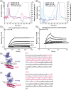
Assaying selectivity of anti‐ApoE‐GAG antibodies for ApoE variants of the novel ApoE antibodies. Titration of anti‐ApoE3 antibodies 7C11 (A), 1H4 (B), a commercially available anti‐ApoE antibody clone D6E10 (C), and a commercially available antibody anti‐His tag antibody (D). The assay was performed using a constant concentration of recombinant His‐tagged ApoE variants (ApoE2 in black; ApoE3Ch in cyan; ApoE3 in red; ApoE4 in purple). Data are expressed as binding percentage normalized to the maximum absorbance over logarithmic nM concentration and suggests that the 7C11 and 1H4 anti‐ApoE3‐HSPGs antibodies are more selective for ApoE2 and ApoE4 variants. (A‐D) All data are expressed as average binding percentage obtained from three independent experiments. (E‐H) Biolayer interferometry (BLI) binding analysis of ApoE3 (E), ApoE4 (F), ApoE2 (G), ApoE3Ch (H), and to immobilized 7C11 antibody to an AMC chip showing that 7C11 has the highest binding affinity for ApoE4, whereas it was not possible to measure binding affinity to ApoE3Ch. n.m. ≡ not measurable.
FIGURE 3
FIGURE 3
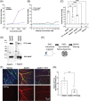
Affinity and in silico analysis of 7C11.mAb antibody. (A, B) Representative chromatograms of ApoE3 1.47 μM (magenta) and ApoE3 1.47 μM incubated with 7C11.mAb 0.6 μM (blue, A) or ApoE4 1.47 μM (purple) and ApoE4 1.47 μM incubated with 7C11.mAb 0.6 μM (light blue, B). Data show that 0.6 μM concentration of 7C11.mAb has a strong inhibitory effect on the heparin binding of ApoE3 and ApoE4, as confirmed by the shift of pmax at lower retention times and reduction of the peak intensity as compared to ApoE alone. All chromatograms are expressed as normalized intensities to the maximum intensity of emission over time in minutes (min) and are representative of n = 3 independent measurements. To compare the changes in ApoE, the 7C11.mAb chromatographic profile was subtracted. The percentage of 0.8 M NaCl over the salt gradient is represented by the dotted line. (C) BLI binding analysis of ApoE4 to immobilized 7C11.chIgG1 antibody to an anti‐human IgG Fc (AHC) chip showing that 7C11.chIgG1 binding affinity for ApoE4 with a KD < 10‐12 M. (D) Binding analysis via surface plasmon resonance (SPR) of the Fab fragment of the 7C11.mAb antibody expressed as response difference over time of increasing concentrations of ApoE4 tested with immobilized Fab from antibody 7C11.mAb. (E, F) Representative in silico docking analysis of 7C11‐Fab antibody (in purple) to ApoE3 (E) or ApoE3Ch (F) N‐terminal region (in magenta). Polar contacts between the antibody and ApoE are represented in cyan. ApoE sequence for both ApoE3 and ApoE3Ch used for in silico analysis is reported next to the structures and highlighted in magenta; polar contacts within the sequence are highlighted in cyan.
FIGURE 4
FIGURE 4
Introducing the Christchurch (Ch) mutation in ApoE4 or mimicking reduced GAG binding rescues cytotoxicity in vitro and tau pathology in vivo. (A) Cytotoxicity percentage of increasing concentrations of ApoE4 (purple) and ApoE4Ch (blue) used to treat for 24 h SH‐SY5Y cells showing the loss of toxicity via LDH assay in the presence of the Ch mutation. (B) Cytotoxicity assay of increasing doses of 7C11 human chimeric antibody (7C11.chIgG1) showing that it is not cytotoxic compared to IgG1 control. (C) Cytotoxicity assay comparing 1 μM ApoE4‐treated cells to 1 μM ApoE4 co‐administered with either 7C11 human chimeric antibody (7C11.chIgG1 6.7 nM) or heparin 13 nM showing that ApoE4‐derived cytotoxicity is significantly reduced in vitro (*p = .0147, unpaired two‐tailed t‐test, n = 4 biological replicates). Neither heparin nor 7C11 were cytotoxic. Data were normalized by considering ApoE4 cytotoxicity as 100%. (D) Representative far‐western blotting of ApoE3 and ApoE4 showing that in the absence of heparin (left blots), 7C11 binds more to ApoE compared to blots probed in the presence of heparin (right blots). Total levels of ApoE were not affected when membranes were probed with anti‐ApoE antibody (botton blots). (E) Pictographic representation of experimental design of in vivo intravitreal injections of ApoE variants or vehicle or ApoE3 + 7C11.mAb performed on day 1, euthanasia and retina dissection from the collected eyes on day 3, and staining on day 4 after injection. (F) Representative immunofluorescence staining of dissected retina injected intravitreally with 2 μL of vehicle (PBS), ApoE3 1.47 μM, or ApoE3 1.47 μM + 7C11.mAb 6 μM, showing reduction of retinal damage and inflammation, as shown by increased levels of isolectin staining (green) and AT8‐tau staining (in red) in the presence of 7C11.mAb antibody. For all images, the top panel is a composite of staining obtained with DAPI to detect nuclei (blue) and AT8‐tau to detect hyperphosphorylated tau accumulation (AT8, red), isolectin B4 (green) to detect vasculature. The red channel is also displayed in the bottom panel. Scale bar = 50 μm. (G) Quantification of the relative AT8‐positive tau levels normalized to retinal ganglion cell (RGC) area in ApoE3 and ApoE3 + 7C11.mAb‐treated retinas showing a significantly reduced AT8‐tau staining in the presence of 7C11.mAb compared to ApoE3 alone (**p = .0042, unpaired two‐tailed t‐test, n = 3, 4 biological replicates).
FIGURE 5
FIGURE 5
In vivo and human neuropathology characterization of new anti‐ApoE‐HSPG antibodies. (A) Pictographic representation of experimental design of intraperitoneal (ip) injections of WT and APOE4 KI mice to determine brain penetration of Alexa‐647‐labeled antibodies (day 1, 50 mg/kg) or WT and APOE4 KI mice to determine changes in pTau levels (days 1 to 4). (B) Representative staining of hippocampal regions of APOE4 KI mice ip injected with either vehicle (PBS), ms.IgG‐Alexa647, or ms.7C11‐Alexa647 for 24 h, 50 mg/kg. Nuclei are stained with DAPI in blue. Scale bar = 200 μm. (C) Image analysis of injected brains shown in panel (B) was performed by measuring fluorescence intensity (FI) on 647‐nm channel. Higher FI of both ms.IgG1‐Alexa647 (p < .001, one‐way ANOVA) and 7C11.mAb‐Alexa647 (p < .001, one‐way ANOVA) injected groups was observed compared to vehicle. (D) Representative images of DAPI and pTau (S396) staining in APOE4 KI mice injected ip with either ms.IgG1 or 7C11.mAb, compared to negative control (secondary only antibody). Scale bar = 50 μm. (E) Representative coronal section stained with DAPI showing with a red square the anatomical region selected for the analysis presented in panel (F). Scale bar = 1 mm. (F) APOE4 KI mice treated with 7C11 antibody showed a reduced number of pTau (S396)‐positive cells (p = .0286, one‐tailed Mann‐Whitney test). (B, D, E) Nuclei are stained with DAPI in blue. (G) Representative immunohistochemistry staining of ApoE levels on paraffin‐embedded temporal cortex specimens from PSEN1 E280A carriers with different APOE genotypes: APOEε3/ε3 (first row), APOEε3/ε3Ch (second row), APOEε3Ch/ε3Ch (third row), APOEε4/ε4 (last row). ApoE staining performed (from left to right) with anti‐ApoE‐E6D7, 1H4.mAb, 7C11.mAb, and 19G.mAb. Comparison of different staining patterns suggesting that, unlike the anti‐ApoE‐E6D7, which homogeneously stains ApoE variants throughout the tissue, 1H4.mAb preferentially stains ApoE3 and ApoE4 located either in the neuronal tissue or surrounding the vessels; 7C11.mAb preferentially detects ApoE3 and ApoE4 localized in deposits and surrounding the vessels, and 19G.mAb selectively stains ApoE3Ch. For each specimen analyzed, the region of the cerebral cortex used for the staining is reported in the bottom left boxes. Scale bar = 100 μm. Regions of interest in panels (B), (D), (G) are pointed with triangles.

References

    1. Jiwaji Z, Tiwari SS, Aviles‐Reyes RX, et al. Reactive astrocytes acquire neuroprotective as well as deleterious signatures in response to Tau and Ass pathology. Nat Commun. 2022;13(1):135. - PMC - PubMed
    1. Neddens J, Temmel M, Flunkert S, et al. Phosphorylation of different tau sites during progression of Alzheimer's disease. Acta Neuropathol Commun. 2018;6(1):52. - PMC - PubMed
    1. Karlawish J, Grill JD. The approval of Aduhelm risks eroding public trust in Alzheimer research and the FDA. Nat Rev Neurol. 2021;17(9):523‐524. - PubMed
    1. Tanzi RE. FDA approval of aduhelm paves a new path for Alzheimer's disease. ACS Chem Neurosci. 2021;12(15):2714‐2715. - PubMed
    1. Sevigny J, Chiao P, Bussière T, et al. The antibody aducanumab reduces Aβ plaques in Alzheimer's disease. Nature. 2016;537(7618):50‐56. - PubMed

Publication types