. 2023 Oct 19;127(41):8736-8748.
doi: 10.1021/acs.jpcb.3c02337. Epub 2023 Oct 4.

Modulation of Structure and Dynamics of Cardiac Troponin by Phosphorylation and Mutations Revealed by Molecular Dynamics Simulations

Affiliations

Modulation of Structure and Dynamics of Cardiac Troponin by Phosphorylation and Mutations Revealed by Molecular Dynamics Simulations

Zeyu Yang et al. J Phys Chem B. .

Abstract

Adrenaline acts on β1 receptors in the heart muscle to enhance contractility, increase the heart rate, and increase the rate of relaxation (lusitropy) via activation of the cyclic AMP-dependent protein kinase, PKA. Phosphorylation of serines 22 and 23 in the N-terminal peptide of cardiac troponin I is responsible for lusitropy. Mutations associated with cardiomyopathy suppress the phosphorylation-dependent change. Key parts of troponin responsible for this modulatory system are disordered and cannot be resolved by conventional structural approaches. We performed all-atom molecular dynamics simulations (5 × 1.5 μs runs) of the troponin core (419 amino acids) in the presence of Ca2+ in the bisphosphorylated and unphosphorylated states for both wild-type troponin and the troponin C (cTnC) G159D mutant. PKA phosphorylation affects troponin dynamics. There is significant rigidification of the structure involving rearrangement of the cTnI(1-33)-cTnC interaction and changes in the distribution of the cTnC helix A/B angle, troponin I (cTnI) switch peptide (149-164) docking, and the angle between the regulatory head and ITC arm domains. The familial dilated cardiomyopathy cTnC G159D mutation whose Ca2+ sensitivity is not modulated by cTnI phosphorylation exhibits a structure inherently more rigid than the wild type, with phosphorylation reversing the direction of all metrics relative to the wild type.


Conflict of interest statement

The authors declare no competing financial interest.

Figures

Figure 1
Figure 1
(A) The effects of phosphorylation and the G159D mutation on Ca2+activation of thin filaments. The plots show the decrease in Ca2+ sensitivity upon phosphorylation and the absence of this shift (uncoupling) due to the G159D mutation, measured by an in vitro motility assay. Dotted lines, open points wild type; solid lines and points, G159D; unphosphorylated TnI, squares; phosphorylated TnI, circles. Mean and SEM are shown. The lines are fits of the data to the Hill equation. Replotted from data of Dyer et al. (B) Representative structure of the backbone of the unphosphorylated troponin core from molecular dynamics simulation. The residues are colored according to their root mean square fluctuation (RMSF), with the highest RMSF as the deepest red. The locations of the phosphorylatable serines, 22 and 23, in NcTnI (magenta arrow), cTnC G159 (blue arrow), and the switch peptide (green arrow) are indicated. Bound Ca2+ ions are colored in green. The N-terminal domains of troponin C (NcTnC) and of the ITC domain are outlined in blue. The ITC domain contains the C-terminus of TnC (94–161), helices H1 and H2 of cTnI (42–136), and helices H1 and H2 of cTnT (201–277).
Figure 2
Figure 2
Heat maps showing the key interactions between troponin peptides related to phosphorylation. The scale represents percentage of interactions observed in 37,500 frames. Selected regions of interest are shown. The complete set of interactions are shown in Supplementary Figure 6. Numerical values for the key interactions are presented in Supporting Information Table 1 with a detailed commentary. (A) Ionic interactions between NcTnI (1–34) and NcTnC (1–84). NcTnI is anchored to NcTnC via Arg9 interacting with NcTnC Gln15 and 19 and Arg 12 interacting with NcTn C Asp25 and Glu32 (orange circle). A strong interaction between NcTnC Lys33 and NcTnI Arg19, 20, and 21 is lost on phosphorylation (blue circle), while interaction with NcTnC Lys39 is gained (green circle). (B) Ionic intramolecular interaction within NcTnI (1–34 vs 1–48). Phosphorylation promotes interaction of phosphoserine 22 with Arg19, 21, and 26 (blue circle). (C–E) Key interactions of CcTnT (C) ionic interactions between CcTnT (280–288) and NcTnC (1–46). There is a medium interaction formed between NcTnC Asp33 and Arg286 upon phosphorylation (blue circle). (D) Ionic interactions between CcTnT (281–288) and NcTnI (1–40). Upon phosphorylation, CcTnT, especially Lys288, interacts with TnI Ser 22 and 23 (blue circle). (E) H-bond interactions between CcTnT (266–288) and NcTnC (1–40). Phosphorylation increases H bonding between NcTnI Arg 19, 20, and 21 and CcTnT Lys280, 287, and 288 (blue circle). (F, G) Ionic and H-bonding interactions of the switch peptide and cTnI interdomain peptides with cTnC. The switch peptide is outlined in black; the “cTnI interdomain” peptide is outlined in blue. The TnC interdomain peptide (86–95) is represented by a gray bar. (F) Ionic interaction between cTnI (126–170) and cTnC (1–109). The switch peptide is anchored by a stable interaction between NcTnC Glu19 and cTnI 161 (green circle). The interaction of NcTnC Lys43 with cTnI 164 and 169 and the interaction of cNcTnc Arg46 with cTnI Ala170 are decreased on phosphorylation (pink circle). Altered interactions of the cTnI interdomain peptide with NcTnC helices C and D are shown in the blue circle: Asp62 and Glu63 interaction with cTnI Arg140 and 145 increases, while interaction of Glu56 with cTnI Arg144 and 145 decreases. Altered interactions of the cTnC linker peptide with the cTnI interdomain peptide upon phosphorylation are indicated by the orange circle. (G) H-bond interactions between cTnI (126–170) and cTnC (1–109). The switch peptide is anchored by a stable interaction between NcTnC Glu19 and cTnI 161 (green circle). Increase of the interactions of cTnC 84 and Ser89 with cTnI Ala150 and Gly159 is highlighted by the blue circle. (H) Comparison of ionic interaction between cTnC (152–161) and cTnC (1–100) in unphosphorylated wild type and G159D. The most significant change from the wild type is the formation of a strong, ∼80%, ionic interaction between Arg83, at the bottom of the D helix and the Asp159.
Figure 3
Figure 3
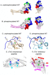
Representative structures of the cardiac troponin core for wild type and G159D in the unphosphorylated and phosphorylated states. (A, B) Representative structure of wild-type cardiac troponin with surface rendering of NcTnC (white), NcTnI (blue), switch peptide (green), and CcTnT (red). Views are from the side and the “top” of troponin. Rotating models are presented as mp4 movies, and the PDB files used to generate the structures are presented in the supplement. (A) Unphosphorylated, (B) phosphorylated. (C, D) Partial model showing the key phosphorylation-dependent interactions between NcTnI and NcTnC only. TnI Arg19–Arg26 and TnC Asp33, Thr38, and Lys39 are shown in stick format. Rotating models are presented in the supplement. (C) Unphosphorylated, (D) phosphorylated. (E) The interdomain peptide of cTnC in unphosphorylated (cyan) and phosphorylated (blue) states. Phosphorylated and unphosphorylated representative structures are aligned to the IT coiled coil. The interdomain peptide (86–95), helix D, and part of helix E are highlighted to illustrate the lengthening and twisting of the peptide upon phosphorylation. Distribution of interdomain peptide lengths is shown in Supplement Figure 5 and quantified in Table 1. (F) The cTnI interdomain peptide (136–148) and switch peptide (149–164) are highlighted in unphosphorylated (magenta) and phosphorylated (red) states. TnC helix C is highlighted in light green (uP) and dark green (P). Phosphorylated and unphosphorylated representative structures are aligned to the IT coiled coil. The model is oriented to show the change in the NcTnc-ITC domain angle.
Figure 4
Figure 4
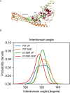
(A) Definition of the interdomain angle. Head: the average of C-alpha coordinates of TnC3–85; fulcrum: the average of C-alpha coordinates of TnC94–157; end of IT arm: the average of C-alpha coordinates of TnT241–251 and TnI69–76. (B) Probability distribution of the angle between the NcTnC and ITC domains.
Figure 5
Figure 5
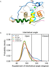
(A) Model of the backbone structure of NcTnC showing the relative disposition of helices A and B in the unphosphorylated state. (B) The distribution of the interhelical angle formed by helices A and B in the unphosphorylated and phosphorylated (SEP) states for the wild-type and cTnC G159D-mutant troponin. The definition of open and closed angles is adopted from Lindert et al.

Similar articles

Cited by

References

    1. Layland J.; Solaro R. J.; Shah A. M. Regulation of cardiac contractile function by troponin I phosphorylation. Cardiovasc. Res. 2005, 66 (1), 12–21. 10.1016/j.cardiores.2004.12.022. - DOI - PubMed
    1. Messer A. E.; Marston S. B. Investigating the role of uncoupling of Troponin I phosphorylation from changes in myofibrillar Ca2+-sensitivity in the pathogenesis of Cardiomyopathy. Front. Physiol. 2014, 5, 315.10.3389/fphys.2014.00315. - DOI - PMC - PubMed
    1. Dong W. J.; Jayasundar J. J.; An J.; Xing J.; Cheung H. C. Effects of PKA phosphorylation of cardiac troponin I and strong crossbridge on conformational transitions of the N-domain of cardiac troponin C in regulated thin filaments. Biochemistry 2007, 46, 9752–9761. 10.1021/bi700574n. - DOI - PMC - PubMed
    1. Little S. C.; Biesiadecki B. J.; Kilic A.; Higgins R. S. D.; Janssen P. M. L.; Davis J. P. The rates of Ca2+ dissociation and cross-bridge detachment from ventricular myofibrils as reported by a fluorescent cardiac Troponin C. J. Biol. Chem. 2012, 287, 27930–27940. 10.1074/jbc.M111.337295. - DOI - PMC - PubMed
    1. Marston S. B. How do mutations in contractile proteins cause the primary familial cardiomyopathies?. J. Cardiovasc. Transl. Res. 2011, 4 (3), 245–255. 10.1007/s12265-011-9266-2. - DOI - PubMed

Publication types