. 2023 Nov 10;51(20):11358-11374.
doi: 10.1093/nar/gkad752.

Systematic sequence engineering enhances the induction strength of the glucose-regulated GTH1 promoter of Komagataella phaffii

Affiliations

Systematic sequence engineering enhances the induction strength of the glucose-regulated GTH1 promoter of Komagataella phaffii

Mirelle Flores-Villegas et al. Nucleic Acids Res. .

Abstract

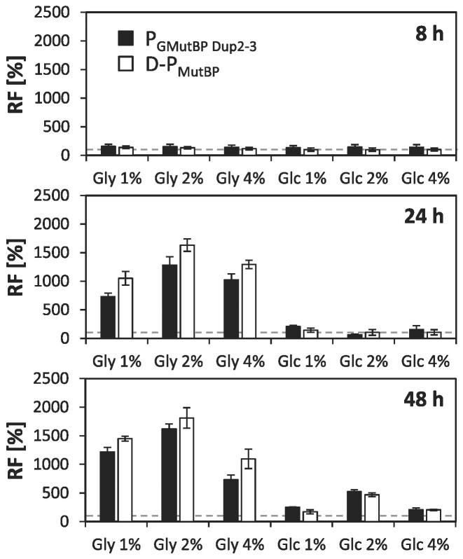
The promoter of the high-affinity glucose transporter Gth1 (PGTH1) is tightly repressed on glucose and glycerol surplus, and strongly induced in glucose-limitation, thus enabling regulated methanol-free production processes in the yeast production host Komagataella phaffii. To further improve this promoter, an intertwined approach of nucleotide diversification through random and rational engineering was pursued. Random mutagenesis and fluorescence activated cell sorting of PGTH1 yielded five variants with enhanced induction strength. Reverse engineering of individual point mutations found in the improved variants identified two single point mutations with synergistic action. Sequential deletions revealed the key promoter segments for induction and repression properties, respectively. Combination of the single point mutations and the amplification of key promoter segments led to a library of novel promoter variants with up to 3-fold higher activity. Unexpectedly, the effect of gaining or losing a certain transcription factor binding site (TFBS) was highly dependent on its context within the promoter. Finally, the applicability of the novel promoter variants for biotechnological production was proven for the secretion of different recombinant model proteins in fed batch cultivation, where they clearly outperformed their ancestors. In addition to advancing the toolbox for recombinant protein production and metabolic engineering of K. phaffii, we discovered single nucleotide positions and correspondingly affected TFBS that distinguish between glycerol- and glucose-mediated repression of the native promoter.


Figures

Graphical Abstract
Graphical Abstract
Figure 1.
Figure 1.
Schematic workflow of the random mutagenesis approach leading to the K. phaffii GTH1 promoter library. (A) Random mutagenesis strategy: EP-PCR was conducted on the main regulatory region of PGTH1 in three consecutive steps. Subsequently, the EP-PCR product was re-cloned into the appropriate position in the PGTH1-EGFP expression vector, resulting in the PGTH1-mut-EGFP plasmid library, which was then transformed into K. phaffii X33. (B) The average error rate and nucleotide exchange profile of the generated K. phaffii PGTH1-mut library 2 is based on sequencing of 56 randomly selected clones. Error bars represent the 95% confidence interval.
Figure 2.
Figure 2.
Relative EGFP fluorescence intensity and schematic representation of the identified point mutations. (A) Relative fluorescence levels (RF) of the top seven promoter variants re-cloned into Golden PiCS and transformed into K. phaffii CBS2612. Six independent clones per variant were analyzed and compared to four replicates of the PGTH1 control strain. Outliers were excluded from the analysis. Bars represent mean values and closed circles the calculated RF values of the individual clones. Cells were grown in the 24-DWP format in inducing (limiting glucose) and repressing (excess glycerol) conditions and EGFP levels measured on a flow cytometer. Relative fluorescence levels were calculated by normalizing to the PGTH1 control. The horizontal dotted line highlights the average expression level of the native PGTH1 control strain (set to 100% for each condition). Statistical analysis was done employing Student t-test (**P≤ 0.05; ***P≤ 0.005). (B) Schematic representation of the point mutations and their respective location identified in the five promoter variants for which an increased induction strength could be confirmed based on the results shown in (A). The red colored mutations are located outside of a TFBS. Further information regarding the identified mutations as well as sequence alignment of the relevant region is provided in Supplementary Table S4 and Supplementary Figure S3 in Supporting File 1.
Figure 3.
Figure 3.
Impact of single point mutations creating additional CSRE or reducing MIG-TFBS on expression properties of PGTH1. (A) Sequence information on the point mutations identified in the PGS1, PGS4 and PGS5 variant in the −453 to −445 bp region which lead to the creation of an additional F$CSRE binding site and their recreation by point mutations MutA and MutB. (B) Relative EGFP fluorescence levels (RF) of at least seven independent clones per point mutation variant as compared to at least four replicates of the PGTH1 control strain. Outliers were excluded from the analysis. Bars represent mean values and closed circles the calculated RF values of individual clones. Cells were cultivated in the 24-DWP screening format in either inducing (limiting glucose) or repressing (excess glycerol) conditions. Fluorescence was measured on a flow cytometer. Statistical analysis was done employing Student t-test (**P≤ 0.05; ***P≤ 0.005). The horizontal dotted line highlights the average expression level of native PGTH1 (set to 100% for each condition). Vertical lines separate point mutations that create an additional CSRE and the deletion of one MIG site, respectively.
Figure 4.
Figure 4.
Duplication of the main regulatory promoter region increases induction strengths of all promoter variants. (A) Schematic representation of the duplication of the main regulatory region of PGTH1 and its mutated variants. To indicate the duplication, the prefix ‘D-’ was added to the promoter variant name. (B) Relative EGFP fluorescence levels (RF) of D-PGTH1 compared to native PGTH1 and (C) RF of the ‘duplicated’ sorting (D-PGSn) and ‘duplicated’ single point mutation (D-PGMutX) variants compared to the ‘duplicated’ control D-PGTH1. Cells were cultivated in the 24-DWP screening format in either inducing (X, limiting glucose) or repressing (G, excess glycerol) conditions. Fluorescence was measured on a flow cytometer. Per variant at least four independent clones were analyzed, while for the D-PGTH1 control at least six independent clones per screening were tested. Outliers were excluded from the analysis. Bars represent mean values and closed circles the calculated RF values of individual clones. Statistical analysis was done employing Student t-test (**P≤ 0.05; ***P≤ 0.005). The dotted line highlights the average expression level of the appropriate control (PGTH1 for (B) and D-PGTH1 for (C), respectively; set to 100% for each condition).
Figure 5.
Figure 5.
Sliding window deletions guide promoter engineering by segmental duplications. (A) Schematic representation of the segmental deletions introduced to the −200 to −400 bp region of PGTH1. Each deletion is 30 bp long and overlaps with the respective contiguous variants for 10 bps on each side. (B) Relative EGFP fluorescence levels (RF) of the different segmental deletion variants compared to PGTH1. (C) Schematic representation of the duplications or triplication of the key regions Del2-3 as well as Del5-7 and (D) respective relative EGFP fluorescence levels (RF) of the corresponding promoter variants compared to PGTH1. Cells for expression screenings were cultivated in the 24-DWP format in either inducing (limiting glucose) or repressing (excess glycerol) conditions. Per variant at least five independent clones were screened and compared to at least three independent cultivations of the PGTH1 control strain. Outliers were excluded from the analysis. Bars represent mean values and closed circles the calculated RF values of individual clones. Fluorescence was measured by flow cytometry. Statistical analysis was done employing Student t-test (**P≤ 0.05; ***P≤ 0.005). The dotted line highlights the average expression level of the PGTH1 control (set to 100% for each condition).
Figure 6.
Figure 6.
Combinatorial promoter variants. (A) Schematic representation of the promoter variants for which the segmental duplications and triplications as well as the most successful point mutations were combined. (B) Relative EGFP fluorescence levels (RF) of D-PGTH1 as well as the different combinatorial variants compared to PGTH1. Per variant at least seven independent clones were compared to at least nine individual cultivations of the PGTH1 control strain. Outliers were excluded from the analysis. Bars represent mean values and closed circles the calculated RF values of individual clones. Cells for expression screenings were cultivated in the 24-DWP format in either inducing (limiting glucose) or repressing (excess glycerol) conditions. Fluorescence was measured by flow cytometry. Statistical analysis was done employing Student t-test (***P≤ 0.005). The dotted line highlights the average expression level of the PGTH1 control (set to 100% for each condition).
Figure 7.
Figure 7.
De-repression of promoter variants was evident in glycerol excess but not in glucose excess conditions. Relative EGFP fluorescence levels (RF) of the promoter variants PGMutBP Dup2-3 and PGMutBP compared to D-PGTH1. Cells were cultivated in 24-deep-well-plates in minimal media supplemented with 1%, 2% or 4% glycerol (Gly) or glucose (Glc), respectively. Fluorescence levels were measured after 8, 24 and 48 h by flow cytometry. Per variant, four independent clones were analyzed. Bars represent mean values and error bars indicate standard deviation of the mean. The dotted line highlights the average expression level of the D-PGTH1 control (set to 100% for each condition).
Figure 8.
Figure 8.
The novel promoter variants increase the production of secreted recombinant proteins in small scale screenings. Secreted protein yield fold-changes obtained for different promoter variants driving the expression of human serum albumin (HSA) or a single chain variable fragment (scFv) in comparison to the respective D-PGTH1 control. Per promoter variant at least four (typically six or more) independent clones were compared against at least four independent cultivations of the D-PGTH1 control strain. Outliers were excluded from the analysis. Bars represent mean values and open circles the calculated secreted protein yield of individual clones. Yields are based on extracellular recombinant protein titer and WCW measurements after 48 hours of incubation under limiting glucose (inducing) conditions in the 24-DWP format. Statistical analysis was done employing Student t-test (**P≤ 0.05; ***P≤ 0.005). The dotted line highlights the average yield of the respective D-PGTH1 control (set to 100%).
Figure 9.
Figure 9.
Secreted protein production by different promoter variants in glucose-based fed batch cultivations. Representative profiles of the measured (A) HSA and (B) scFv titers and biomass (YDM) concentrations throughout the glucose-limited fed-batch production phase as well as the titer fold-change (titer-FC) of the secreted protein calculated between the respective D-PGTH1 control and the various promoter variants at the corresponding feed-phase sampling point. Secreted protein titers were measured in technical duplicates and corrected for the biomass concentration. If multiple fed-batch cultivations were conducted for a given variant, mean values were used to calculate the titer fold-change. The dotted horizontal line highlights the titers obtained for the respective D-PGTH1 control.

Similar articles

Cited by

References

    1. Guarente L., Yocum R.R., Gifford P.. A GAL10-CYC1 hybrid yeast promoter identifies the GAL4 regulatory region as an upstream site. Proc. Natl. Acad. Sci. U.S.A. 1982; 79:7410–7414. - PMC - PubMed
    1. Cazier A.P., Blazeck J.. Advances in promoter engineering: novel applications and predefined transcriptional control. Biotechnol. J. 2021; 16:e2100239. - PubMed
    1. Alper H., Fischer C., Nevoigt E., Stephanopoulos G.. Tuning genetic control through promoter engineering. Proc. Natl. Acad. Sci. U.S.A. 2005; 102:12678–12683. - PMC - PubMed
    1. Xu N., Wei L., Liu J.. Recent advances in the applications of promoter engineering for the optimization of metabolite biosynthesis. World J. Microbiol. Biotechnol. 2019; 35:33. - PubMed
    1. He S., Zhang Z., Lu W.. Natural promoters and promoter engineering strategies for metabolic regulation in Saccharomyces cerevisiae. J. Ind. Microbiol. Biotechnol. 2023; 50:kuac029. - PMC - PubMed

Publication types

MeSH terms

Supplementary concepts