. 2023 Nov 10;51(20):e100.
doi: 10.1093/nar/gkad784.

A generalizable scaffold-based approach for structure determination of RNAs by cryo-EM

Affiliations

A generalizable scaffold-based approach for structure determination of RNAs by cryo-EM

Conner J Langeberg et al. Nucleic Acids Res. .

Abstract

Single-particle cryo-electron microscopy (cryo-EM) can reveal the structures of large and often dynamic molecules, but smaller biomolecules (≤50 kDa) remain challenging targets due to their intrinsic low signal to noise ratio. Methods to help resolve small proteins have been applied but development of similar approaches to aid in structural determination of small, structured RNA elements have lagged. Here, we present a scaffold-based approach that we used to recover maps of sub-25 kDa RNA domains to 4.5-5.0 Å. While lacking the detail of true high-resolution maps, these maps are suitable for model building and preliminary structure determination. We demonstrate this method helped faithfully recover the structure of several RNA elements of known structure, and that it promises to be generalized to other RNAs without disturbing their native fold. This approach may streamline the sample preparation process and reduce the optimization required for data collection. This first-generation scaffold approach provides a robust system to aid in RNA structure determination by cryo-EM and lays the groundwork for further scaffold optimization to achieve higher resolution.


Figures

Graphical Abstract
Graphical Abstract
Figure 1.
Figure 1.
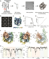
Workflow of the scaffold-based approach and identification of a suitable scaffold.(A) The construct has the RNA of interest (red) appended to the larger group I intron RNA. After transcription and purification, the pure RNA is subjected to a standard RNA folding protocol, applied to cryo-EM grids and vitrified. Data collection and processing yields a map for model building and refinement. (B) Representative 2D class averages of the TetP6b construct. (C) Local resolution map of the recovered TetP6b construct. (D) Fourier Shell correlation graph of the TetP6b construct with tight mask (dashed line) and noise subtracted (solid line). (E) Henderson-Rosenthal plot of the TetP6b construct. (F) Recovered map of the TetP6b construct. (G) Resulting atomic model of the TetP6b construct. (H) Per-nucleotide Q-score graph of the TetP6b construct. (I) Map-to-model cross-correlation score graph of the TetP6b construct.
Figure 2.
Figure 2.
Engineering of the Zika Virus xrRNA-containing test RNA.(A) Secondary structures of the TetP6b RNA and the Zika Virus xrRNA (shaded blue) used in this study. The two RNAs were appended at the locations boxed in grey and designated with red lines. In the xrRNA, P2 can be altered without affecting xrRNA function, therefore the apical loop of this stem was removed and became the point of attachment. (B) Cartoon secondary structure depictions of the chimeric RNA, with the xrRNA in blue. (C) Close-up of the engineered region.
Figure 3.
Figure 3.
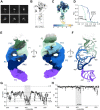
Cryo-EM map of the Zika Virus xrRNA appended onto a scaffold. (A) Representative 2D class averages of the Zika Virus xrRNA + TetP6b. (B) Cryo-EM map indicating the location of the Zika Virus xrRNA in the overall initial map. (C) Local resolution map of the chimeric Zika Virus xrRNA construct. (D) Fourier Shell correlation graph of the Zika Virus xrRNA construct with global resolution (blue) and the local xrRNA resolution (teal) using tight mask (dashed line) and noise subtracted (solid line). (E) Recovered map of the chimeric Zika Virus xrRNA construct. (F) Resulting atomic model of the chimeric Zika Virus xrRNA construct. A visible magnesium ion is shown as a bright green sphere. (G) Per-nucleotide Q-score graph of the TetP6b construct. (H) Map-to-model cross-correlation score graph of the TetP6b construct.
Figure 4.
Figure 4.
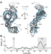
Comparison of cryo-EM-derived model with the X-ray crystallography structure of the Zika Virus xrRNA. (A) Overlay of the cryo-EM-based model of the Zika Virus xrRNA (grey) and the crystallography-based model of the Zika Virus xrRNA (cyan). (B) Per-nucleotide backbone RMSD graph of both Zika Virus xrRNA structures. The average backbone RMSD across the entire RNA was calculated at 3.0 Å. When P4 is excluded this decreases to 2.1 Å. The RMSD of the L2 loop is omitted as the analogous nucleotides are not present in the cryo-EM model.
Figure 5.
Figure 5.
Additional examples of two ∼17 kDa RNA structures recovered using cryo-EM. (A) Secondary structure depiction of the TABV xrRNA appended onto TetP6b. (B) Local resolution map of the recovered chimeric TABV xrRNA construct. (C) Recovered map of the chimeric TABV xrRNA construct. (D) Resulting atomic model of the chimeric Zika Virus xrRNA construct. (E) Secondary structure depiction of the chimeric Thermotoga petrophila Fluoride riboswitch appended onto TetP6b. (F) Local resolution map of the recovered Fluoride riboswitch construct. (G) Recovered map of the chimeric Fluoride riboswitch construct. (H) Resulting atomic model of the chimeric Fluoride riboswitch construct.

Update of

References

    1. He W., Henning-Knechtel A., Kirmizialtin S.. Visualizing RNA structures by SAXS-driven MD simulations. Front. Bioinform. 2022; 2:781949. - PMC - PubMed
    1. Chen Y., Pollack L.. SAXS studies of RNA: structures, dynamics, and interactions with partners. Wiley Interdiscip. Rev. RNA. 2016; 7:512–526. - PMC - PubMed
    1. Christy T.W., Giannetti C.A., Houlihan G., Smola M.J., Rice G.M., Wang J., Dokholyan N.V., Laederach A., Holliger P., Weeks K.M.. Direct mapping of higher-order RNA interactions by SHAPE-JuMP. Biochemistry. 2021; 60:1971–1982. - PMC - PubMed
    1. Yang S., Parisien M., Major F., Roux B.. RNA structure determination using SAXS data. J. Phys. Chem. B. 2010; 114:10039–10048. - PMC - PubMed
    1. Nakane T., Kotecha A., Sente A., McMullan G., Masiulis S., Brown P., Grigoras I.T., Malinauskaite L., Malinauskas T., Miehling J.et al.. Single-particle cryo-EM at atomic resolution. Nature. 2020; 587:152–156. - PMC - PubMed

Publication types