. 2023 Dec 12;11(6):e0197523.
doi: 10.1128/spectrum.01975-23. Epub 2023 Oct 4.

Common scab disease: structural basis of elicitor recognition in pathogenic Streptomyces species

Affiliations

Common scab disease: structural basis of elicitor recognition in pathogenic Streptomyces species

Frédéric Kerff et al. Microbiol Spectr. .

Abstract

Common scab is a disease caused by a few Streptomyces species that affects important root and tuber crops including potato, beet, radish, and parsnip, resulting in major economic losses worldwide. In this work, we unveiled the molecular basis of host recognition by these pathogens by solving the structure of the sugar-binding protein CebE of Streptomyces scabiei in complex with cellotriose, the main elicitor of the pathogenic lifestyle of these bacteria. We further revealed that the signaling pathway from CebE-mediated transport of cellotriose is conserved in all pathogenic species except Streptomyces ipomoeae, which causes soft rot disease in sweet potatoes. Our work also provides the structural basis of the uptake of cellobiose and cellotriose in saprophytic Streptomyces species, the first step activating the expression of the enzymatic system degrading the most abundant polysaccharide on earth, cellulose.

Keywords: Streptomyces; carbohydrate metabolism; elicitor binding; host-pathogen interaction; ligand-protein interaction; plant pathogens; sugar transport.


Conflict of interest statement

The authors declare no conflict of interest.

Figures

Fig 1
Fig 1
Overall structure of the CebE/cellotriose complex. Cartoon representation of CebEscab with Domains 1 and 2 in blue and green, respectively. The three hinge regions are colored in magenta. The closed cavity occupied by cellotriose (gray sticks) is shown as a yellow transparent surface.
Fig 2
Fig 2
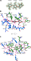
Substrate binding site of the CebE:cellotriose complex. (A) OMIT electron density map displayed at 1 σ level around cellotriose. (B) Interactions stabilizing cellotriose (gray sticks), residues from Domains 1 and 2 are displayed as blue and green sticks, respectively, water molecules as small red spheres, and H-bonds as magenta dashed lines. (C) Same as panel (B) with a 90° rotation around a horizontal axis.
Fig 3
Fig 3
Conformational change of CebE upon cellotriose binding. Superimposition of the CebEscab:cellotriose structure (colored with a gradient from the white N-terminus to the blue C-terminus) to the KfSBP structure in an open conformation (colored with a gradient from the white N-terminus to the red C-terminus); only Domain 1 residues were used to calculate the superimposition. Note the ±40° angle corresponding to the movement of Domain 2 upon cellotriose binding.
Fig 4
Fig 4
Type of cellotriose/CebE-mediated signaling pathways to thaxtomin production. 1, the values refer to the amino acid identity expressed in percentage compared to the proteins of S. scabiei 87–22 used as reference sequences. 2, the values refer to the amino acid identity expressed in percentage compared to the CebE proteins of S. scabiei 87–22 (WP_013003368.1), S. reticuli (CAB46342.1), and S. griseus (WP_012379731.1). 3, the values refer to the score obtained for a 14-nt sequence according to the position weight matrix generated from the experimentally validated CebR-binding sites (23 is the maximum score corresponding to the 14-nt TGGGACGCGTCCCA palindromic sequence).
Fig 5
Fig 5
Substrate binding site of the CebE:cellotriose complex superimposed to the CebEreti model. (A) Interactions of residues from Domain 1 (blue sticks) stabilizing cellotriose (gray sticks) superimposed to their equivalent in CebEreti (yellow sticks), water molecules are displayed as small red spheres, and H-bonds as magenta dashed lines. (B) Same as panel (A) for Domain 2 (green sticks) with a rotation of approximately 180° around a horizontal axis.
Fig 6
Fig 6
Sequence alignment of CebE proteins from model Streptomyces pathogenic species with amino acid numbering of CebEscab. Secondary structure elements of the CebEscab:cellotriose structure are schematized above the alignment with the same domain coloring code as in Fig. 1B (Domain 1 in blue and Domain 2 in green), whereas hinge regions are identified with magenta ellipses below the alignment. Residues strictly conserved are shown in red, and those directly interacting with cellotriose or indirectly interacting with cellotriose via a water molecule are highlighted in yellow (or orange when the short and bulky side chains are switched). Stars indicate direct interaction with the ligand. CebE proteins reference IDs: scabiei_87–22 (C9Z451; WP_013003368.1); griseiscabiei_NRRL_B-2795 (MBZ3900963.1); brasiliscabiei_IBSBF2867 (WP_216591689); europaeiscabiei_NRRL_B-24443 (WP_046704818.1); stelliscbiei_NRRL_B-24447 (WP_046918411.1); acidiscabieis_98–48 (WP_075734941.1); niveiscabies_NRRL_B-24457 (WP_055721858.1); acidiscabieis_NRRL_B16521 (WP_029183343.1); acidiscabieis_NCPPB4445 (WP_050369574); caniscabiei_NRRL_B-24093 (WP_060884585.1); AMCC400023 (WP_045557721.1); reticuli_CebE1 (Q9 × 9R7; CAB46342.1); reticuliscabiei_NRRL_B-24446 (WP_059073075.1); and turgidiscabies_Car8 (ELP70267.1).

References

    1. Liu H, Carvalhais LC, Crawford M, Singh E, Dennis PG, Pieterse CMJ, Schenk PM. 2017. Inner plant values: diversity, colonization and benefits from endophytic bacteria. Front Microbiol 8:2552. doi:10.3389/fmicb.2017.02552 - DOI - PMC - PubMed
    1. Berlemont R, Martiny AC. 2013. Phylogenetic distribution of potential cellulases in bacteria. Appl Environ Microbiol 79:1545–1554. doi:10.1128/AEM.03305-12 - DOI - PMC - PubMed
    1. Book AJ, Lewin GR, McDonald BR, Takasuka TE, Wendt-Pienkowski E, Doering DT, Suh S, Raffa KF, Fox BG, Currie CR, Hillis DM. 2016. Evolution of high cellulolytic activity in symbiotic Streptomyces through selection of expanded gene content and coordinated gene expression. PLOS Biol 14:e1002475. doi:10.1371/journal.pbio.1002475 - DOI - PMC - PubMed
    1. Book AJ, Lewin GR, McDonald BR, Takasuka TE, Doering DT, Adams AS, Blodgett JAV, Clardy J, Raffa KF, Fox BG, Currie CR. 2014. Cellulolytic Streptomyces strains associated with herbivorous insects share a phylogenetically linked capacity to degrade lignocellulose. Appl Environ Microbiol 80:4692–4701. doi:10.1128/AEM.01133-14 - DOI - PMC - PubMed
    1. Saini A, Aggarwal NK, Sharma A, Yadav A. 2015. Actinomycetes: a source of lignocellulolytic enzymes. Enzyme Res 2015:279381. doi:10.1155/2015/279381 - DOI - PMC - PubMed

MeSH terms

LinkOut - more resources