

Radiomics-Based Prediction of Anti-VEGF Treatment Response in Neovascular Age-Related Macular Degeneration With Pigment Epithelial Detachment
- PMID: 37792693
- PMCID: PMC10565708
- DOI: 10.1167/tvst.12.10.3
Radiomics-Based Prediction of Anti-VEGF Treatment Response in Neovascular Age-Related Macular Degeneration With Pigment Epithelial Detachment
Abstract
Purpose: Machine learning models based on radiomic feature extraction from clinical imaging data provide effective and interpretable means for clinical decision making. This pilot study evaluated whether radiomics features in baseline optical coherence tomography (OCT) images of eyes with pigment epithelial detachment (PED) associated with neovascular age-related macular degeneration (nAMD) can predict treatment response to as-needed anti-vascular endothelial growth factor (VEGF) therapy.
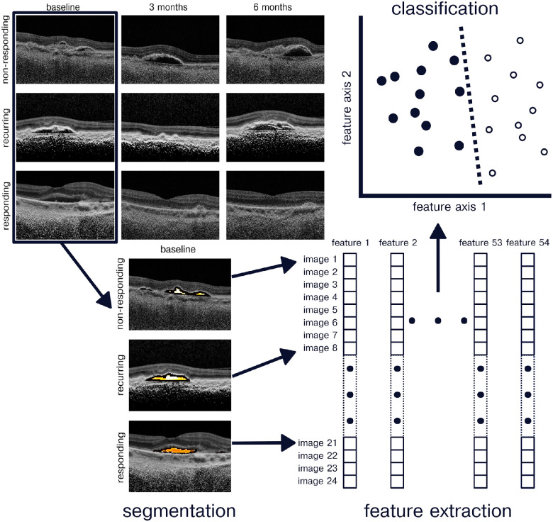
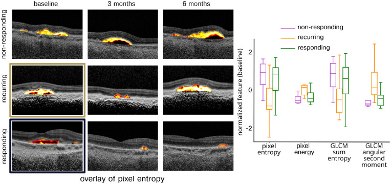
Methods: Thirty-nine eyes of patients with PED undergoing anti-VEGF therapy were included. All eyes underwent a loading dose followed by as-needed therapy. OCT images at baseline, month 3, and month 6 were analyzed. Images were manually separated into non-responding, recurring, and responding eyes based on the presence or absence of subretinal fluid at month 6. PED radiomics features were then extracted from each image and images were classified as responding or recurring using a machine learning classifier applied to the radiomics features.
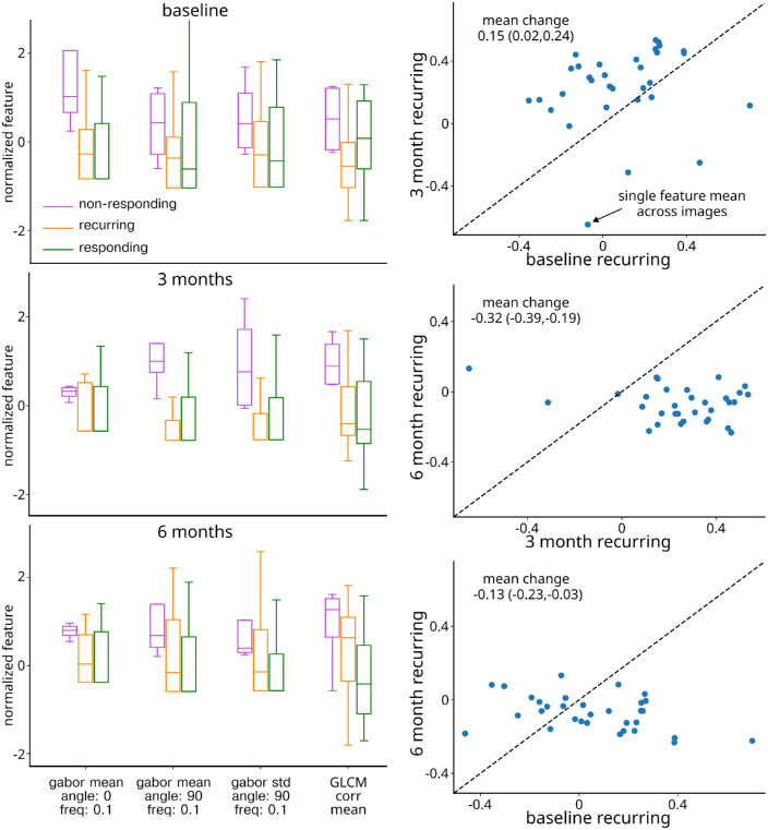
Results: Linear discriminant analysis classification of baseline features as responsive versus recurring resulted in classification performance of 64.0% (95% confidence interval [CI] = 0.63-0.65), area under the curve (AUC = 0.78, 95% CI = 0.72-0.82), sensitivity 0.79 (95% CI = 0.63-0.87), and specificity 0.58 (95% CI = 0.50-0.67). Further analysis of features in recurring eyes identified a significant shift toward non-responding mean feature values over 6 months.
Conclusions: Our results demonstrate the use of radiomics features as predictors for treatment response to as-needed anti-VEGF therapy. Our study demonstrates the potential for radiomics feature in clinical decision support for personalizing anti-VEGF therapy.
Translational relevance: The ability to use PED texture features to predict treatment response facilitates personalized clinical decision making.
Conflict of interest statement
Disclosure:
Figures
Similar articles
-
OCT-Derived Radiomic Features Predict Anti-VEGF Response and Durability in Neovascular Age-Related Macular Degeneration.Ophthalmol Sci. 2022 May 18;2(4):100171. doi: 10.1016/j.xops.2022.100171. eCollection 2022 Dec. Ophthalmol Sci. 2022. PMID: 36531588 Free PMC article.
-
Texture-Based Radiomic SD-OCT Features Associated With Response to Anti-VEGF Therapy in a Phase III Neovascular AMD Clinical Trial.Transl Vis Sci Technol. 2024 Jan 2;13(1):29. doi: 10.1167/tvst.13.1.29. Transl Vis Sci Technol. 2024. PMID: 38289610 Free PMC article. Clinical Trial.
-
Brolucizumab for recalcitrant macular neovascularization in age-related macular degeneration with pigment epithelial detachment.Eur J Ophthalmol. 2024 Mar;34(2):487-496. doi: 10.1177/11206721231187663. Epub 2023 Jul 18. Eur J Ophthalmol. 2024. PMID: 37461836
-
OPTIMAL MANAGEMENT OF PIGMENT EPITHELIAL DETACHMENTS IN EYES WITH NEOVASCULAR AGE-RELATED MACULAR DEGENERATION.Retina. 2018 Nov;38(11):2103-2117. doi: 10.1097/IAE.0000000000002195. Retina. 2018. PMID: 29697591 Free PMC article. Review.
-
Non-ICGA treatment criteria for Suboptimal Anti-VEGF Response for Polypoidal Choroidal Vasculopathy: APOIS PCV Workgroup Report 2.Ophthalmol Retina. 2021 Oct;5(10):945-953. doi: 10.1016/j.oret.2021.04.002. Epub 2021 Apr 16. Ophthalmol Retina. 2021. PMID: 33866022
Cited by
-
Role of traditional Chinese medicine in age-related macular degeneration: exploring the gut microbiota's influence.Front Pharmacol. 2024 Jan 25;15:1356324. doi: 10.3389/fphar.2024.1356324. eCollection 2024. Front Pharmacol. 2024. PMID: 38333011 Free PMC article. Review.
-
Bioinformatics Analysis of Lactylation-related Biomarkers and Potential Pathogenesis Mechanisms in Age-related Macular Degeneration.Curr Genomics. 2025;26(3):191-209. doi: 10.2174/0113892029291661241114055924. Epub 2025 Jan 2. Curr Genomics. 2025. PMID: 40433417 Free PMC article.
-
Radiomics-Based OCT Analysis of Choroid Reveals Biomarkers of Central Serous Chorioretinopathy.Transl Vis Sci Technol. 2025 Apr 1;14(4):23. doi: 10.1167/tvst.14.4.23. Transl Vis Sci Technol. 2025. PMID: 40266602 Free PMC article.
References
-
- Rosenfeld PJ, Brown DM, Heier JS, et al. .. Ranibizumab for neovascular age-related macular degeneration. N Engl J Med. 2006; 355(14): 1419–1431. - PubMed
-
- Brown DM, Kaiser PK, Michels M, et al. .. Ranibizumab versus verteporfin for neovascular age-related macular degeneration. N Engl J Med. 2006; 355(14): 1432–1444. - PubMed
-
- Regillo CD, Brown DM, Abraham P, et al. .. Randomized, double-masked, sham-controlled trial of ranibizumab for neovascular age-related macular degeneration: PIER Study Year 1. Am J Ophthalmol. 2008; 145(2): 239–248.e5. - PubMed
-
- Lalwani GA, Rosenfeld PJ, Fung AE, et al. .. A variable-dosing regimen with intravitreal ranibizumab for neovascular age-related macular degeneration: year 2 of the PrONTO Study. Am J Ophthalmol. 2009; 148(1): 43–58.e1. - PubMed
-
- Yim J, Chopra R, Spitz T, et al. .. Predicting conversion to wet age-related macular degeneration using deep learning. Nat Med. 2020; 26(6): 892–899. - PubMed
MeSH terms
Substances
Grants and funding
LinkOut - more resources
Full Text Sources
Medical
