. 2023 Oct 4;15(716):eadh3839.
doi: 10.1126/scitranslmed.adh3839. Epub 2023 Oct 4.

A humanized chemogenetic system inhibits murine pain-related behavior and hyperactivity in human sensory neurons

Affiliations

A humanized chemogenetic system inhibits murine pain-related behavior and hyperactivity in human sensory neurons

Jimena Perez-Sanchez et al. Sci Transl Med. .

Abstract

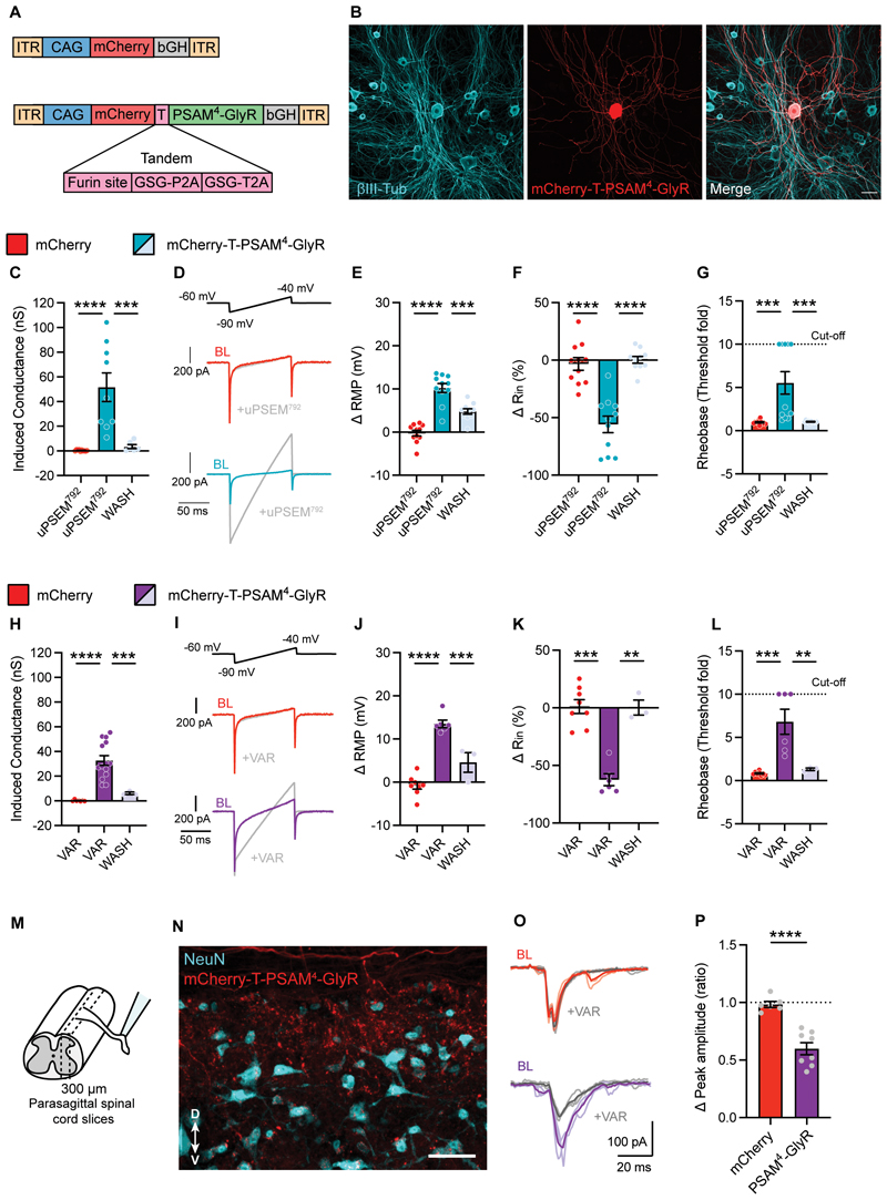
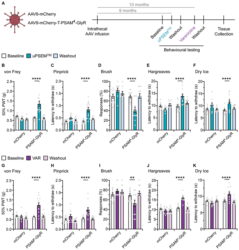
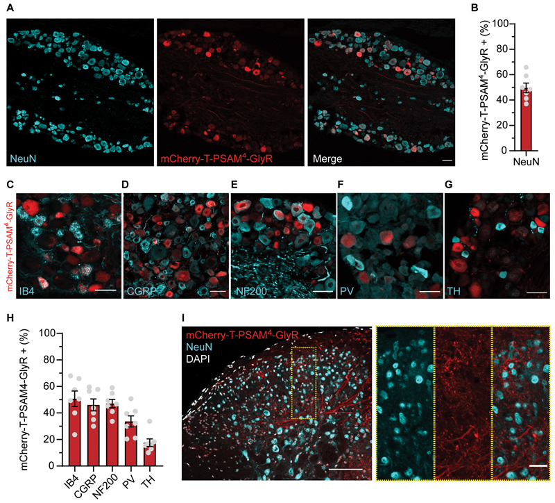
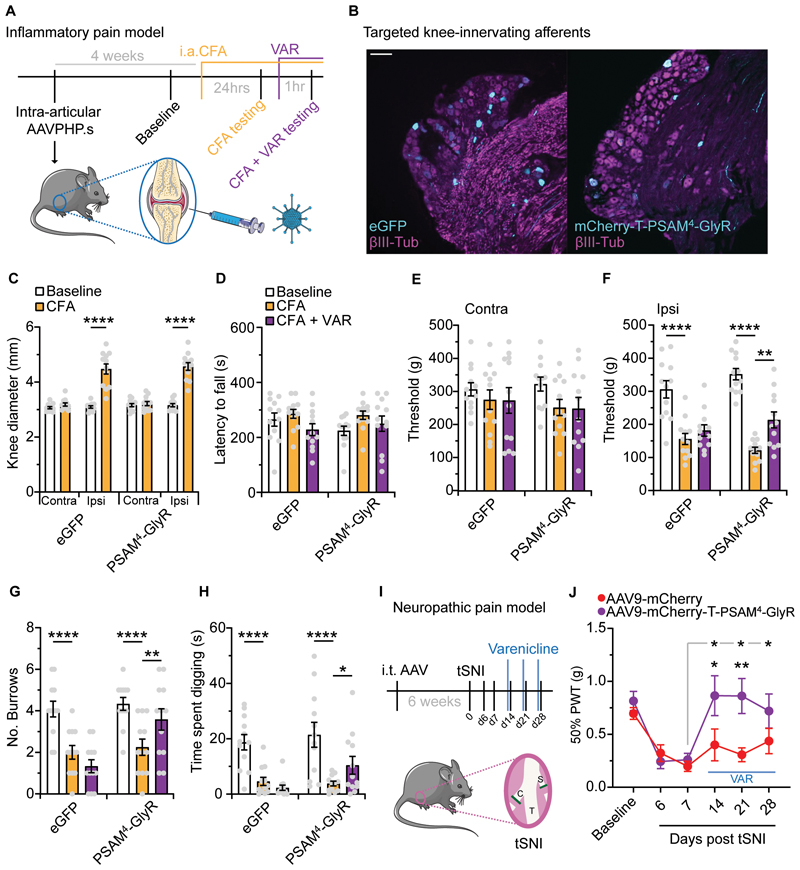
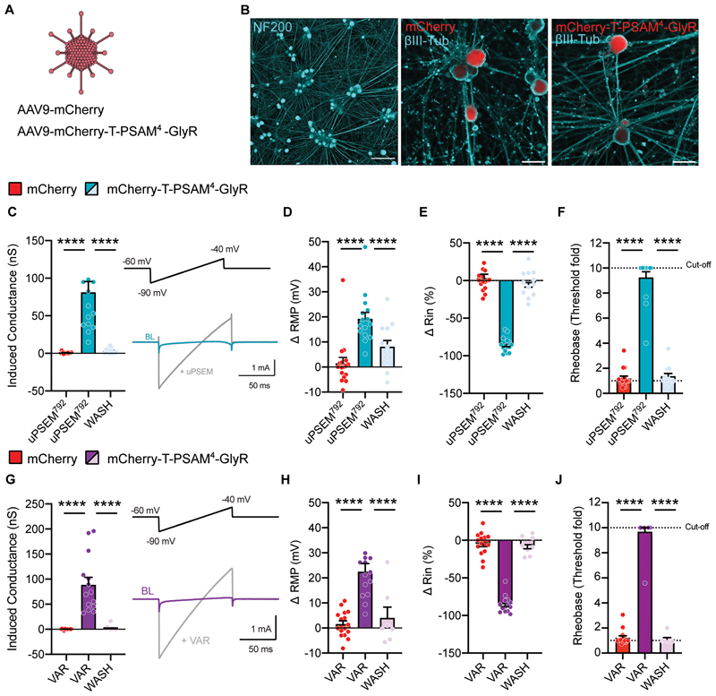
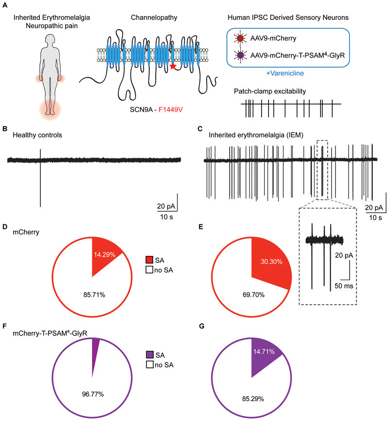
Hyperexcitability in sensory neurons is known to underlie many of the maladaptive changes associated with persistent pain. Chemogenetics has shown promise as a means to suppress such excitability, yet chemogenetic approaches suitable for human applications are needed. PSAM4-GlyR is a modular system based on the human α7 nicotinic acetylcholine and glycine receptors, which responds to inert chemical ligands and the clinically approved drug varenicline. Here, we demonstrated the efficacy of this channel in silencing both mouse and human sensory neurons by the activation of large shunting conductances after agonist administration. Virally mediated expression of PSAM4-GlyR in mouse sensory neurons produced behavioral hyposensitivity upon agonist administration, which was recovered upon agonist washout. Stable expression of the channel led to similar reversible suppression of pain-related behavior even after 10 months of viral delivery. Mechanical and spontaneous pain readouts were also ameliorated by PSAM4-GlyR activation in acute and joint pain inflammation mouse models. Furthermore, suppression of mechanical hypersensitivity generated by a spared nerve injury model of neuropathic pain was also observed upon activation of the channel. Effective silencing of behavioral hypersensitivity was reproduced in a human model of hyperexcitability and clinical pain: PSAM4-GlyR activation decreased the excitability of human-induced pluripotent stem cell-derived sensory neurons and spontaneous activity due to a gain-of-function NaV1.7 mutation causing inherited erythromelalgia. Our results demonstrate the contribution of sensory neuron hyperexcitability to neuropathic pain and the translational potential of an effective, stable, and reversible humanized chemogenetic system for the treatment of pain.


Conflict of interest statement

Competing interests:

D.L.B. has acted as a consultant on behalf of Oxford Innovation for Abide, Amgen, Combigene, G Mitsubishi, Lilly, Tanabe, GSK, TEVA, Biogen, Lilly, Orion, Third Rock and Theranexus.

Figures

Fig. 1
Fig. 1. PSAM4-GlyR can reversibly silence mouse sensory neurons and reduce synaptic transmission.
(A) Schematic representation of the constructs used in this study: mCherry and mCherry-T-PSAM4-GlyR. (B) Example images of dissociated sensory neurons transduced with mCherry-T-PSAM4-GlyR after 2 days in vitro (Scale bar = 50 μm). (C) Voltage-clamp recordings of membrane conductance in the presence of uPSEM792 (10 nM) and after agonist washout (mCherry: n = 10 cells; mCherry-T-PSAM4-GlyR: n = 9 cells). (D) Representative current traces obtained by the application of voltage ramps used to determine membrane conductance. BL: Baseline. (E-G) Change in resting membrane potential (RMP) (E) input resistance (Rin) (F) and rheobase (G) after uPSEM792 administration and washout. The cut-off rheobase value was defined as 10 times the threshold rheobase before uPSEM792 treatment. (mCherry: n = 11 cells; mCherry-T-PSAM4-GlyR: n = 11 cells). (H) Membrane conductance measurements in PSAM4-GlyR neurons after varenicline (VAR; 20 nM) administration and agonist washout (mCherry: n = 7 cells; mCherry-T-PSAM4-GlyR: n = 14 cells). (I) Representative current traces obtained by the application of voltage ramps used to determine membrane conductance. (J–L) Changes in resting membrane potential (RMP) (J) input resistance (K) and rheobase (L) after varenicline administration and washout (mCherry: n = 8 cells; mCherry-T-PSAM4-GlyR: n = 6 cells). C-L: One-way ANOVA with Tukey Post-hoc, ** P < 0.01, *** P < 0.001, **** P < 0.0001). (M) Schematic representation of the experimental procedure for spinal cord slices. (N) Example image of a parasagittal slice used for recording, showing mCherry-T-PSAM4-GlyR expressing terminals in the dorsal horn of the spinal cord. D: dorsal; V: ventral (Scale bar = 50 μm). (O) Representative postsynaptic currents evoked by dorsal root stimulation (ePSCs) recorded from superficial dorsal horn neurons obtained from mCherry-transduced animals (red) or mCherry-T-PSAM4-GlyR animals (purple) before and after application of varenicline (20 nM; grey). (P) Change in ePSC amplitude after application of varenicline (mCherry: n = 6 cells; mCherry-T-PSAM4-GlyR: n = 8 cells. Unpaired t-test, **** P<0.0001). All data are expressed as mean ± S.E.M.
Fig. 2
Fig. 2. Robust, reversible, and repeatable silencing of acute sensory behaviors via agonist-induced activation of PSAM4-GlyR.
(A) Timeline of the experimental design. Intrathecal infusion of AAV-mCherry or mCherry-T-PSAM4-GlyR followed by baseline sensory testing, and re-testing during or post uPSEM792 or varenicline. (B–D) Mechanical sensory testing in mCherry or PSAM4-GlyR expressing mice pre (baseline), during and post (washout) uPSEM792; von Frey (B), Pinprick (C) and Brush (D). (E and F) Thermal sensory in mCherry or PSAM4-GlyR expressing mice pre (baseline), during and post (washout) uPSEM792; Hargreaves (E) and Dry Ice (F). (G–I) Mechanical sensory testing in mCherry or PSAM4-GlyR expressing mice pre (baseline), during and post (washout) varenicline; von Frey (G), Pinprick (H) and Brush (I). (J and K) Thermal sensory testing in mCherry or PSAM4-GlyR expressing mice pre (baseline), during and post (washout) varenicline; Hargreaves (J) and Dry Ice (K) (mCherry: n = 10 mice, PSAM4-GlyR: n = 12 mice, all data sets RM-two way ANOVA, post-hoc Bonferroni test, ** P < 0.01, **** P < 0.0001). Data expressed as mean ± S.E.M.
Fig. 3
Fig. 3. Long-term, stable expression of AAV9-mCherry-T-PSAM4-GlyR selectively in sensory neurons.
(A) Example image of DRG neurons transduced by AAV9-mCherry-T-PSAM4-GlyR 11-months post i.t. injection (Scale bar 50 um) (B) Quantification of NeuN positive neurons that are also mCherry-T-PSAM4-GlyR positive (n = 7 mice, 1631/3229 neurons) (C–G) Example images of DRG neuron subpopulation markers, isolectin-B4 (IB4) (C), calcitonin gene-related peptide (CGRP) (D), neurofilament 200 (NF200) (E), parvalbumin (PV) (F), tyrosine hydroxylase (TH) (G), and their co-expression with mCherry-T-PSAM4-GlyR (Scale bars 25 um). (H) Percentage of each DRG neuron subpopulation that co-express mCherry-T-PSAM4-GlyR (n = 7 mice, (IB4: 480/973 neurons, CGRP: 435/967 neurons, NF200: 479/1014 neurons, PV: 69/219 neurons, TH: 51/261 neurons). (I) Example image of mCherry-T-PSAM4-GlyR positive afferents entering and terminating in the dorsal horn of the spinal cord (Scale Bar 100 μm, insert scale bar 25 μm). Data mean ± S.E.M.
Fig. 4
Fig. 4. PSAM4-GlyR mediated silencing of inflammatory-joint and neuropathic pain.
(A) Schematic of the experimental timeline of targeting joint afferents with AAVPHP.S, baseline testing, i.a. CFA and behavioral testing pre- and post-varenicline. (B) Example images of L4 DRG neurons transduced with either AAVPHP.S-eGFP or AAVPHP.S-mCherry-T-PSAM4-GlyR following i.a. injection (scale bar = 100 μm). (C) Evaluation of knee swelling following i.a. CFA (eGFP: n = 12 mice, PSAM4-GlyR = 12 mice, RM two-way ANOVA, post hoc Bonferroni test, **** P<0.0001). Quantification of (D) motor function, mechanical sensitivity of the contralateral knee (E), and mechanical sensitivity of the ipsilateral knee (F) pre CFA (baseline), post CFA and post CFA and varenicline. (G and H) Analysis of spontaneous behaviors, burrowing (G) and time spent digging (H) pre CFA (baseline), post CFA and post CFA and varenicline. eGFP: n = 12 mice, PSAM4-GlyR = 12 mice, RM two-way ANOVA, post hoc Bonferroni tests compared to CFA,* P < 0.05, ** P < 0.01, **** P<0.0001). (I) Illustration of the tibial spared-nerve injury (tSNI) neuropathic pain model and experimental timeline. Intrathecal targeting of sensory neurons, followed by tSNI and mechanical testing pre- and post-varenicline. C: common peroneal; T: tibial; S: sural. (J) Mechanical testing of mCherry or PSAM4-GlyR expressing mice before (baseline), post tSNI and post tSNI with varenicline treatment (mCherry: n = 10 mice, PSAM4-GlyR = 12 mice, RM two-way ANOVA, post hoc Bonferroni tests compared to D7 (grey line) or between groups, * P < 0.05, ** P < 0.01). Data expressed as mean ± S.E.M.
Fig. 5
Fig. 5. PSAM4-GlyR mediated silencing of human iPSC derived sensory neurons.
(A) AAV9-mCherry or AAV9-mCherry-T-PSAM4-GlyR were used to virally transduce sensory neurons derived from human induced pluripotent stem cells (hiPSC-SNs). (B) Example images showing mature hiPSC-SNs (left) and mCherry labeling in AAV-mCherry (middle) and AAV-mCherry-T-PSAM4-GlyR (right) transduced neurons. (C) Quantification of membrane conductance after uPSEM792 application and washout. Inset shows representative traces of voltage clamp recordings used to measure membrane conductance. BL: Baseline. (D-F) Changes in resting membrane potential (RMP) (B) input resistance (Rin) (E) and rheobase (F) after uPSEM792 administration and washout (mCherry: n = 16 cells; mCherry-T-PSAM4-GlyR: n = 15 cells. One-way ANOVA with Tukey post-hoc, **** P<0.0001). (G) Quantification of membrane conductance after varenicline application and washout. Inset shows representative currents, used to measure membrane conductance, at baseline (BL) and post varenicline administration. (H-J) Changes in resting membrane potential (RMP) (H) input resistance (Rin) (I) and rheobase (J) after varenicline treatment and washout. (mCherry: n = 15 cells; mCherry-T-PSAM4-GlyR: n = 14 cells. One-way ANOVA with Tukey post-hoc, **** P<0.0001). Rheobase cut-off was defined as 10 times the baseline rheobase threshold. All data are expressed as mean ± S.E.M.
Fig. 6
Fig. 6. Silencing of spontaneous activity in a human neuropathic pain model.
(A) Schematic representation of the experimental design. Patients with inherited erythromelalgia (IEM) exhibit pain and erythema in the extremities (left). Illustration of the point mutation (F1449V) in the Nav1.7 channel α-subunit identified in a person living with IEM (middle). Transduction of hiPSC-SN derived from healthy control participants and from a patient with IEM-F1449V with AAV9-mCherry and AAV9-mCherry-T-PSAM4-GlyR (right). (B-C) Example traces of spontaneous activity (SA) recorded in cell-attached configuration from healthy control hiPSC-SNs (B) and from IEM hiPSC-SNs (C). Inset shows magnification of a bursting event. (D) Proportion of AAV9-mCherry transduced healthy control hiPSC-SNs exhibiting SA; no SA (n = 24, white) and SA (n = 4, red). (E) Proportion of AAV9-mCherry transduced IEM hiPSCs exhibiting SA; no SA (n = 23, white) and SA (n = 10, red). χ2 = 6.9, P = 0.009 (F) Proportion of AAV9-mCherry-T-PSAM4-GlyR transduced healthy control hiPSC-SNs showing SA; no SA (n = 30, white) and SA (n = 1, purple). (G) Proportion of AAV-mCherry-T-PSAM4-GlyR transduced IEM hiPSC-SNs with SA; no SA (n = 29, white) and SA (n = 5, purple). χ2 = 3.9, P = 0.04.

Comment in

References

    1. Breivik H, Collett B, Ventafridda V, Cohen R, Gallacher D. Survey of chronic pain in Europe: prevalence, impact on daily life, and treatment. Eur J Pain. 2006;10:287–333. - PubMed
    1. Colloca L, et al. Neuropathic pain. Nat Rev Dis Primers. 2017;3:17002. - PMC - PubMed
    1. Walsh DA, McWilliams DF. Mechanisms, impact and management of pain in rheumatoid arthritis. Nat Rev Rheumatol. 2014;10:581–592. - PubMed
    1. Finnerup NB, et al. Pharmacotherapy for neuropathic pain in adults: a systematic review and meta-analysis. Lancet Neurol. 2015;14:162–173. - PMC - PubMed
    1. Chakrabarti S, et al. Acute inflammation sensitizes knee-innervating sensory neurons and decreases mouse digging behavior in a TRPV1-dependent manner. Neuropharmacology. 2018;143:49–62. - PMC - PubMed

Publication types