. 2023 Oct 4;11(10):e23.00041.
doi: 10.2106/JBJS.RVW.23.00041. eCollection 2023 Oct 1.

Team Approach: Hip Preservation Surgery

Affiliations

Team Approach: Hip Preservation Surgery

Andrew W Kuhn et al. JBJS Rev. .

Abstract

The evaluation and treatment of adolescents and young adults with hip pain has seen tremendous growth over the past 20 years. Labral tears are well established as a common cause of hip pain but often occur because of underlying bony abnormalities. Femoroacetabular impingement (FAI) and acetabular dysplasia are now well-established causes of hip osteoarthritis and are increasingly treated in the prearthritic stage in hopes of improving symptoms and prolonging the longevity of the native hip. Beyond FAI and acetabular dysplasia, this patient population can present with a complex and variable group of underlying conditions that need to be taken into account. Expertise in the conservative management of this population, including physical therapy, is valuable to maximize the success. Preoperative, surgical, and postoperative decision-making and care in this population is complex and evolving. A comprehensive, multidisciplinary approach to the care of this patient population has been used for over 20 years by our institution with great success. The purpose of this article is to review the "team-based approach" necessary for successful management of the spectrum of adolescent and young adult hip disorders.


Conflict of interest statement

Disclosure: The Disclosure of Potential Conflicts of Interest forms are provided with the online version of the article (http://links.lww.com/JBJSREV/B12).

Figures
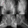
Figure 1A-D.
Figure 1A-D.
Plain radiographic imaging of a 17 year old female with left hip pain. (A) Standing AP Pelvis with inset view demonstrating the posterior (red) and anterior (navy) acetabular walls (B) 45 degree Dunn view, (C) False Profile, and (D) Frog-Leg Lateral views of the left hip demonstrating Tonnis Grade 0 OA and signs of acetabular retroversion including a positive cross-over sign and ischial spine sign. The lateral center edge angle is 33 degrees, but there is about 5 degrees of pelvic obliquity resulting from leg length discrepancy. The posterior wall sign is negative but the superior portion of the posterior wall projects in a very vertical position not meeting the anterior wall at the lateral sourcil. A mild leg length discrepancy results in pelvic obliquity with increased inclination of the acetabulum on the longer left leg. There is also slight loss of femoral head neck offset with an alpha angle of 55 degrees on the Dunn view.
Figure 2A-E.
Figure 2A-E.
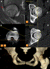
Advanced imaging including (A) sagittal MRI demonstrating an anterosuperior labral tear (red arrow) and low dose CT scans of the (B) distal femur demonstrating measurement of femoral anteversion and of the hip to calculate (C) central acetabular version and (D) cranial acetabular version. CT scans can be reformatted into (E) 3D models to help with visualization of the complex pathoanatomy of the proximal femur and/or acetabulum.
Figure 3.
Figure 3.
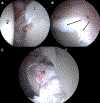
Intraoperative arthroscopic photos demonstrating an (a) anterior and (B) anterosuperior labral tear with chondromalacia which was (C) repaired with suture anchor fixation. FH = femoral head; L = labrum; LR = labral repair.
Figure 4A-D.
Figure 4A-D.
Intraoperative fluoroscopic images demonstrating the steps involved in PAO including (A) osteotomies of the ischium, pubis, and ilium, (B) provisional fixation and correction, and (C) definitive fixation with (D) correction of the decreased femoral head neck offset after proximal femoral osteochondroplasty.
Figure 5A-B.
Figure 5A-B.
(A) Pre- and (B) post-operative AP Pelvis at 5 months after PAO with insets demonstrating orientation of the posterior (red) and anterior (navy) acetabular walls.\
Figure 6.
Figure 6.. Flowchart demonstrating the multidisciplinary team approach to the patient with prearthritic hip disease.

Similar articles

References

    1. Bonazza NA, Homcha B, Liu G, Leslie DL, Dhawan A. Surgical Trends in Arthroscopic Hip Surgery Using a Large National Database. Arthroscopy. 2018. Jun;34(6):1825–30. . - PubMed
    1. Erickson BJ, Cvetanovich GL, Frank RM, Bhatia S, Bush-Joseph CA, Nho SJ, et al. International trends in arthroscopic hip preservation surgery-are we treating the same patient? J Hip Preserv Surg. 2015. Jan;2(1):28–41. - PMC - PubMed
    1. Adler KL, Cook PC, Yen YM, Giordano BD. Current Concepts in Hip Preservation Surgery: Part I. Sports Health. 2015. Nov-Dec;7(6):518–26. . - PMC - PubMed
    1. Bedi A, Kelly BT, Khanduja V. Arthroscopic hip preservation surgery: current concepts and perspective. Bone Joint J. 2013. Jan;95-B(1):10–9. - PubMed
    1. Bedi A, Kelly BT. Femoroacetabular impingement. J Bone Joint Surg Am. 2013. Jan 2;95(1):82–92. - PubMed