. 2023 Oct 3;35(10):1688-1703.e10.
doi: 10.1016/j.cmet.2023.09.004.

Neutrophils resist ferroptosis and promote breast cancer metastasis through aconitate decarboxylase 1

Affiliations

Neutrophils resist ferroptosis and promote breast cancer metastasis through aconitate decarboxylase 1

Yun Zhao et al. Cell Metab. .

Abstract

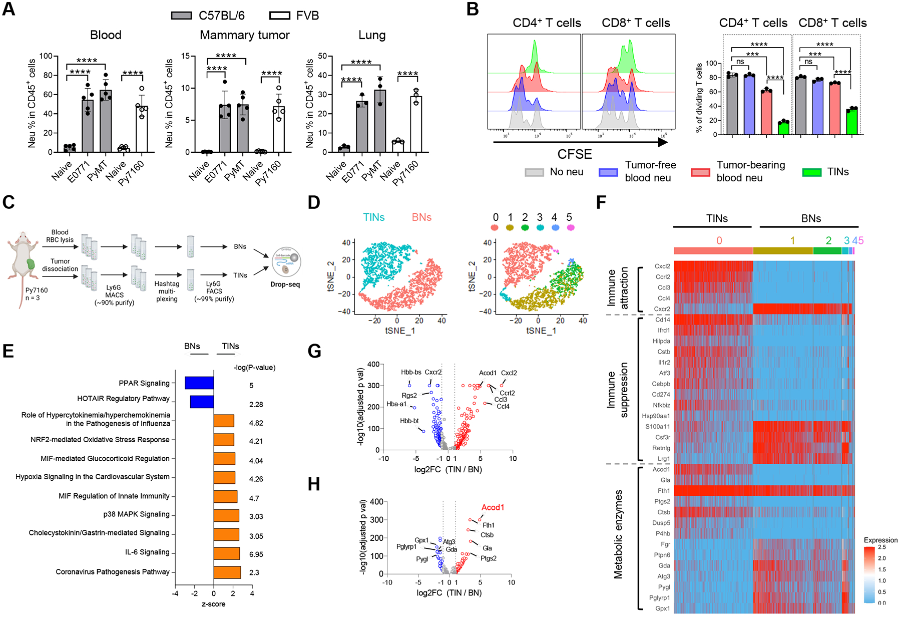
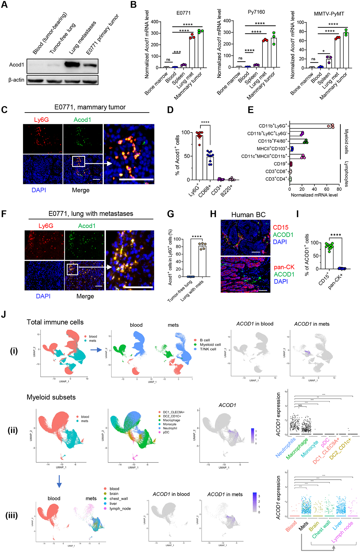
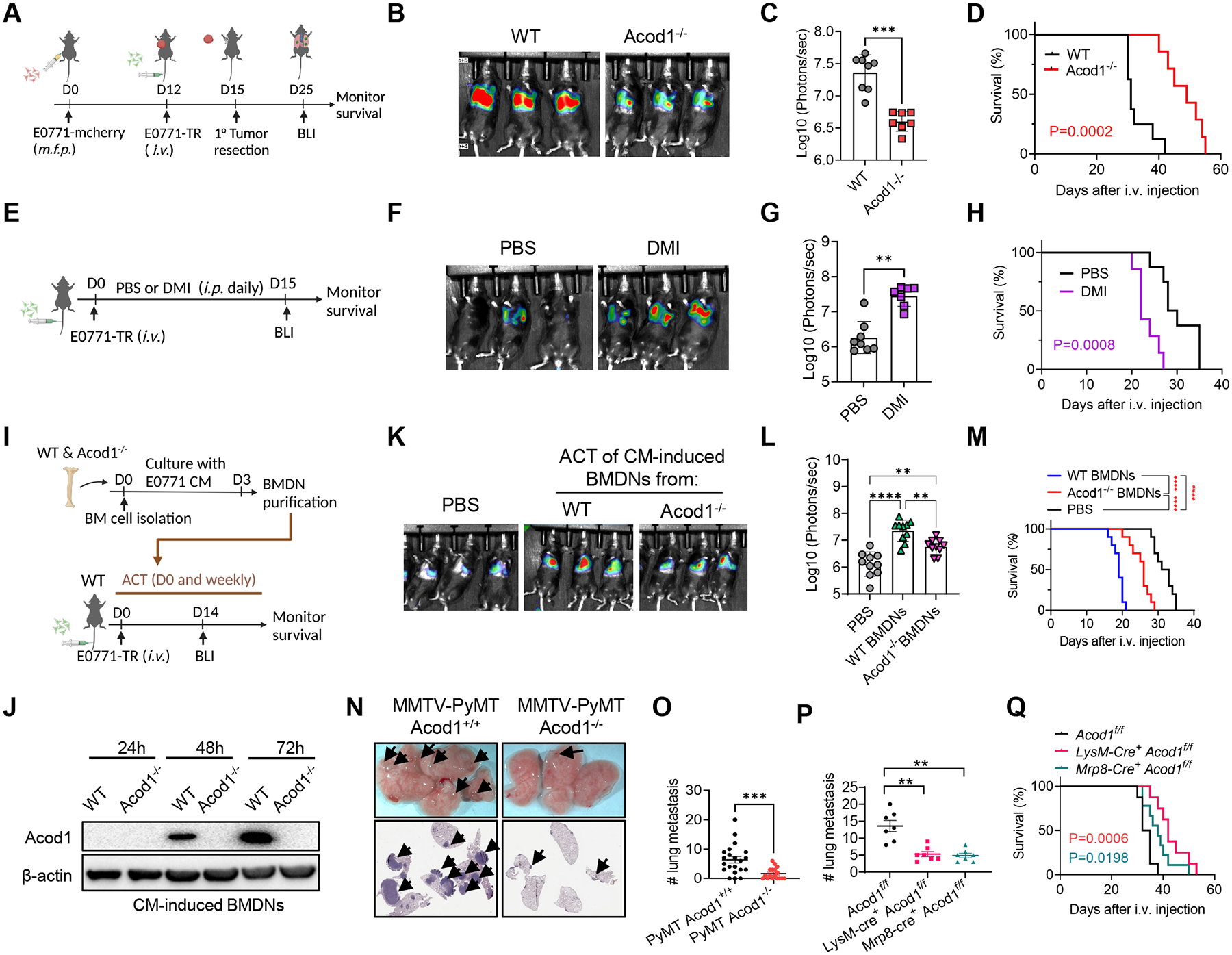
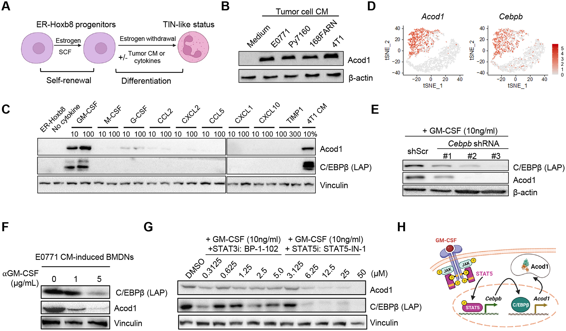
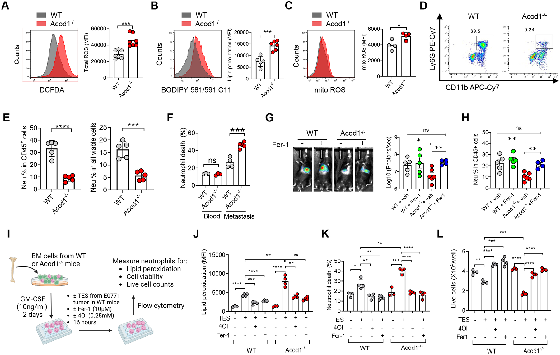
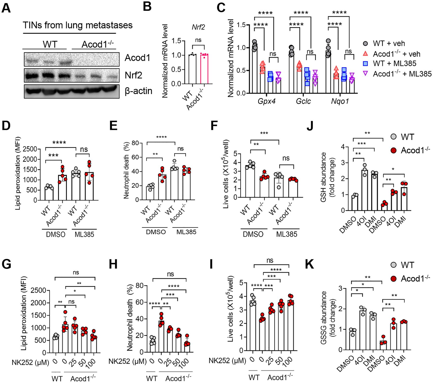
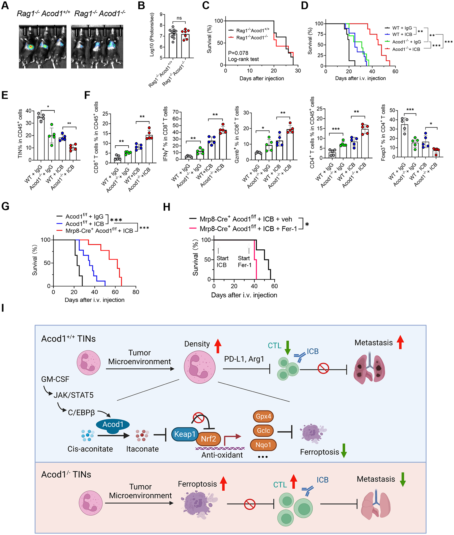
Metastasis causes breast cancer-related mortality. Tumor-infiltrating neutrophils (TINs) inflict immunosuppression and promote metastasis. Therapeutic debilitation of TINs may enhance immunotherapy, yet it remains a challenge to identify therapeutic targets highly expressed and functionally essential in TINs but under-expressed in extra-tumoral neutrophils. Here, using single-cell RNA sequencing to compare TINs and circulating neutrophils in murine mammary tumor models, we identified aconitate decarboxylase 1 (Acod1) as the most upregulated metabolic enzyme in mouse TINs and validated high Acod1 expression in human TINs. Activated through the GM-CSF-JAK/STAT5-C/EBPβ pathway, Acod1 produces itaconate, which mediates Nrf2-dependent defense against ferroptosis and upholds the persistence of TINs. Acod1 ablation abates TIN infiltration, constrains metastasis (but not primary tumors), bolsters antitumor T cell immunity, and boosts the efficacy of immune checkpoint blockade. Our findings reveal how TINs escape from ferroptosis through the Acod1-dependent immunometabolism switch and establish Acod1 as a target to offset immunosuppression and improve immunotherapy against metastasis.

Keywords: Acod1; MDSC; breast cancer; ferroptosis; immune checkpoint blockade; immune metabolism; itaconate; metastasis; neutrophil; single-cell RNA sequencing.


Conflict of interest statement

Declaration of interests The authors declare no competing interests.

Figures

Figure 1.
Figure 1.. Neutrophils in the mammary TME demonstrate potent immunosuppression and distinct transcriptomic features.
(A) Flow cytometry analysis of neutrophil populations (CD11b+Ly6G+) in blood (n=5), mammary tumors (n=5) and lung metastases (n=3) of mouse BC models. (B) CFSE dilution histograms and proliferating proportions of αCD3/αCD28-stimulated spleen T cell subsets cocultured with neutrophils (1:1 ratio) isolated from tumor-free or E0771 tumor-bearing mice (n=3). (C) Schematic for sample processing, neutrophil sorting and Drop-seq of BNs and TINs from Py7160-bearing mice (n=3). (D) t-distributed stochastic neighbor embedding (t-SNE) plots colored by tissue of origin (left) or cell clusters (right). (E) Top-ranked IPA pathways upregulated in TINs or BNs. (F) Heatmap of most differentially expressed genes by TINs and BNs associated with immune attraction, immune suppression, and metabolic enzymes. (G) Volcano plot of differentially expressed genes (adjusted p values < 0.05). (H) Volcano plot of differentially expressed genes encoding metabolic enzymes (adjusted p values < 0.05). For A and B, data represent mean ± s.e.m.; ns, not significant, ***P<0.001, ****P<0.0001, unpaired two-tailed Student’s t-test. See also Figure S1 and Table S1–S3.
Figure 2.
Figure 2.. Acod1 is highly expressed in TINs of primary and metastatic BC in mice and patients.
(A) Western blot to assess Acod1 expression in blood or tissues of tumor-free and E0771-bearing mice. (B) qRT-PCR to assess Acod1 expression in neutrophils isolated from tumor-bearing mice of syngeneic and GEM models (n=3). (C) IF co-staining of Acod1 and Ly6G in E0771 primary tumors. (D) Percentage of Acod1+ cells in different immune cells (neutrophils, Ly6G+; macrophages, CD68+; T cells, CD3+; B cells, B220+) based on IF co-staining of E0771 primary tumors (n=10). (E) qRT-PCR quantification of Acod1 in lymphocyte subsets and myeloid subsets sorted from E0771 tumors (n=3). (F) IF co-staining of Acod1 and Ly6G in E0771 lung metastases. (G) Percentage of Acod1+ neutrophils in tumor-free lung or lung with E0771 metastases (n=5). (H-I) Representative image and quantitative result of IF co-staining of ACOD1 and CD15 or pan-Cytokeratin in human BC tissues (n=10). (J) Analysis of scRNA-seq dataset GSE169246 of blood and metastasized tissues of human BC. for ACOD1 expression pattern. (i) UMAP of total CD45+ cells segregated into three main immune populations for blood and metastases (mets) featuring ACOD1 expression (rightmost). (ii) UMAP of myeloid cells segregated into six subsets plotting ACOD1 expression (rightmost). (iii) UMAP of myeloid cells for blood and mets separately featuring ACOD1 expression in different sites (rightmost). In B, D, E, G, I and J, data represent mean ± s.e.m.; ns, not significant, *P<0.05, **P<0.01,***P<0.001, ****P<0.0001, unpaired two-tailed Student’s t-test. In C, F, H, scale bar 100μm. See also Figure S2.
Figure 3.
Figure 3.. Neutrophil Acod1 promotes lung metastasis in mouse BC models.
(A) Experimental lung metastasis assay scheme based on E0771. (B-C) Representative BLI images (n=3) and quantification (WT n=8, Acod1‒/‒ n=7) at day 15 after i.v. injection of E0771-TR. (D) Survival curves (WT n=8, Acod1‒/‒ n=7). (E) Assessing the effect of PBS or DMI (100mg kg‒1, daily i.p.) on E0771-TR lung metastasis grown in WT mice. (F-G) Representative BLI images (n=3) and quantification (PBS n= 8, DMI n=7) at day 15 after i.v. injection of E0771-TR. (H) Survival curves (PBS n= 8, DMI n=7). (I) Schematic of ACT of WT or Acod1‒/‒ BMDNs cultured with E0771 CM into WT cohorts i.v. injected of E0771-TR. (J) Western blot of Acod1 for WT or Acod1‒/‒ BMDNs cultured with E0771 CM for 24h, 48h or 72h. (K-L) Representative BLI images and quantification (n=10) at day 14 after i.v. injection of E0771-TR. (M) Survival curves (n=10). (N) Representative photographs and HE staining of lungs in MMTV-PyMT Acod1+/+ and MMTV-PyMT Acod1‒/‒ cohorts when mice died from primary tumors. Arrows denote metastases. (O) Quantification of lung metastasis nodules (MMTV-PyMT Acod1+/+ n=21, MMTV-PyMT Acod1‒/‒ n=17). (P) Quantification of lung metastasis nodules in cohorts Acod1 f/f, LysM-Cre+ Acod1f/f and Mrp8-Cre+ Acod1f/f, 2 weeks after i.v. injection of E0771 (n=7/group). (Q) Survival of Acod1 f/f (n=8), LysM-Cre+ Acod1f/f (n=9) and Mrp8-Cre+ Acod1f/f (n=9) mice after i.v. injection of E0771. For C, G, O and P, data represent mean ± s.e.m.; **P<0.01, ***P<0.001, Mann-Whitney test. For L, data represent mean ± s.e.m.; **P<0.01, ****P<0.0001, one-way ANOVA with Tukey’s multiple comparisons test. For D, H, M and Q, log-rank test with P values labeled or ****P<0.0001. See also Figure S3.
Figure 4.
Figure 4.. Tumor-secreted GM-CSF induces Acod1 in neutrophils through the STAT5-C/EBPβ axis.
(A) ER-Hoxb8-immortalized mouse myeloid progenitors proliferate in the presence of estrogen (β-estradiol) and stem cell factor (SCF). When estrogen is withdrawn, progenitors differentiate to mature neutrophils (ER-Hoxb8-DNs) and can be induced to a TIN-like status with tumor CM or specific cytokines. (B) Western blot to examine Acod1 expression by ER-Hoxb8-DNs induced with CM from murine mammary cancer cell lines. (C) Western blot of Acod1 and C/EBPβ (LAP) in ER-Hoxb8 progenitor cells (first lane) and ER-Hoxb8-DNs (other lanes) treated with 4T1-expressed cytokines individually (unit ng/ml) or 4T1 CM. (D) t-SNE plot of Acod1 and Cebpb from the Drop-seq data. (E) Western blot of Acod1 and C/EBPβ (LAP) for ER-Hoxb8-DNs with Cebpb knockdown by shRNA (three designs) and induced with GM-CSF. (F) Western blot of Acod1 and C/EBPβ (LAP) for E0771-CM-treated BMDNs with or without αGM-CSF. (G) Western blot of Acod1 and C/EBPβ (LAP) for ER-Hoxb8-DNs induced with GM-CSF in the presence of BP-1–102 (STAT3 inhibitor) or STAT5-IN-1 (STAT5 inhibitor). (H) Schematic of Acod1 upregulation in TINs by tumor-secreted GM-CSF. Western blot results were representative of at least three independent experiments showing consistent patterns. See also Figure S4.
Figure 5.
Figure 5.. Acod1 sustains TIN survival by blunting ferroptosis.
(A-C) Flow cytometry for total cellular ROS (DCFDA) (n=7), lipid ROS (BODIPY 581/591 C11) (n=5–6), and mitochondrial ROS (n=4) in TINs isolated from E0771 lung metastases in WT and Acod1‒/‒ mice. (D) Representative flow cytometry plots to quantify the frequency of CD11b+ Ly6G+ cells in all CD45+DAPI singlets in E0771 lung metastases of WT and Acod1‒/‒ mice. (E) Frequency of CD11b+ Ly6G+ cells in the blood and lungs of E0771 metastasis-bearing WT and Acod1‒/‒ mice (n=5). Cells were gated on CD45+ DAPI singlets (left) or all DAPI singlets (right). (F) Cell death (DAPI+) fraction in total CD45+ CD11b+ Ly6G+ singlets (n=3–5). (G) BLI of WT and Acod1‒/‒ mice treated with vehicle or Fer-1 (2mg kg‒1, i.p., twice weekly) and imaged 14 days after i.v. injection of E0771-TR (n=4–6). (H) TIN percentage in lungs of vehicle or Fer-1 treated cohorts (n=4–6). (I-L) Schematic and results of evaluating the effect of TES (from E0771 mammary tumor grown in WT mice, 20% into culture medium), Fer-1 and 4OI on GM-CSF-induced WT and Acod1‒/‒ BMDNs for measures including lipid peroxidation (BODIPY 581/591 C11), cell death fractions, and viable cell counts (n=4/condition). Data represent mean ± s.e.m.; ns, not significant, *P<0.05, **P<0.01, ***P<0.001, ****P<0.0001, unpaired two-tailed Student’s t-test (except for G, which used Mann-Whitney test). Experiments were repeated three times with consistent results. See also Figure S5.
Figure 6.
Figure 6.. Acod1 blunts TIN ferroptosis through activating Nrf2-mediated antioxidant response.
(A) Immunoblot of Acod1 and Nrf2 in TINs isolated from E0771 lung metastases in WT and Acod1‒/‒ mice. (B) qRT-PCR of Nrf2 in TINs isolated from E0771 lung metastases in WT and Acod1‒/‒ mice (n=5). (C) qRT-PCR of Gpx4, Gclc and Nqo1 in TINs isolated from E0771 lung metastases in WT and Acod1‒/‒ mice (n=5) treated with vehicle or ML385 (30mg kg‒1, i.p., daily for 7 days). (D-F) The effect of ML385 (10μM) on E0771 TES-primed WT and Acod1‒/‒ BMDNs for measures including lipid peroxidation (BODIPY 581/591 C11), cell death fractions, and viable cell counts (n=5/condition). (G-I) The effect of NK252 on E0771 TES-primed WT and Acod1‒/‒ BMDNs for measures including lipid peroxidation (BODIPY 581/591 C11), cell death fractions, and viable cell counts (n=5/condition). (J-K) GSH and GSSG abundance in E0771 CM-induced WT and Acod1‒/‒ BMDNs treated with DMSO, 0.25mM 4OI or 0.25mM DMI (n=3). Data represent mean ± s.e.m.; ns, not significant, *P<0.05, **P<0.01, ***P<0.001, ****P<0.0001, unpaired two-tailed Student’s t-test. Experiments were repeated three times with consistent results. See also Figure S6.
Figure 7.
Figure 7.. Acod1 extinction boosts adaptive immunity and enhances immunotherapy.
(A-C) Lung metastasis of E0771-TR in Rag1‒/‒Acod+/+ (n=11) and Rag1‒/‒Acod1‒/‒ (n=7) mice, established with the method illustrated in Figure 3A. Shown are representative BLI images (A), BLI quantification (B) and animal survival (C). (D) Survival of WT and Acod1‒/‒ mice (n=8–10) bearing E0771-TR lung metastasis and treated with isotype IgG or ICB (αPDL1 + αCTLA4, 10mg kg‒1 each, i.p., twice/week). (E-F) Flow cytometry to measure frequencies of TINs and T cell subsets from lungs of WT and Acod1‒/‒ interim cohorts 15 days after i.v. injection of E0771-TR (n=5 for each group). (G) Survival of Acod1f/f and Mrp8-cre+Acod1f/f mice bearing E0771-TR lung metastasis and treated with isotype IgG or ICB (αPDL1 + αCTLA4, 10mg kg‒1 each, i.p., twice/week, started three days after i.v. injection). n=10 for each group. (H) Survival of Mrp-8cre+Acod1f/f mice undergoing ICB treatment further treated with or without Fer-1 (2mg kg-1, i.p., once/2 days). Start timepoints of ICB and Fer-1 treatments were indicated, n=4 for each group. (I) Schematic of the mechanism and function of Acod1 in TINs to promote BC lung metastasis (created with BioRender). In B, E and F, data represent mean ± s.e.m.; ns, not significant, *P<0.05, **P<0.01, ***P<0.001, Mann-Whitney test (for B) or unpaired two-tailed Student’s t-test (for E and F). In C, D, G and H, *P<0.05, **P<0.01, ***P<0.001, log-rank test. See also Figure S7.

References

    1. Latif F, Bint Abdul Jabbar H, Malik H, Sadaf H, Sarfraz A, Sarfraz Z, and Cherrez-Ojeda I (2022). Atezolizumab and pembrolizumab in triple-negative breast cancer: a meta-analysis. Expert review of anticancer therapy 22, 229–235. 10.1080/14737140.2022.2023011. - DOI - PubMed
    1. Corbeau I, Jacot W, and Guiu S (2020). Neutrophil to Lymphocyte Ratio as Prognostic and Predictive Factor in Breast Cancer Patients: A Systematic Review. Cancers (Basel) 12. 10.3390/cancers12040958. - DOI - PMC - PubMed
    1. Gentles AJ, Newman AM, Liu CL, Bratman SV, Feng W, Kim D, Nair VS, Xu Y, Khuong A, Hoang CD, et al. (2015). The prognostic landscape of genes and infiltrating immune cells across human cancers. Nat Med 21, 938–945. 10.1038/nm.3909. - DOI - PMC - PubMed
    1. Quail DF, Amulic B, Aziz M, Barnes BJ, Eruslanov E, Fridlender ZG, Goodridge HS, Granot Z, Hidalgo A, Huttenlocher A, et al. (2022). Neutrophil phenotypes and functions in cancer: A consensus statement. J Exp Med 219. 10.1084/jem.20220011. - DOI - PMC - PubMed
    1. Zhao Y, Rahmy S, Liu Z, Zhang C, and Lu X (2020). Rational targeting of immunosuppressive neutrophils in cancer. Pharmacol Ther 212, 107556. 10.1016/j.pharmthera.2020.107556. - DOI - PubMed

Publication types

LinkOut - more resources