. 2023 Oct;11(10):e007572.
doi: 10.1136/jitc-2023-007572.

SGN-B7H4V, an investigational vedotin ADC directed to the immune checkpoint ligand B7-H4, shows promising activity in preclinical models

Affiliations

SGN-B7H4V, an investigational vedotin ADC directed to the immune checkpoint ligand B7-H4, shows promising activity in preclinical models

Elizabeth Gray et al. J Immunother Cancer. 2023 Oct.

Abstract

Background: SGN-B7H4V is a novel investigational vedotin antibody-drug conjugate (ADC) comprising a B7-H4-directed human monoclonal antibody conjugated to the cytotoxic payload monomethyl auristatin E (MMAE) via a protease-cleavable maleimidocaproyl valine citrulline (mc-vc) linker. This vedotin linker-payload system has been clinically validated in multiple Food and Drug Administration approved agents including brentuximab vedotin, enfortumab vedotin, and tisotumab vedotin. B7-H4 is an immune checkpoint ligand with elevated expression on a variety of solid tumors, including breast, ovarian, and endometrial tumors, and limited normal tissue expression. SGN-B7H4V is designed to induce direct cytotoxicity against target cells by binding to B7-H4 on the surface of target cells and releasing the cytotoxic payload MMAE upon internalization of the B7-H4/ADC complex.

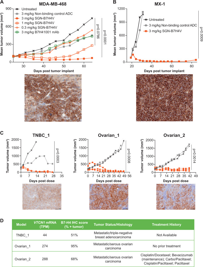
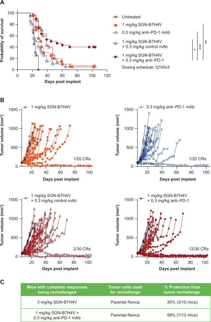
Methods: B7-H4 expression was characterized by immunohistochemistry across multiple solid tumor types. The ability of SGN-B7H4V to kill B7-H4-expressing tumor cells in vitro and in vivo in a variety of xenograft tumor models was also evaluated. Finally, the antitumor activity of SGN-B7H4V as monotherapy and in combination with an anti-programmed cell death-1 (PD-1) agent was evaluated using an immunocompetent murine B7-H4-expressing Renca tumor model.

Results: Immunohistochemistry confirmed B7-H4 expression across multiple solid tumors, with the highest prevalence in breast, endometrial, and ovarian tumors. In vitro, SGN-B7H4V killed B7-H4-expressing tumor cells by MMAE-mediated direct cytotoxicity and antibody-mediated effector functions including antibody-dependent cellular cytotoxicity and antibody-dependent cellular phagocytosis. In vivo, SGN-B7H4V demonstrated strong antitumor activity in multiple xenograft models of breast and ovarian cancer, including xenograft tumors with heterogeneous B7-H4 expression, consistent with the ability of vedotin ADCs to elicit a bystander effect. In an immunocompetent murine B7-H4-expressing tumor model, SGN-B7H4V drove robust antitumor activity as a monotherapy that was enhanced when combined with an anti-PD-1 agent.

Conclusion: The immune checkpoint ligand B7-H4 is a promising molecular target expressed by multiple solid tumors. SGN-B7H4V demonstrates robust antitumor activity in preclinical models through multiple potential mechanisms. Altogether, these preclinical data support the evaluation of SGN-B7H4V as a monotherapy in the ongoing phase 1 study of SGN-B7H4V in advanced solid tumors (NCT05194072) and potential future clinical combinations with immunotherapies.

Keywords: Drug Evaluation, Preclinical; Drug Therapy, Combination; Therapies, Investigational; Translational Medical Research; Tumor Microenvironment.


Conflict of interest statement

Competing interests: EG, MU, AE, PY, DS, KH, SA, L-YH, JH, KG, PMT, EST, JJG, RT, SW, KS, EJH, KSn, DM, MP, AJS, CF, JPS, NN, and SJG were employees of and had equity ownership in Seagen Inc. at the time of this work.

Figures

Figure 1
Figure 1
B7-H4 expression is elevated on multiple solid tumor types, including breast, ovarian, and endometrial tumors. (A) Quantification of membrane and/or apical B7-H4 staining on various tumor cores or full tumor sections as indicated. (B) Quantification of total (membrane, apical, or cytoplasmic) B7-H4 staining on full tumor sections as indicated. (C, D) Images of formalin-fixed, paraffin-embedded triple-negative breast cancer (TNBC, membrane B7-H4 staining on 100% of tumor cells with overall intensity of 3+) (C) and endometrioid carcinoma (apical cytoplasmic and some true membrane B7-H4 staining on 100% of tumor cells with overall intensity of 3+) tumors (D) stained for B7-H4 by immunohistochemistry using monoclonal antibody clone D1M8I. Tumor tissue in panels A and B were stained on a BOND-III autostainer and IntelliPATH automated stainer, respectively, and scored by two different pathologists as follows: Intensity: 0=none, 1=weak, 2=moderate, 3=strong. For prevalence calculations, tumors were considered positive if staining was observed on greater than 25% of tumor cells. Tumors scored with intensities “1–2” or “2–3” were plotted as the lower intensity score number. BC, breast cancer; HER2+, human epidermal growth factor receptor 2-positive; HR+, hormone receptor-positive; IHC, immunohistochemistry; NSCLC, non-small cell lung cancer; TMA, tissue microarray.
Figure 2
Figure 2
SGN-B7H4V is internalized following binding to B7-H4 and kills tumor cells via MMAE-mediated direct cytotoxicity. (A) Quantification of unquenched fluorescence over time following incubation of MX-1 cells with B7H41001 mAb or non-binding control mAb conjugated to a quenched fluorophore using the same maleimidocaproyl valine-citrulline linker as in SGN-B7H4V. (B) Summary of mean half maximal cytotoxic concentration values (defined as the concentration of ADC required to reduce cell viability to 50%) of MX-1, SKBR3, MDA-MB-468, and MDA-MB-231 tumor cell spheroids 96 hours following incubation with SGN-B7H4V or a non-binding control ADC. B7-H4 surface expression levels on each cell line as determined by quantitative flow cytometry are indicated in the top panel. (C) Quantification of cell viability of MX-1 tumor cell spheroids 96 hours following incubation with the indicated doses of SGN-B7H4V or a non-binding control ADC. ADC, antibody–drug conjugate; mAb, monoclonal antibody; MMAE, monomethyl auristatin E; RFU, relative fluorescence units.
Figure 3
Figure 3
SGN-B7H4V kills tumor cells by ADCC and ADCP in vitro. (A) Quantification of tumor cell lysis 4 hours after incubation of purified human natural killer cells with MX-1 cells treated with SGN-B7H4V, B7H41001 mAb, a non-binding control ADC, or a non-binding mAb. (B) Quantification of tumor cell phagocytosis (plotted as the PKH26 gMFI on CD14+/CD45+ cells) following overnight incubation of monocytes/macrophages with PKH26-labeled SKBR3 cells treated with SGN-B7H4V, B7H41001 mAb, or a non-binding mAb. ADC, antibody–drug conjugate; ADCC, antibody-dependent cellular cytotoxicity; ADCP, antibody-dependent cellular phagocytosis; gMFI, geometric mean fluorescence intensity; mAb, monoclonal antibody.
Figure 4
Figure 4
SGN-B7H4V demonstrates robust activity in vivo in multiple xenograft models of breast and ovarian cancer. (A,B) Mean tumor volume over time of MDA-MB-468 tumor-bearing NSG mice following treatment. (B) Mean tumor volume over time of MX-1 tumor-bearing nude mice following treatment. (C) Tumor volume over time of PDX tumor-bearing nude mice following treatment; tumor volume for individual mice (SGN-B7H4V) or mean tumor volume (untreated and non-binding control ADC) is plotted. Representative images of IHC staining for B7-H4 on untreated xenograft tumors is shown in the bottom of panels (A–C). PDX model metadata are shown in panel (D). All CDX and PDX tumor-bearing mice were treated with 3 weekly doses of SGN-B7H4V, B7H41001 mAb, and/or non-binding control ADC as indicated. Statistical analysis of antitumor activity was performed as described in online supplemental table 4. ADC, antibody–drug conjugate; CDX, cell-line derived xenograft; IHC, immunohistochemistry; mAb, monoclonal antibody; mRNA, messenger RNA; PDX, patient-derived xenograft; TNBC, triple-negative breast cancer.
Figure 5
Figure 5
SGN-B7H4V demonstrates robust antitumor activity accompanied by immunomodulatory changes in vivo in an immunocompetent murine tumor model. (A) Mean tumor volume over time of mB7-H4-Renca tumor-bearing Balb/c mice following treatment with SGN-B7H4V, non-binding control ADC, B7H41001 mAb, or the non-fucosylated B7H41001 mAb (3 mg/kg, 3 weekly doses). (B) Immunohistochemical staining of F4/80 and CD11c in tumor sections 6–7 days following treatment with a single 3 mg/kg dose of SGN-B7H4V, non-binding control ADC, or B7H41001 mAb (top panel, representative IHC images; bottom panel, quantification of the % positive tissue in tumor bed). (C) Quantification of the following transcripts in tumors treated as in (B): Cd86 and Icosl (encode the co-stimulatory molecules CD86 and ICOS-L), H2-Aa and H2-eb1 (encode major histocompatibility complex class II molecules), as well as Itgax (encodes CD11c) and Batf3 (encodes BatF3, which is involved in antigen cross-presentation). (D) Immunohistochemical staining of CD3, CD4, CD8, and PD-1 in tumors treated as in (B) (left panel, representative IHC images; right panel, quantification of the % positive cells in tumor bed). (E) Quantification of the following transcripts in tumors treated as in (B): Cd3e, Cd4, Cd8a, Pdcd1 (encodes PD-1) and Icos (encodes ICOS). Statistical analysis for IHC was performed using an unpaired t-test with Holm-Sidak’s multiple comparisons test. *p<0.05, **p<0.01, ***p<0.001. Statistical differences for RNA-sequencing were determined based on the raw read counts between groups of samples using DESeq2 V.1.28.1. Adjusted p value: **p<0.01; ***p<0.001. ADC, antibody–drug conjugate; IHC, immunohistochemistry; mAb, monoclonal antibody; PD-1, programmed cell death-1; TPM, transcripts per million reads; ICOS, inducible costimulator.
Figure 6
Figure 6
SGN-B7H4V combines with an anti-PD-1 agent and induces long-term immune memory. (A) Per cent survival of mB7-H4-Renca tumor-bearing mice treated with 3 weekly doses of 1 mg/kg ADC and 0.3 mg/kg anti-PD-1 antibody alone or in combination. (B) Individual tumor volume of tumor-bearing mice that were treated as in (A). (C) Mice with complete responses were rechallenged with parental (non-B7-H4-expressing) Renca tumor cells. Seven out of 12 mice (58%) previously treated with SGN-B7H4V in combination with an anti-PD-1 mAb were protected, compared with only 3 out of 10 mice (30%) treated with SGN-B7H4V alone. Statistical analysis of overall survival (A) was performed using a log-rank (Mantel-Cox) test. *p<0.05, **p<0.01, ***p<0.001. See online supplemental table 3 for statistical analysis of tumor growth described in panel B. Anti-PD-1, anti-programmed cell death-1; CR, complete response; mAb, monoclonal antibody; Q1W, weekly.

References

    1. Gray E, Epp A, Ulrich M, et al. . SGN-B7H4V, a novel, investigational Vedotin antibody-drug conjugate directed to the T cell Checkpoint ligand B7-H4, shows promising activity in Preclinical models. J Immunother Cancer 2021;9:A895. 10.1136/jitc-2021-SITC2021.854 - DOI - PMC - PubMed
    1. ADCETRIS . Brentuximab Vedotin (ADCETRIS). FDA Prescribing Information,
    1. PADCEV . Enfortumab Vedotin (PADCEV). FDA Prescribing Information,
    1. Tivdak . Tisotumab vedotin (Tivdak). FDA Prescribing Information,
    1. ClinicalTrials.gov . A study of SGN-B7H4V in advanced solid tumors. 2022. Available: https://www.clinicaltrials.gov/ct2/show/NCT05194072 [Accessed 14 Apr 2023].

Publication types