. 2023 Oct 12;186(21):4567-4582.e20.
doi: 10.1016/j.cell.2023.08.041. Epub 2023 Oct 3.

Mitigation of chromosome loss in clinical CRISPR-Cas9-engineered T cells

Affiliations

Mitigation of chromosome loss in clinical CRISPR-Cas9-engineered T cells

Connor A Tsuchida et al. Cell. .

Abstract

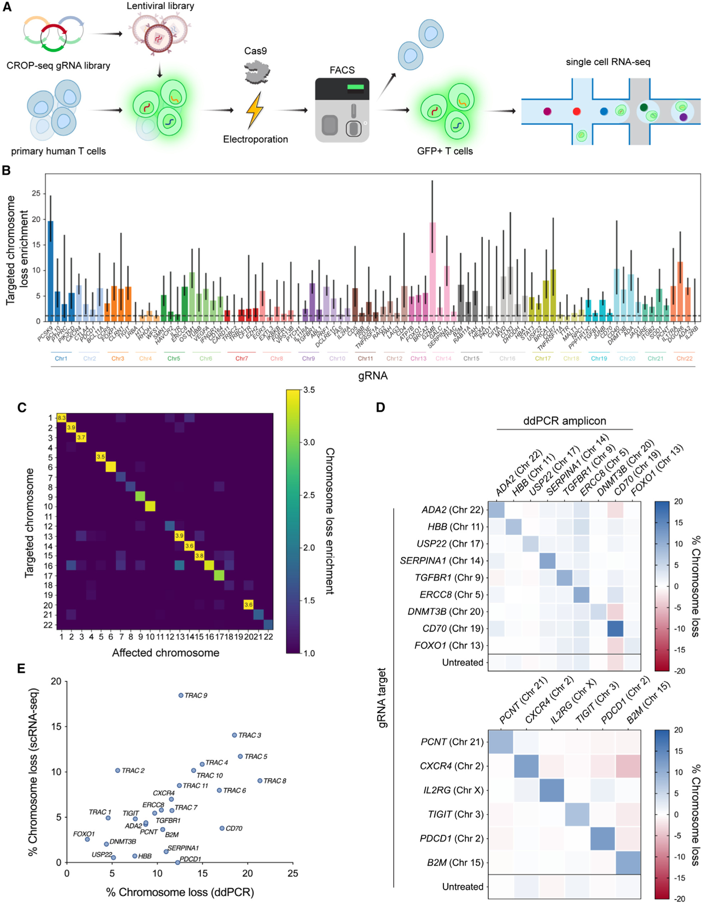
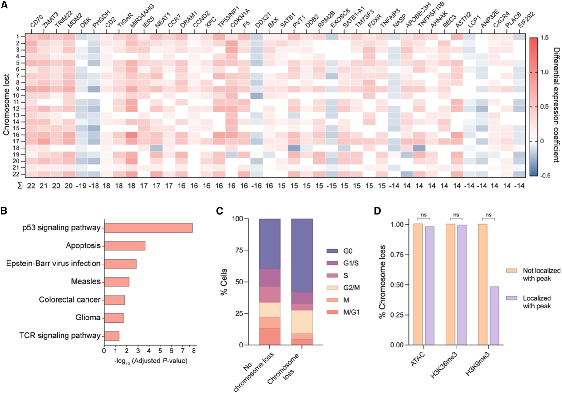
CRISPR-Cas9 genome editing has enabled advanced T cell therapies, but occasional loss of the targeted chromosome remains a safety concern. To investigate whether Cas9-induced chromosome loss is a universal phenomenon and evaluate its clinical significance, we conducted a systematic analysis in primary human T cells. Arrayed and pooled CRISPR screens revealed that chromosome loss was generalizable across the genome and resulted in partial and entire loss of the targeted chromosome, including in preclinical chimeric antigen receptor T cells. T cells with chromosome loss persisted for weeks in culture, implying the potential to interfere with clinical use. A modified cell manufacturing process, employed in our first-in-human clinical trial of Cas9-engineered T cells (NCT03399448), reduced chromosome loss while largely preserving genome editing efficacy. Expression of p53 correlated with protection from chromosome loss observed in this protocol, suggesting both a mechanism and strategy for T cell engineering that mitigates this genotoxicity in the clinic.

Keywords: CAR T cells; CRISPR screen; CRISPR-Cas9; DNA repair; T cells; aneuploidy; chromosome loss; clinical trial; genome editing; immunoengineering.


Conflict of interest statement

Declaration of interests C.A.T., J.A.D., and the Regents of the University of California have patents pending or issued related to the use of CRISPR genome editing technologies. R.B. is an employee of BioMarin Pharmaceutical Inc., J.L. is an employee of Altos Labs, and K.R.P. is a co-founder and employee of Cartography Biosciences. A.T.S. is a co-founder of Immunai and Cartography Biosciences. A.T.S. has received research support from Arsenal Biosciences, Allogene Therapeutics, and 10x Genomics. J.H.D.C. is a co-founder of Initial Therapeutics. J.E. is a co-founder of Mnemo Therapeutics, a scientific advisory board member of Cytovia Therapeutics, and a consultant for Casdin Capital, Resolution Therapeutics, IndeeLabs, and Treefrog Therapeutics. J.E. has received research support from Cytovia Therapeutics, Mnemo Therapeutics, and Takeda Pharmaceutical Company. J.A.F. has received research support from Tmunity. C.H.J. and the University of Pennsylvania have patents pending or issued related to the use of gene modification in T cells for adoptive T cell therapy. C.H.J. is a co-founder of Tmunity. H.Y.C. is a co-founder of Accent Therapeutics, Boundless Bio, Cartography Biosciences, and Orbital Therapeutics, and an advisor to 10x Genomics, Arsenal Biosciences, Chroma Medicine, Spring Discovery, and Vida Ventures. C.J.Y. is a co-founder of Survey Genomics, and a scientific advisory board member of Related Sciences and Immunai. C.J.Y. is a consultant for Maze Therapeutics, TReX Bio, ImYoo, and Santa Ana Bio. C.J.Y. has received research support from the Chan Zuckerberg Initiative, Chan Zuckerberg Biohub, Genentech, BioLegend, ScaleBio, and Illumina. J.A.D. is a co-founder of Editas Medicine, Intellia Therapeutics, Caribou Biosciences, Mammoth Biosciences, and Scribe Therapeutics, and a scientific advisory board member of Intellia Therapeutics, Caribou Biosciences, Mammoth Biosciences, Scribe Therapeutics, Vertex Pharmaceuticals, Felix Biosciences, The Column Group, Inari, and Isomorphic Labs. J.A.D. is the Chief Science Advisor at Sixth Street and a Director at Johnson & Johnson, Tempus, and Altos Labs. J.A.D. has sponsored research projects through Apple Tree Partners, Genentech, and Roche.

Figures

Figure 1.
Figure 1.. CRISPR-Cas9 genome editing of TRAC results in whole and partial chromosome loss
(A) Cas9 gRNA target sites tiled across the first exon of TRAC on chromosome 14. (B) Gene dosage from transcriptome-wide scRNA-seq of T cells treated with Cas9 and a non-targeting gRNA (top heatmap) or TRAC-targeting gRNA (bottom heatmap). Each individual row corresponds to a single cell and each column corresponds to a specific gene and its genomic position, grouped by chromosome (outlined in black). Red represents increase in gene dosage, while blue represents decrease in gene dosage. Rows outlined in black represent cells treated with different TRAC-targeting gRNAs. Blue arrows highlight chromosome 14, where TRAC is located. (C) Distribution of computationally predicted chromosome 14 breakpoints in cells predicted to have a chromosomal loss event. The distribution is an aggregate of 11 different TRAC-targeting gRNAs (all within ~300 bp) in cells with partial chromosome loss. (D) Representative single cell chromosome 14 gene dosage plots illustrating a cell with no chromosome loss (left), whole chromosome loss (middle), or partial chromosome loss (right). Gene dosage was normalized to non-targeting samples. Gray shaded area (gene dosage of 0.95–1.05) represents normal gene dosage (2 copies). Blue shaded area (gene dosage of <0.95) represents reduction in gene dosage (1 copy). Dotted lines represent the centromere, black lines represent the Cas9 target site, and the red line represents the computationally predicted breakpoint. (E) Quantification of whole and partial chromosome 14 loss across all gRNAs from scRNA-seq. NT indicates non-targeting gRNA. (F) Schematic of ddPCR assay to measure partial chromosome loss. The yellow lightning bolt represents the Cas9 target site. The detection of both HEX and FAM probes indicates no DSB or repaired DSB (top illustration). The detection of the HEX probe but not the FAM probe indicates a non-ligated DSB that represents partial chromosome loss (bottom illustration). Primers and probes were positioned ~30–120 bp from the Cas9 cut site (see Figure S1G). (G) Quantification of partial chromosome loss at the Cas9 target site across all gRNAs from ddPCR (n = 3, n = 2 biological donors). Numbers next to each point represent the TRAC-targeting gRNA. NT indicates non-targeting gRNA and represents samples from four different ddPCR amplicons. Error bars represent the standard deviation from the mean. Dashed line represents linear regression line of best fit and shaded region represent 95% confidence intervals (slope = 1.082; R2 = 0.9853). (H) Relationship between genome editing efficacy and partial chromosome loss for two TRAC-targeting gRNAs. 5-fold dilutions of Cas9 RNP were electroporated into T cells (n = 3). Genome editing efficacy was measured at the genomic level, via next-generation sequencing (NGS), or at the protein expression level, via flow cytometry, while chromosome loss was measured via ddPCR. Error bars represent the standard deviation from the mean. See also Figures S1 and S7.
Figure 2.
Figure 2.. Genome-scale CRISPR-Cas9 screen reveals target-specific chromosome loss
(A) Workflow for a CRISPR-Cas9 screen to estimate chromosome loss in T cells. Primary human T cells were transduced with a CROP-seq lentiviral library expressing one of 384 gRNAs. Cells were then electroporated with Cas9 protein, before GFP+ cells (co-expressed on the CROP-seq gRNA vector) were enriched via fluorescence-activated cell sorting. Enriched cells were subject to scRNA-seq and downstream analysis. (B) Quantification of targeted chromosome loss enrichment for each target gene. Each of the 92 bars represents the combination of four unique gRNAs targeting the same gene. Chromosome loss enrichment was calculated relative to the baseline loss per chromosome in cells containing a gRNA targeting a different chromosome. Error bars represent 95% confidence intervals. (C) Chromosomal loss enrichment at each somatic chromosome across all gRNAs. Rows represent the chromosome targeted by the Cas9 gRNA. Columns represent the chromosome analyzed for chromosome loss. Heatmap values are based on multiple gRNAs targeting multiple genes on a specified chromosome (all target genes appear in B). (D) Partial chromosome loss measured by ddPCR at 15 different Cas9 target sites across the genome. Row titles indicate the identity of the gRNA used. Column titles indicate the site in the genome that was analyzed via ddPCR. Heatmap values represent the mean of replicates (n = 3, except n = 2 for B2M target column). (E) Correlation between chromosome loss from 25 gRNAs as measured by scRNA-seq and ddPCR. Spearman’s correlation = 0.59; **p = 0.0017 (two-tailed). See also Figures S2, S3, and S7.
Figure 3.
Figure 3.. Genetic and epigenetic factors influence Cas9-induced chromosome loss
(A) Heatmap of differentially expressed genes in cells with chromosome loss compared with cells without chromosome loss. Cells with chromosome loss were divided into 22 groups depending on which somatic chromosome was lost (rows), and differentially expressed genes were individually investigated (columns). Upregulated genes are shown in red while downregulated genes are shown in blue. Genes were given a score of 1 (upregulated), —1 (downregulated), or 0 (no difference) for each chromosome loss group. Summed gene scores across all chromosome loss groups are shown below; genes with a score >|13| are displayed. (B) Gene ontology analysis based on differential gene expression. The most significantly upregulated modules are displayed. (C) Cell cycle analysis based on expression profiles. The percentage of cells in each cell cycle phase were quantified for cells with no chromosome loss or cells with chromosome loss. (D) Influence of epigenetic marks on chromosome loss. The gRNA sequence for cells with or without chromosome loss was analyzed for localization within ±75 bp of an epigenetic marker peak. p values were calculated using a two-sided Fisher’s exact test and are from left to right (ATAC) 0.365496, (H3K36me3) 0.789824, and (H3K9me3) 0.305706. ns = not significant. See also Figures S4 and S7.
Figure 4.
Figure 4.. Cas9-induced chromosome loss persists for weeks but results in reduced fitness and proliferation
(A) ddPCR measurements of partial chromosome loss at the Cas9 TRAC target site over 14 days. Error bars represent the standard deviation from the mean (n = 3). Day 3 results were additional used as the donor 2 (female) results shown in Figure 1G. (B) ddPCR measurements of partial chromosome loss for 15 different gRNAs targeted to sites across the genome over 14 days. Error bars represent the standard deviation from the mean (n = 3, except n = 2 for B2M). Day 3 results were additionally used for the diagonal values in the heatmaps of Figure 2D. (C) Measurement of chromosome loss across T cells of varying proliferative capacity. T cells were stained with CellTrace Violet (CTV) and cultured for 5 days before sorting the top and bottom quartile (top panel). ddPCR was used to measure partial chromosome loss in lowly proliferative (CTV high) and highly proliferative (CTV low) populations (bottom panel). NT = non-targeting gRNA. Non-targeted samples evaluated for chromosome loss at the gRNA 2 or gRNA 8 amplicon were combined into a single column (n = 3 for each of the two different ddPCR amplicons). p values are from Welch’s unpaired t tests and from left to right are 0.002970, 0.002970, and 0.275572. See also Figure S4.
Figure 5.
Figure 5.. Preclinical CAR T cell production via homology-directed repair results in chromosome loss
(A) Strategy to generate CAR T cells via HDR with Cas9. AAV6 encoding a 1928ζ CAR between left and right homology arms (LHA and RHA, respectively) serves as a template for HDR after Cas9 cleavage (yellow lightning bolt) of TRAC. (B) Three potential genomic outcomes after Cas9 HDR: indels that disrupt TCR expression (top), insertion of the CAR transgene that simultaneously disrupts TCR expression (middle), and chromosome loss that disrupts TCR expression (bottom). (C) Quantification of chromosome 14 loss enrichment across two TRAC-targeting gRNAs with or without an AAV HDR template from scRNA-seq (n = 2 biological donors). Two separate batches of CAR T cells were manufactured, before being subjected to scRNA-seq 4 or 7 days after generation. Chromosome 14 loss enrichment was calculated relative to T cells treated with Cas9 and a non-targeting gRNA. (D) Chromosome 14 loss enrichment over time, normalized to Cas9 editing efficacy (n = 2 biological donors). Editing efficacy was determined by the percentage of TCR negative cells as measured via flow cytometry (see Figure S5E). See also Figures S5 and S7.
Figure 6.
Figure 6.. Clinical CRISPR-Cas9 genome editing protocol in patient T cells mitigates chromosome loss
(A) Strategy to investigate chromosome loss in two clinical trial patients with CRISPR-edited T cells. Two patients with refractory cancer had T cells isolated, electroporated with TRAC, TRBC, and PDCD1-targeting Cas9 RNPs, and transduced with a lentivirus encoding an NY-ESO-1 TCR (NYCE cells). Cells were subjected to scRNA-seq prior to infusion (day 0) as well as at different time points postinfusion (days 10, 28, and/or 113). (B) Chromosome loss enrichment on chromosome 14 (TRAC), chromosome 7 (TRBC), or chromosome 2 (PDCD1) at different time points for both patients. Enrichment was calculated relative to non-targeted chromosomes (all chromosomes but 2, 7, and 14). Day 0 represents NYCE cells prior to infusion, while other later time points represent NYCE cells that were collected after circulation in vivo. (C) Diagram of two different protocols for Cas9 genome editing of primary human T cells. The activated T cell editing protocol (top) consisted of activating/stimulating cells prior to Cas9 electroporation and was used throughout this study along with most clinical trials. The non-activated T cell editing protocol (bottom) consisted of electroporating cells with Cas9 prior to activating/stimulating and is representative of our unique clinical trial. (D) Relative partial chromosome loss with 11 different TRAC-targeting gRNAs using the activated T cell editing or non-activated T cell editing protocol in primary human T cells. Partial chromosome loss was normalized to the indel efficacy (see Figure S6B) (n = 3 biological donors). (E) Fold TP53 mRNA expression during the activated T cell editing or non-activated T cell editing protocols for Cas9 genome editing of primary human T cells (n = 5 biological donors). Data points are the mean of two technical replicates. x-axis letters correspond to time points in (C). Cas9 DSBs represents the timepoints in the two protocols where Cas9 was electroporated into T cells to generate DSBs. See also Figures S6 and S7.

Update of

References

    1. Tsai SQ, Nguyen NT, Malagon-Lopez J, Topkar VV, Aryee MJ, and Joung JK (2017). CIRCLE-seq: a highly sensitive in vitro screen for genome-wide CRISPR–Cas9 nuclease off-targets. Nat. Methods 14, 607–614. 10.1038/nmeth.4278. - DOI - PMC - PubMed
    1. Wienert B, Wyman SK, Richardson CD, Yeh CD, Akcakaya P, Porritt MJ, Morlock M, Vu JT, Kazane KR, Watry HL, et al. (2019). Unbiased detection of CRISPR off-targets in vivo using DISCOVER-Seq. Science 364, 286–289. 10.1126/science.aav9023. - DOI - PMC - PubMed
    1. Bae S, Park J, and Kim J-S (2014). Cas-OFFinder: a fast and versatile algorithm that searches for potential off-target sites of Cas9 RNA-guided endonucleases. Bioinformatics 30, 1473–1475. 10.1093/bioinformatics/btu048. - DOI - PMC - PubMed
    1. Kleinstiver BP, Pattanayak V, Prew MS, Tsai SQ, Nguyen NT, Zheng Z, and Joung JK (2016). High-fidelity CRISPR–Cas9 nucleases with no detectable genome-wide off-target effects. Nature 529, 490–495. 10.1038/nature16526. - DOI - PMC - PubMed
    1. Chen JS, Dagdas YS, Kleinstiver BP, Welch MM, Sousa AA, Harrington LB, Sternberg SH, Joung JK, Yildiz A, and Doudna JA (2017). Enhanced proofreading governs CRISPR–Cas9 targeting accuracy. Nature 550, 407–410. 10.1038/nature24268. - DOI - PMC - PubMed

Publication types

Associated data

LinkOut - more resources