Review
. 2023 Sep 19:14:1230049.
doi: 10.3389/fimmu.2023.1230049. eCollection 2023.

Iatrogenic air embolism: pathoanatomy, thromboinflammation, endotheliopathy, and therapies

Affiliations
Review

Iatrogenic air embolism: pathoanatomy, thromboinflammation, endotheliopathy, and therapies

Phillip L Marsh et al. Front Immunol. .

Erratum in

  • Corrigendum: Iatrogenic air embolism: pathoanatomy, thromboinflammation, endotheliopathy, and therapies.
    Marsh PL, Moore EE, Moore HB, Bunch CM, Aboukhaled M, Condon SM 2nd, Al-Fadhl MD, Thomas SJ, Larson JR, Bower CW, Miller CB, Pearson ML, Twilling CL, Reser DW, Kim GS, Troyer BM, Yeager D, Thomas SG, Srikureja DP, Patel SS, Añón SL, Thomas AV, Miller JB, Van Ryn DE, Pamulapati SV, Zimmerman D, Wells B, Martin PL, Seder CW, Aversa JG, Greene RB, March RJ, Kwaan HC, Fulkerson DH, Vande Lune SA, Mollnes TE, Nielsen EW, Storm BS, Walsh MM. Marsh PL, et al. Front Immunol. 2024 Feb 6;15:1378003. doi: 10.3389/fimmu.2024.1378003. eCollection 2024. Front Immunol. 2024. PMID: 38380313 Free PMC article.

Abstract

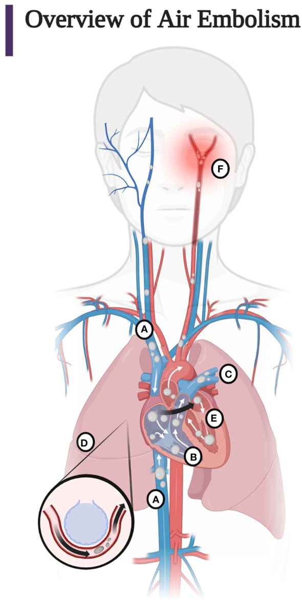
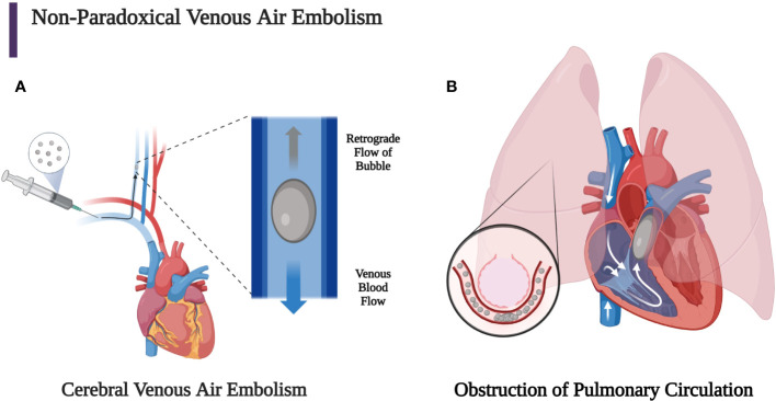
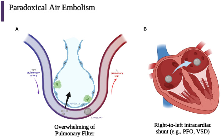
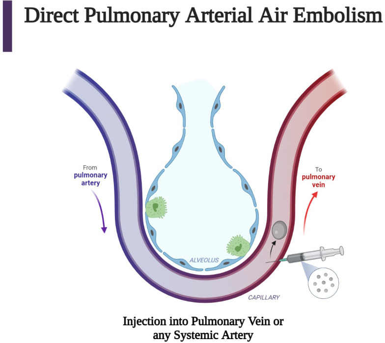
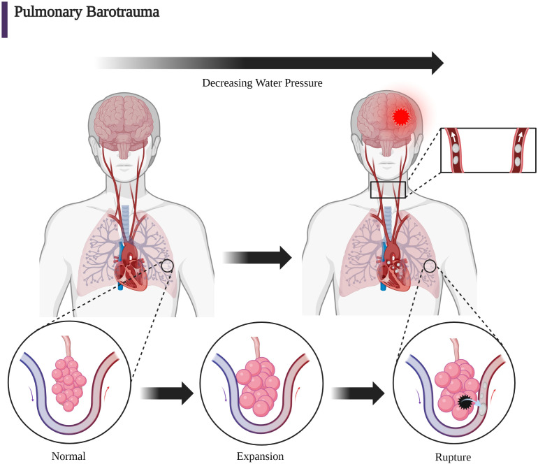
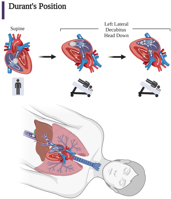
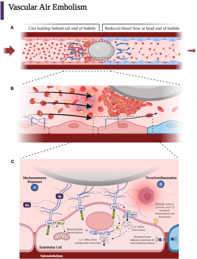
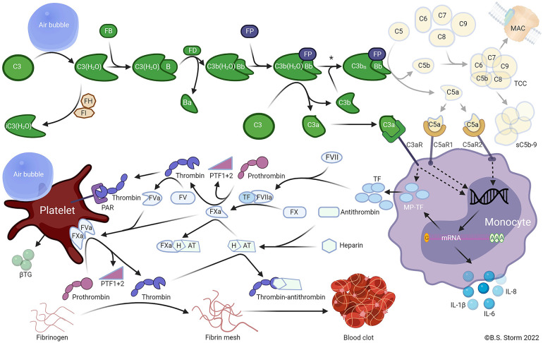
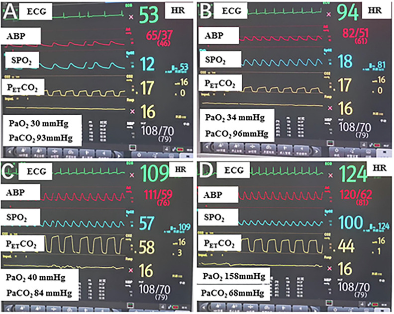
Iatrogenic vascular air embolism is a relatively infrequent event but is associated with significant morbidity and mortality. These emboli can arise in many clinical settings such as neurosurgery, cardiac surgery, and liver transplantation, but more recently, endoscopy, hemodialysis, thoracentesis, tissue biopsy, angiography, and central and peripheral venous access and removal have overtaken surgery and trauma as significant causes of vascular air embolism. The true incidence may be greater since many of these air emboli are asymptomatic and frequently go undiagnosed or unreported. Due to the rarity of vascular air embolism and because of the many manifestations, diagnoses can be difficult and require immediate therapeutic intervention. An iatrogenic air embolism can result in both venous and arterial emboli whose anatomic locations dictate the clinical course. Most clinically significant iatrogenic air emboli are caused by arterial obstruction of small vessels because the pulmonary gas exchange filters the more frequent, smaller volume bubbles that gain access to the venous circulation. However, there is a subset of patients with venous air emboli caused by larger volumes of air who present with more protean manifestations. There have been significant gains in the understanding of the interactions of fluid dynamics, hemostasis, and inflammation caused by air emboli due to in vitro and in vivo studies on flow dynamics of bubbles in small vessels. Intensive research regarding the thromboinflammatory changes at the level of the endothelium has been described recently. The obstruction of vessels by air emboli causes immediate pathoanatomic and immunologic and thromboinflammatory responses at the level of the endothelium. In this review, we describe those immunologic and thromboinflammatory responses at the level of the endothelium as well as evaluate traditional and novel forms of therapy for this rare and often unrecognized clinical condition.

Keywords: air embolism; arterioles; decompression sickness; hyperbaric oxygenation; microbubbles; thromboinflammation.


Conflict of interest statement

EM and HM have received research grants from Haemonetics Corporation outside the submitted work. The remaining authors declare that the research was conducted in the absence of any commercial or financial relationships that could be construed as a potential conflict of interest.

Figures

Figure 1
Figure 1
(A) Entry of air into the superior and inferior vena cava. Most air follows the superior or inferior vena cava into the right ventricle. Less commonly, air injected into the venous circulation will rise in a retrograde fashion into the cerebral circulation due to the natural buoyancy of the air bubble and cause protean symptoms of venous occlusion of the cerebral vasculature. This scenario occurs, for example, with the injection of air during the insertion or withdrawal of a central line in the sitting position. Note that due to the competing effects of buoyancy and drag, larger bubbles are more likely than smaller bubbles to become retrograde (60, 61). (B) Air in the right ventricle may totally occlude the pulmonary outflow tract causing an “air lock” or “vapor lock” with resultant shock. (C) Air may pass through the pulmonary artery, may diffuse into the alveoli, or be trapped in the pulmonary filter causing inflammation with impairment of gas exchange. (D) Air may overwhelm the pulmonary circulation and enter the left heart and systemic circulation or traverse a patent foramen ovale or a right-to-left shunt (arrow). (E) Air may enter the left heart and systemic circulation directly by injection into the pulmonary vein or from the right heart via the lung, a right-to-left shunt, or a patent foramen ovale. (F) Systemic air embolism, whether from the right heart or injected directly into the arterial circulation causes end organ damage, most commonly cerebral and cardiac. (Adapted from Storm 2022) (Created with BioRender.com).
Figure 2
Figure 2
(A) Bubbles that stay in the venous circulation may proceed retrogradely towards the head if the patient remains in a sitting position since the buoyancy of the bubble can overcome slow antegrade venous blood flow. Larger bubbles are more buoyant than smaller bubbles, so they are more likely to experience retrograde flow, which can allow the bubble to reach the brain and cause a venous cerebral air embolism. This can lead to mental status change, seizure, focal neurological deficit, and shock. (B) Venous air emboli may also travel through the superior or inferior vena cava into the heart, which can lead to obstruction of the right ventricular outflow tract if the bubble is large enough. Smaller bubbles may obstruct pulmonary capillaries, leading to pulmonary vasoconstriction, obstruction, and capillary leak (2, 3, 29, 60, 67). (Created with BioRender.com).
Figure 3
Figure 3
Iatrogenic paradoxical air embolism can occur when air enters the venous circulation with subsequent entrance into the systemic arterial circulation which then causes end-artery occlusion. (A) Venous air may overwhelm the pulmonary circulation either through a large bolus of gas or small continuous amounts. (B) Another mechanism for entrance of venous air into the arterial circulation is the passage of air through a patent foramen ovale (PFO), a ventricular septal defect (VSD), or another right-to-left cardiac or pulmonary shunt. Every venous air embolism has the potential to evolve into an arterial air embolism (2, 3, 84). (Created with BioRender.com).
Figure 4
Figure 4
Direct air embolism is caused by the entry of gas into the pulmonary veins or directly into the arteries of the systemic circulation. This figure describes the injection of air into pulmonary venous circulation, which can occur after a procedure such as a biopsy. Subsequent arterial air embolization can occur immediately after the biopsy (63). (Created with BioRender.com).
Figure 5
Figure 5
The mechanical forces due to increasing barometric pressure expressed on the alveolar wall can cause infiltration into the pulmonary venous circulation. With respect to DCI, Boyle’s law (P1V1 = P2V2) can give mathematical insight to the etiology of baro-rupture. As pressure decreases during ascent, volume of air in the lungs increases. This is why rapid ascension coupled with slow exhalation (or holding one’s breath) can lead to a disastrous scenario (63, 90). (Created with BioRender.com).
Figure 6
Figure 6
Coronal views of the right ventricle and pulmonary outflow tract based on the open chest observations by Durant after injection of large quantities of air into dogs in the supine position and after Durant’s maneuver. (Left) Supine position: “Air lock” caused by large air bubbles obstructing pulmonary outflow tract in the superior right ventricle. (Middle) Left lateral decubitus position: Immediately after assumption of Durant’s maneuver, air is no longer in the pulmonary outflow tract due to an inferior position in right ventricular outflow tract. (Right) Minutes after assumption of Durant’s maneuver, there is right ventricular compression of the bubble in the inferior aspect of the right ventricle undergoing mechanical induced “frothing” and movement of the smaller bubbles into the pulmonary circulation for absorption. (Bottom) In addition, air in the atrium may also migrate into the vena cava due to the increased mobility of the smaller bubbles, which may result in the microbubbles also migrating upward and distally into the hepatic vein and vena cava circulation (1, 15, 62, 81). (Adapted from Storm 2022) (Created with BioRender.com).
Figure 7
Figure 7
(A) As the bubble reaches smaller vessels, it may experience “slug flow” as it deforms to form a Taylor bubble—an elongated bubble that takes up the circumference of the vessel it occupies. The bubble may intermittently stop blood flow when it stops and starts, leading to an increase in pressure that causes the bubble to move again. This process continues so that the bubble stops and starts due to the pulsatile driving pressure behind it. A clot forms at the tail end of the air embolus. Flow recirculation causes fibrinogen, platelets, red blood cells, and white blood cells to accumulate at the tail end of the bubble surface, while the forward flow in front of the bubble moves the cellular components away from the bubble. (B) Accumulation of platelets, fibrin, activated white blood cells, and red blood cells around the gas bubble, which with further progression upstream into smaller microvasculature, can lead to hypoxia, penumbra, and terminal ischemia leading to cytotoxic edema and necrosis mediated at the endothelial bubble interface by leukocyte activation, which enhances inflammation causing increased blood viscosity and a reduction in blood flow. Blue endothelial cells represent post-obstructive induced hypoxia. Slug flow can contribute to thromboinflammation due to the buildup of thrombin and fibrin when the bubble slows down. (C) Heparan sulfates (HSs) of the endothelial luminal layer have been shown to interact with the air embolism. HS may also interact with the incipient clot at the tail of the embolus as well as with the fibrin, platelets, red blood cells, and activated neutrophils within the lubricant space between the endothelium and bubble. These HS are covalently bound and part of proteoglycans termed syndecans. Syndecans confer mechanosensing capabilities to the endothelium. Syndecan 4 (Syn4) transmits the extracellular shear stress exerted on the HS to the intracellular domain to phosphorylate and activate protein kinase C, which is thought to increase apoptotic signaling through mitochondrial depolarization within the endothelial cell. Inositol triphosphate (IP3) signaling molecules open Ca2+ channels on the endoplasmic reticulum to cause a transient increase in endothelial cytoplasmic calcium. The increase in cytoplasmic calcium is thought to disrupt adherens junctions, leading to loss of tight junctions between endothelial cells, increased endothelial permeability, and perivascular edema. Moreover, air emboli have been demonstrated to increase tissue factor levels and complement component 3 (C3) activation, leading to thromboinflammation (37, 121). (Created with BioRender.com).
Figure 8
Figure 8
Air emboli activate the alternative pathway and trigger a C3-dependent thromboinflammation. In plasma, C3 undergoes spontaneous slow-rate hydrolysis of the internal thioester to form C3(H2O). The C3 hydrolysis is accelerated by contact with foreign surfaces, e.g., an air embolus. C3(H2O) is inactivated to iC3(H2O) by factor I (FI) in the presence of a cofactor such as factor H (FH). C3(H2O) may bind FB (FB) to form C3(H2O)B. Catalyzed by factor D (FD), the small Ba fragment is cleaved from C3(H2O)B to form the fluid-phase C3 convertase C3(H2O)Bb. The C3 convertase is stabilized by properdin (FP) binding to form C3b(H2O)BbP. The stabilized C3 convertase cleaves additional C3 molecules to C3b and C3a or forms the C5 convertase C3bnBbP by binding to one or more C3b fragments deposited on foreign surfaces. The C5 convertase then cleaves C5 into C5a and C5b. C5b combines with C6, C7, C8, and C9 to form C5b-9, the terminal complement complex (TCC). TCC may become anchored in cell or bacterial membranes, forming a pore termed the membrane attack complex (MAC), which can cause the lysis of sensitive cells. Alternatively, the TCC may form a soluble complex (sC5b-9) in plasma. The C3a and C5a anaphylatoxins bind receptors for C3a and C5a (C3aR and C5aR1 or C5aR2, respectively) on various cells, including monocytes and granulocytes. The activation of anaphylatoxin receptors on monocytes stimulates de novo synthesis and surface expression of tissue factor (TF), extracellular release of microparticles expressing TF (MP-TF), and various inflammatory cytokines, including IL-1β, IL-6, and IL-8. TF binds to coagulation factor VIIa (FVIIa), subsequently activating FX to FXa. FXa catalyzes the cleavage of prothrombin to PTF1 + 2 and thrombin. Thrombin then catalyzes the cleavage of FV to FVa. FVa and FXa combine to form the prothrombinase complex on the surface of activated platelets. The prothrombinase complex cleaves prothrombin into prothrombin fragment 1 + 2 (PTF1 + 2) and thrombin. Platelets can be activated by several mechanisms, including thrombin binding to protease-activated receptors (PAR) 1 and 4, and possibly by direct contact with air emboli, whereby β-thromboglobulin (βTG) and many other mediators are released. Thrombin cleaves fibrinogen to fibrin, which crosslinks and forms a fibrin mesh leading to the formation of a blood clot. The affinity of antithrombin for thrombin is enhanced by binding heparin to form the heparin-antithrombin complex (HAT), which binds to and inactivates FXa. Footnote: The asterisk (*) indicated between the two C3 convertases (upper right) indicates that air emboli-activated C3 is less likely to form an active C5 convertase than is C3 activated by solid substances when C3b is bound to the surface, and the C5a-C5aR axis thus plays only a minor role in air-induced C3-driven thromboinflammation. Note: only key components relevant to the model are included in the figure (36). (Used with Permission from Mollnes et al., 2022) (Created with BioRender.com).
Figure 9
Figure 9
The changes in a patient’s vital signs during operation after treatment. (A) Patient’s vital signs when venous air embolism was considered; (B) Patient’s vital signs after Durant’s maneuver, sealing the operative field with saline, using pure oxygen, fluid resuscitation, and hemodynamics support. (C) Patient’s vital signs after an aggressive manual pulmonary recruitment maneuver was repeated for five minutes. (D) Patient’s vital signs after an aggressive pulmonary recruitment maneuver repeated for 10 minutes. ECG, Electrocardiograph; HR, heart rate; ABP, arterial blood pressure; SPO2, oxygen saturation; PETCO2, end-tidal carbon dioxide partial pressure; PaO2, arterial oxygen pressure; PaCO2, arterial carbon dioxide pressure (110). (Used with Permission from Zheng et al., 2022).

References

    1. Storm BS. Venous Air Embolism and Complement-driven Thrombo inflammation. In vitro human whole blood studies and in vivo porcine studies on the effect of air emboli on the complement system, cytokine network, and the hemostasis. (2022). Available at: https://hdl.handle.net/10037/25922.
    1. Muth CM, Shank ES. Gas embolism. N Engl J Med (2000) 342(7):476–82. doi: 10.1056/NEJM200002173420706 - DOI - PubMed
    1. van Hulst RA, Klein J, Lachmann B. Gas embolism: pathophysiology and treatment. Clin Physiol Funct Imag (2003) 23(5):237–46. doi: 10.1046/j.1475-097X.2003.00505.x - DOI - PubMed
    1. Mirski MA, Lele AV, Fitzsimmons L, Toung TJ. Diagnosis and treatment of vascular air embolism. Anesthesiology (2007) 106(1):164–77. doi: 10.1097/00000542-200701000-00026 - DOI - PubMed
    1. Senn NII. An experimental and clinical study of air-embolism. Ann Surg (1885) 1(6):517–49. doi: 10.1097/00000658-188501000-00002 - DOI - PMC - PubMed