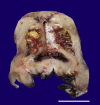
Xanthogranulomatous Endometritis with calculus formation in setting of prolapsed uterus
- PMID: 37795252
- PMCID: PMC10546669
- DOI: 10.4322/acr.2023.439
Xanthogranulomatous Endometritis with calculus formation in setting of prolapsed uterus
Abstract
Xanthogranulomatous inflammation is a rare benign inflammatory lesion characterized by sheets of lipid-laden foamy histiocytes. It has been reported in various organs, mainly the kidney and gall bladder. Xanthogranulomatous endometritis (XGE) is sporadic, with only a few cases reported in the English medical literature. Herein, we report a case of xanthogranulomatous endometritis with the formation of stones in a 50-year-old female patient with a prolapsed uterus. Grossly the endometrium was irregular, and the uterine cavity was filled with a yellow friable material, a polypoid growth, and yellowish stones. The microscopy showed sheets of histiocytes with few preserved endometrial glands. In this case, the xanthogranulomatous inflammation may mimic a clear cell carcinoma involving the endometrium and myometrium. One of the important differential diagnoses is malakoplakia. Immunohistochemistry and special stains are helpful in diagnosis.
Keywords: Endometritis; Endometrium; Histiocytes; Uterine Prolapse.
Copyright © 2023 The Authors.
Conflict of interest statement
Conflict of interest: None.
Figures


References
-
- Russack V, Lammers RJ. Xanthogranulomatous endometritis. Report of six cases and a proposed mechanism of development. Arch Pathol Lab Med. 1990;114(9):929–932. - PubMed

