. 2023 Oct;16(10):e015735.
doi: 10.1161/CIRCIMAGING.123.015735. Epub 2023 Oct 5.

Atrial Myopathy Quantified by Speckle-tracking Echocardiography in Mice

Affiliations

Atrial Myopathy Quantified by Speckle-tracking Echocardiography in Mice

Michael J Zhang et al. Circ Cardiovasc Imaging. 2023 Oct.

Abstract

Background: Emerging evidence suggests that atrial myopathy may be the underlying pathophysiology that explains adverse cardiovascular outcomes in heart failure (HF) and atrial fibrillation. Lower left atrial (LA) function (strain) is a key biomarker of atrial myopathy, but murine LA strain has not been described, thus limiting translational investigation. Therefore, the objective of this study was to characterize LA function by speckle-tracking echocardiography in mouse models of atrial myopathy.

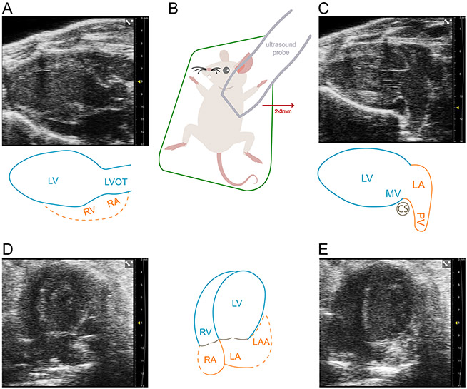
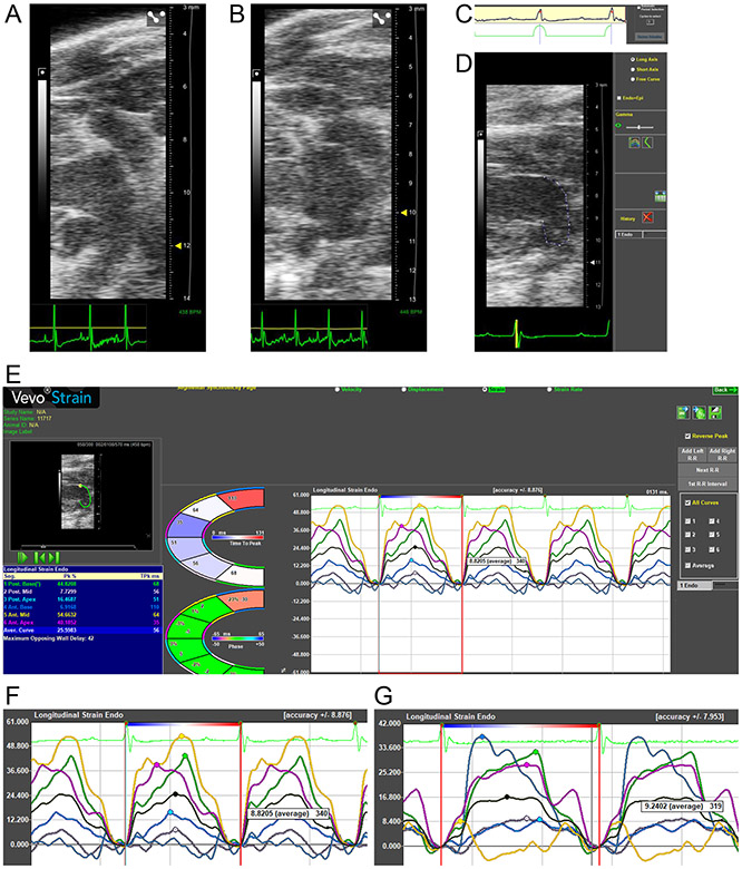
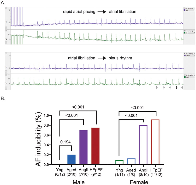
Methods: We used 3 models of atrial myopathy in wild-type male and female C57Bl6/J mice: (1) aged 16 to 17 months, (2) Ang II (angiotensin II) infusion, and (3) high-fat diet+Nω-nitro-L-arginine methyl ester (HF with preserved ejection fraction, HFpEF). LA reservoir, conduit, and contractile strain were measured using speckle-tracking echocardiography from a modified parasternal long-axis window. Left ventricular systolic and diastolic function, and global longitudinal strain were also measured. Transesophageal rapid atrial pacing was used to induce atrial fibrillation.

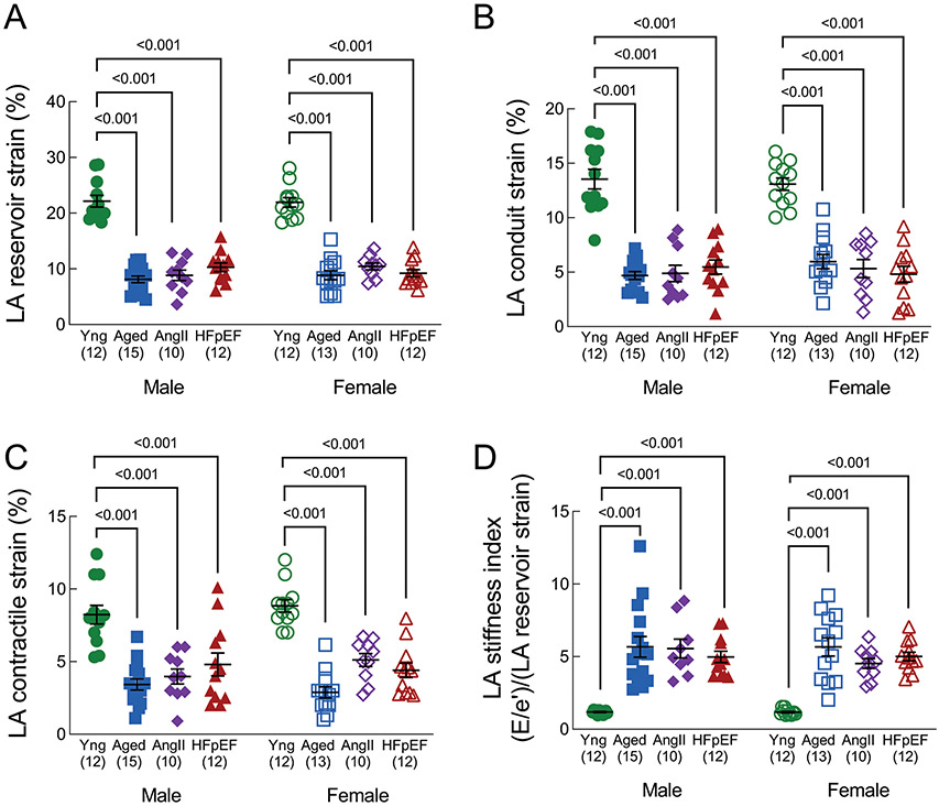
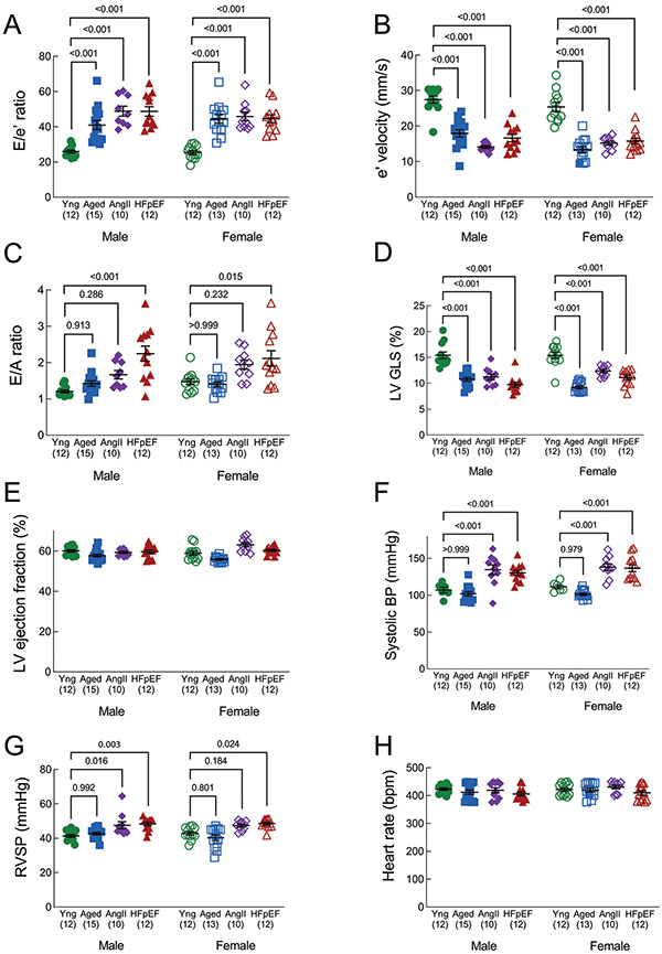
Results: LA reservoir, conduit, and contractile strain were significantly reduced in aged, Ang II and HFpEF mice compared with young controls. There were no sex-based interactions. Left ventricular diastolic function and global longitudinal strain were lower in aged, Ang II and HFpEF, but left ventricular ejection fraction was unchanged. Atrial fibrillation inducibility was low in young mice (5%), moderately higher in aged mice (20%), and high in Ang II (75%) and HFpEF (83%) mice.

Conclusions: Using speckle-tracking echocardiography, we observed reduced LA function in established mouse models of atrial myopathy with concurrent atrial fibrillation inducibility, thus providing the field with a timely and clinically relevant platform for understanding the pathophysiology and discovery of novel treatment targets for atrial myopathy.

Keywords: aging; atrial fibrillation; cardiomyopathies; echocardiography; heart failure; mice.


Conflict of interest statement

Disclosures None.

Figures

Figure 1.
Figure 1.. Current methods of examining the murine left atrium are inadequate
(A) Parasternal long axis view of the left ventricle (LV), LV outflow tract, and with partial view of the right ventricle (RV) and right atrium (RA). This is where left atrial (LA) size is commonly measured. (B) The modified parasternal long axis view, revealing the LA proper, can be achieved by moving the ultrasound probe laterally to the left by 2-3 mm. (C) Modified parasternal long axis view that displays the LA proper and one of three pulmonary veins. (D) Apical four chamber view during atrial diastole displaying the LA and partially obscured left atrial appendage (LAA). Optimization of the probe angle is difficult in this probe position due to the size of the ultrasound probe compared to the mouse thorax. (E) Apical four chamber view during atrial systole displaying a further obscured view of the LA and LAA. The obscured view of the LA and LAA limits the ability to track speckles in this echocardiographic window. Dotted lines in the figure indicate indeterminate anatomical borders. Scale marks indicate 1 millimeter.
Figure 2.
Figure 2.. Measurement of LA function in mice by speckle-tracking echocardiography
(A) Representative image and Video 1 of a left atrium (LA) from a young mouse. (B) Representative image and Video 2 of an LA from an AngII mouse. (C) In the software package to measure strain the period selection is kept at the default R-R interval so that LA reservoir strain is entirely positive through one cardiac cycle. (D) The long axis curve tool is utilized to draw a line between the anterior and posterior mitral valve annulus. This adapts the software, originally designed to measure LV strain, to calculate LA strain instead. (E) The Time-to-Peak software package is selected, with the “Reverse Peak” box checked, to calculate LA strain. The LA is divided automatically into six strain segments, but only the black-colored average curve values are utilized. LA contractile strain can be identified by the second peak following the P wave on ECG. LA conduit strain is calculated as the difference between LA reservoir and contractile strain. (F) Representative LA strain curves from a young mouse. (G) Representative LA strain curves from a HFpEF mouse.
Figure 3.
Figure 3.. Left atrial function
(A) LA reservoir strain, (B) LA conduit strain, (C) LA contractile strain, and (D) LA stiffness index, defined as E/e’ divided by LA reservoir strain %. A lower strain % represents worse mechanical function. LA conduit and contractile strain are presented as absolute values. Data in are mean±SEM and outcomes for each sex were analyzed by one-way ANOVA with Tukey’s multiple comparisons test. A primary interaction P value < 0.05 was considered significant, and significant differences identified by the post-test are indicated.
Figure 4.
Figure 4.. Left ventricular function
(A) E/e’ ratio (septal); higher E/e’ indicates worse diastolic function. (B) e’ velocity (mm/s); lower e’ is associated with worse diastolic function. (C) E/A ratio; higher E/A ratio indicates worse diastolic function. (D) LV global longitudinal strain, presented as an absolute value. Higher strain indicates higher function. (E) LV ejection fraction (systolic function). (F) Right ventricular systolic pressure, calculated from pulmonary artery acceleration and ejection times. (G) Systolic blood pressure, collected by tail cuff. (H) Heart rate, measured by ECG during echocardiography (parasternal long axis view). Data are mean±SEM and outcomes for each sex were analyzed by one-way ANOVA with Tukey’s multiple comparisons test. A primary interaction P value < 0.05 was considered significant, and significant differences identified by the post-test are indicated.
Figure 5.
Figure 5.. Rapid transesophageal atrial pacing to induce atrial fibrillation
(A) Example surface ECG tracing of a mouse that underwent transesophageal rapid atrial pacing to induce AF, followed by subsequent reversion to sinus rhythm (P wave denoted by red arrows). (B) Percentage and number of mice with inducible AF by transesophageal rapid atrial pacing among the mouse groups. P values were calculated by Fisher’s exact test.

Comment in

References

    1. Hoit BD. Left atrial size and function: role in prognosis. J Am Coll Cardiol. 2014;63:493–505. doi: 10.1016/j.jacc.2013.10.055 - DOI - PubMed
    1. Hoit BD, Shao Y, Gabel M, Walsh RA. In vivo assessment of left atrial contractile performance in normal and pathological conditions using a time-varying elastance model. Circulation. 1994;89:1829–1838. doi: 10.1161/01.cir.89.4.1829 - DOI - PubMed
    1. Goette A, Kalman JM, Aguinaga L, Akar J, Cabrera JA, Chen SA, Chugh SS, Corradi D, D'Avila A, Dobrev D, et al. EHRA/HRS/APHRS/SOLAECE expert consensus on atrial cardiomyopathies: Definition, characterization, and clinical implication. Heart Rhythm. 2017;14:e3–e40. doi: 10.1016/j.hrthm.2016.05.028 - DOI - PMC - PubMed
    1. Kamel H, Bartz TM, Elkind MSV, Okin PM, Thacker EL, Patton KK, Stein PK, deFilippi CR, Gottesman RF, Heckbert SR, et al. Atrial Cardiopathy and the Risk of Ischemic Stroke in the CHS (Cardiovascular Health Study). Stroke. 2018;49:980–986. doi: 10.1161/STROKEAHA.117.020059 - DOI - PMC - PubMed
    1. Carlisle MA, Fudim M, DeVore AD, Piccini JP. Heart Failure and Atrial Fibrillation, Like Fire and Fury. JACC Heart Fail. 2019;7:447–456. doi: 10.1016/j.jchf.2019.03.005 - DOI - PubMed

Publication types