. 2023 Oct;12(10):e12371.
doi: 10.1002/jev2.12371.

Engineered small extracellular vesicle-mediated NOX4 siRNA delivery for targeted therapy of cardiac hypertrophy

Affiliations

Engineered small extracellular vesicle-mediated NOX4 siRNA delivery for targeted therapy of cardiac hypertrophy

Ji-Young Kang et al. J Extracell Vesicles. 2023 Oct.

Abstract

Small-interfering RNA (siRNA) therapy is considered a powerful therapeutic strategy for treating cardiac hypertrophy, an important risk factor for subsequent cardiac morbidity and mortality. However, the lack of safe and efficient in vivo delivery of siRNAs is a major challenge for broadening its clinical applications. Small extracellular vesicles (sEVs) are a promising delivery system for siRNAs but have limited cell/tissue-specific targeting ability. In this study, a new generation of heart-targeting sEVs (CEVs) has been developed by conjugating cardiac-targeting peptide (CTP) to human peripheral blood-derived sEVs (PB-EVs), using a simple, rapid and scalable method based on bio-orthogonal copper-free click chemistry. The experimental results show that CEVs have typical sEVs properties and excellent heart-targeting ability. Furthermore, to treat cardiac hypertrophy, CEVs are loaded with NADPH Oxidase 4 (NOX4) siRNA (siNOX4). Consequently, CEVs@siNOX4 treatment enhances the in vitro anti-hypertrophic effects by CEVs with siRNA protection and heart-targeting ability. In addition, the intravenous injection of CEVs@siNOX4 into angiotensin II (Ang II)-treated mice significantly improves cardiac function and reduces fibrosis and cardiomyocyte cross-sectional area, with limited side effects. In conclusion, the utilization of CEVs represents an efficient strategy for heart-targeted delivery of therapeutic siRNAs and holds great promise for the treatment of cardiac hypertrophy.

Keywords: NADPH oxidase 4; cardiac hypertrophy; cardiac-targeting peptide; small extracellular vesicles; small-interfering RNA.


Conflict of interest statement

The authors declare no conflict of interest.

Figures

FIGURE 1
FIGURE 1
Characterization of CEVs and its toxicity in vitro. (a) Schematic diagram of CEVs generation by a two‐step reaction. (b) Transmission electron micrograph of SEVs and CEVs. Scale bar = 100 nm. (c) Size distribution of SEVs and CEVs, as determined by NTA. (d) Zeta potential analysis of SEVs and CEVs. (e) Representative blots of Alix, TSG101 and CD81 in both types of sEVs. The supernatant was used as a negative control. Uncropped blots are shown in Figure S11. (f and g) The analysis of cell viability and LDH release in iPSC‐vCMs treated with SEVs or CEVs. (h and i) Mean diameter of SEVs and CEVs stored at 4°C (h) and −80°C (i), determined by NTA.
FIGURE 2
FIGURE 2
In vitro and in vivo heart‐targeting ability of CEVs. (a–d) Representative immunofluorescence images and quantified data showing HEK293 cells, iPSC‐vCMs, AC16 and H9C2 cells treated with PKH67 (green)‐labelled SEVs and CEVs. Nuclei were stained with Hoechst 33342 (blue). Red fluorescence indicates Cy5.5‐azide conjugated with the sEVs. Scale bar = 50 μm. (e) Representative IVIS images and quantified data showing fluorescence intensity in mice heart tissues at 24 h after intravenous injection of PBS, SEVs, or CEVs in untreated mice; n = 3 per group. (f) Representative immunofluorescence images of SEVs and CEVs internalized by cTnI+ cardiomyocytes, vWF+ endothelial cells, or vimentin+ cardiac fibroblasts. Nuclei were stained with DAPI (blue); n = 3 per group. Scale bar = 20 μm. *P < 0.05.
FIGURE 3
FIGURE 3
Investigation of a molecular target for treating cardiac hypertrophy. (a) Schematic diagram of in silico experimental design and analysis. (b) Volcano plot for a comparison of gene expression profiles between patients with SR and AF. The x‐axis indicates the differential expression profiles, plotting the log2 (fold change). The y‐axis indicates the statistical significance of differences. (c) Venn diagram showing overlapping areas between DEGs and AF‐associated genes identified from DisGeNET. (d) GO and KEGG pathway enrichment analyses of selected 36 DEGs. (e) STRING PPI network of NOX4. (f and g) qRT‐PCR analysis of NOX4 levels in iPSC‐vCMs treated with Ang II at different concentrations and times. (h) qRT‐PCR analysis of NOX4, ANP, BNP and β‐MHC levels in the indicated groups. Data are normalized to GAPDH. *P < 0.05, **P < 0.01, ***P < 0.001, ****P < 0.0001.
FIGURE 4
FIGURE 4
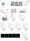
Characterization of CEVs@siNOX4 and its therapeutic effects in vitro. (a) Schematic diagram of siNOX4 loading onto CEVs. (B‐D) Transmission electron micrograph (b), size distribution (c) and zeta potential analysis (d) of CEVs@siCtrl, SEVs@siNOX4 and CEVs@siNOX4. Scale bar = 100 nm. (e) Representative blots of Alix, TSG101 and CD81 in three types of sEVs. Uncropped blots are shown in Figure S11. (f) Representative IVIS images and quantified data showing fluorescence intensity in mice heart tissues 24 h after intravenous injection of PBS, CEVs@siCtrl, SEVs@siNOX4 or CEVs@siNOX4. (g) qRT‐PCR analysis of NOX4 levels in iPSC‐vCMs. Data are normalized to GAPDH. (h) Representative blots and quantified data showing NOX4 protein levels in the indicated groups. β‐actin served as a loading control. Uncropped blots are shown in Figure S11. (i) qRT‐PCR analysis of ANP, BNP and β‐MHC levels in the indicated groups. Data are normalized to GAPDH. (j and k) Representative immunofluorescence images of α‐actinin (green)‐ and Hoechst 33342 (blue)‐stained iPSC‐vCMs along with quantified data showing cardiomyocyte size. Scale bar = 50 μm. * Indicates comparison with the control group, *P < 0.05, **P < 0.01, ***P < 0.001, ****P < 0.0001; # indicates comparison with the Ang II‐treated group, # P < 0.05, ## P < 0.01.
FIGURE 5
FIGURE 5
Therapeutic effects of CEVs@siNOX4 in vivo. (a) Schematic diagram of in vivo experimental design and analysis. (b) qRT‐PCR analysis of NOX4 levels in mice heart tissues; n = 4 per group. Data are normalized to GAPDH. (c and d) Representative images of H&E‐stained heart sections and quantified data showing HW/BW; n = 4 per group. Scale bar = 1 mm. (e and f) Representative images of MT‐stained heart sections and quantified data showing fibrotic area (%); n = 4 per group. Scale bar = 50 μm. (g and h) Representative immunofluorescence images of WGA‐stained heart sections and quantified data showing CSA; n = 4 per group. Scale bar = 50 μm. (i and j) Representative M‐mode images (i) and quantified data showing LVEF and LVFS (j) in the indicated groups; n = 4 per group. (k) qRT‐PCR analysis of ANP, BNP and β‐MHC levels in the indicated groups; n = 4 per group. Data are normalized to GAPDH. * indicates comparison with the control group, *P < 0.05, **P < 0.01, ***P < 0.001, ****P < 0.0001; # indicates comparison with the Ang II‐treated group, # P < 0.05, ## P < 0.01.

References

    1. Alvarez‐Erviti, L. , Seow, Y. , Yin, H. , Betts, C. , Lakhal, S. , & Wood, M. J. (2011). Delivery of siRNA to the mouse brain by systemic injection of targeted exosomes. Nature biotechnology, 29(4), 341–345. - PubMed
    1. Armstrong, J. P. , Holme, M. N. , & Stevens, M. M. (2017). Re‐engineering extracellular vesicles as smart nanoscale therapeutics. ACS Nano, 11(1), 69–83. - PMC - PubMed
    1. Avula, U. M. , Kim, G. , Lee, Y. E. , Morady, F. , Kopelman, R. , & Kalifa, J. (2012). Cell‐specific nanoplatform‐enabled photodynamic therapy for cardiac cells. Heart Rhythm, 9(9), 1504–1509. - PMC - PubMed
    1. Avula, U. M. , Yoon, H. K. , Lee, C. H. , Kaur, K. , Ramirez, R. J. , Takemoto, Y. , Ennis, S. R. , Morady, F. , Herron, T. , Berenfeld, O. , Kopelman, R. , & Kalifa, J. (2015). Cell‐selective arrhythmia ablation for photomodulation of heart rhythm. Science Translational Medicine, 7(311), 311ra172. - PubMed
    1. Balaj, L. , Lessard, R. , Dai, L. , Cho, Y. J. , Pomeroy, S. L. , Breakefield, X. O. , & Skog, J. (2011). Tumour microvesicles contain retrotransposon elements and amplified oncogene sequences. Nature Communications, 2, 180. - PMC - PubMed

Publication types

LinkOut - more resources