

Keratocyte-Derived Myofibroblasts: Functional Differences With Their Fibroblast Precursors
- PMID: 37796488
- PMCID: PMC10561788
- DOI: 10.1167/iovs.64.13.9
Keratocyte-Derived Myofibroblasts: Functional Differences With Their Fibroblast Precursors
Abstract
Purpose: In this study, we aim to elucidate functional differences between fibroblasts and myofibroblasts derived from a keratocyte lineage to better understand corneal scarring.
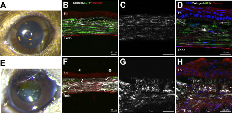
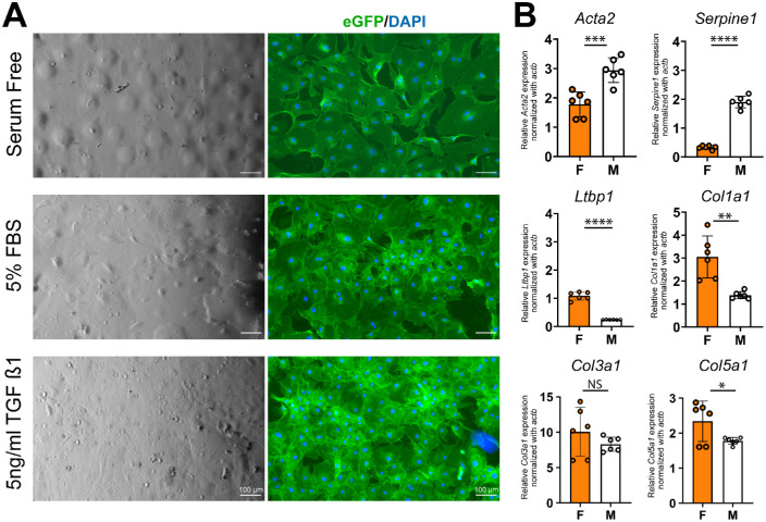
Methods: Corneal fibroblasts, derived from a novel triple transgenic conditional KeraRT/tetO-Cre/mTmG mouse strain that allows isolation and tracking of keratocyte lineage, were expanded, and transformed by exposure to transforming growth factor (TGF)-β1 to myofibroblasts. The composition and organization of a fibroblast-built matrix, deposited by fibroblasts in vitro, was analyzed and compared to the composition of an in vitro matrix built by myofibroblasts. Second harmonic generation microscopy (SHG) was used to study collagen organization in deposited matrix. Different extracellular matrix proteins, expressed by fibroblasts or myofibroblasts, were analyzed and quantified. Functional assays compared latent (TGF-β) activation, in vitro wound healing, chemotaxis, and proliferation between fibroblasts and myofibroblasts.
Results: We found significant differences in cell morphology between fibroblasts and myofibroblasts. Fibroblasts expressed and deposited significantly higher quantities of fibril forming corneal collagens I and V. In contrast, myofibroblasts expressed and deposited higher quantities of fibronectin and other non-collagenous matrix components. A significant difference in the activation of latent TGF-β activation exists between fibroblasts and myofibroblasts when measured with a functional luciferase assay. Fibroblasts and myofibroblasts differ in their morphology, extracellular matrix synthesis, and deposition, activation of latent TGF-β, and chemotaxis.
Conclusions: The differences in the expression and deposition of extracellular matrix components by fibroblasts and myofibroblasts are likely related to critical roles they play during different stages of corneal wound healing.
Conflict of interest statement
Disclosure:
Figures


References
-
- Fini ME. Keratocyte and fibroblast phenotypes in the repairing cornea. Prog Retin Eye Res. 1999; 18: 529–551. - PubMed
-
- Poole CA, Brookes NH, Clover GM.. Keratocyte networks visualised in the living cornea using vital dyes. J Cell Sci. 1993; 106(Pt 2): 685–691. - PubMed
-
- Yam GHF, Riau AK, Funderburgh ML, Mehta JS, Jhanji V. Keratocyte biology. Exp Eye Res. 2020; 196: 108062. - PubMed
-
- Liu CY, Birk DE, Hassell JR, Kane B, Kao WW.. Keratocan-deficient mice display alterations in corneal structure. J Biol Chem. 2003; 278: 21672–21677. - PubMed
Publication types
MeSH terms
Substances
Grants and funding
LinkOut - more resources
Full Text Sources
Medical
