. 2023 Oct 19;83(20):3679-3691.e8.
doi: 10.1016/j.molcel.2023.09.015. Epub 2023 Oct 4.

Crucial roles of the BRCA1-BARD1 E3 ubiquitin ligase activity in homology-directed DNA repair

Affiliations

Crucial roles of the BRCA1-BARD1 E3 ubiquitin ligase activity in homology-directed DNA repair

Meiling Wang et al. Mol Cell. .

Abstract

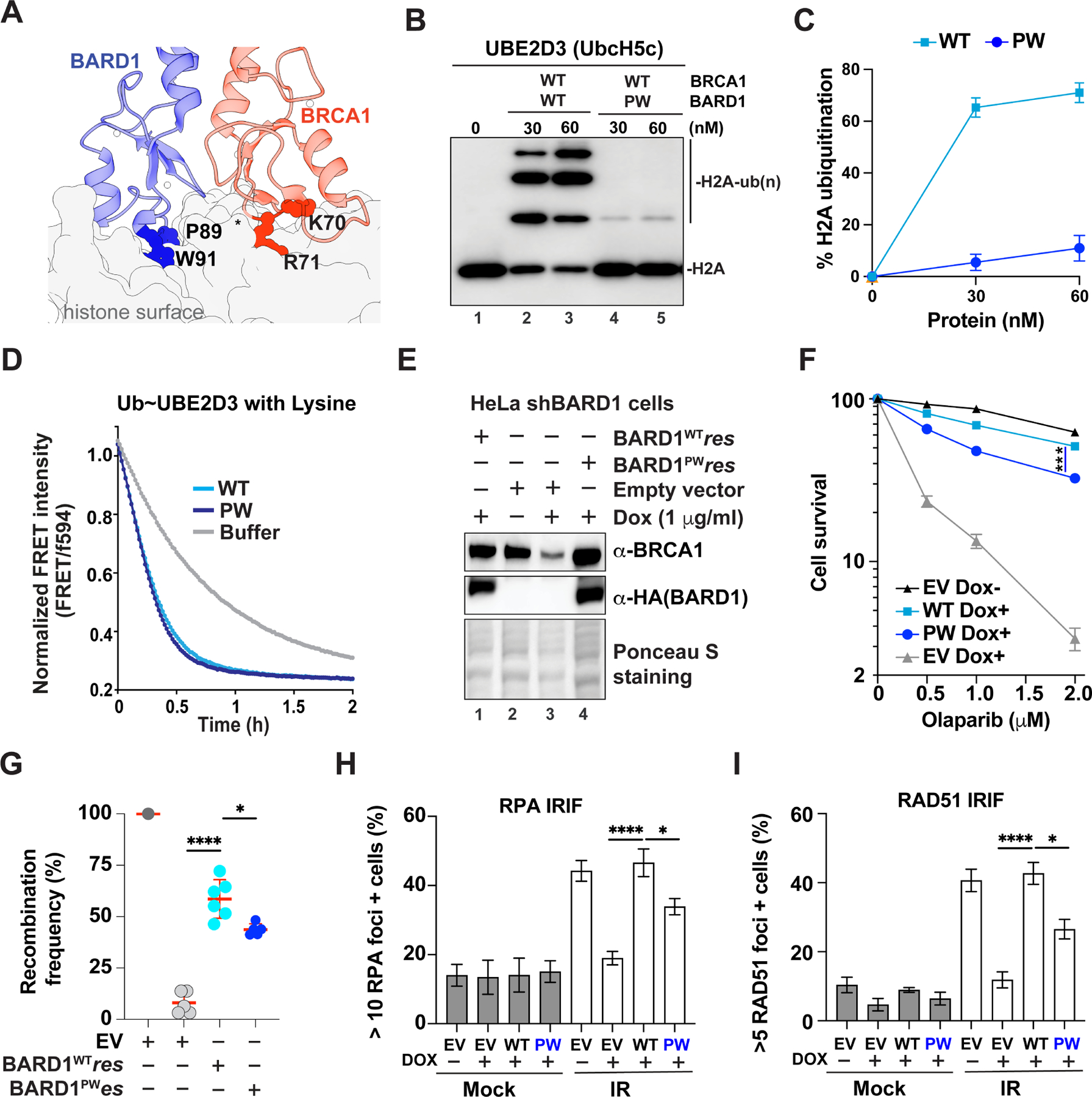
The tumor-suppressor breast cancer 1 (BRCA1) in complex with BRCA1-associated really interesting new gene (RING) domain 1 (BARD1) is a RING-type ubiquitin E3 ligase that modifies nucleosomal histone and other substrates. The importance of BRCA1-BARD1 E3 activity in tumor suppression remains highly controversial, mainly stemming from studying mutant ligase-deficient BRCA1-BARD1 species that we show here still retain significant ligase activity. Using full-length BRCA1-BARD1, we establish robust BRCA1-BARD1-mediated ubiquitylation with specificity, uncover multiple modes of activity modulation, and construct a truly ligase-null variant and a variant specifically impaired in targeting nucleosomal histones. Cells expressing either of these BRCA1-BARD1 separation-of-function alleles are hypersensitive to DNA-damaging agents. Furthermore, we demonstrate that BRCA1-BARD1 ligase is not only required for DNA resection during homology-directed repair (HDR) but also contributes to later stages for HDR completion. Altogether, our findings reveal crucial, previously unrecognized roles of BRCA1-BARD1 ligase activity in genome repair via HDR, settle prior controversies regarding BRCA1-BARD1 ligase functions, and catalyze new efforts to uncover substrates related to tumor suppression.

Keywords: BARD1; BRCA1; DNA damage response; DNA end resection; HDR; histone; homology-directed repair; later stages for HDR completion; substrates; tumor suppression; ubiquitin E3 ligase.


Conflict of interest statement

Declaration of interests D.N.I. is a co-founder and a shareholder of E3 Bioscience LLC, a commercial entity that manufactures FRET-active E2∼Ub conjugates used in this study.

Figures

Figure 1.
Figure 1.. Specificity and regulation of BRCA1-BARD1 mediated ubiquitylation in vitro. (See also Figure S1–4)
A. Domain organization of BRCA1 and BARD1. The domain names are indicated below the cartoons (RING, really interesting new gene; CC, coiled-coil; BRCT, BRCA1 C-terminal; ANK, Ankyrin repeat domain). The amino acid boundaries of exon 11 are labeled above. The findings that auto-ubiquitylation (Auto-ub) promotes the E3 ligase activity towards substrates (NCP/pCtIP) (black arrows), that polypeptides derived from the BRCA1 region encoded by exon 11 inhibits E3 activity (red block), and that NCP/pCtIP substrates require distinct domains of BRCA1-BARD1 for efficient ubiquitylation (black arrows) are summarized below. Double-direction cyan arrows show the known interactions between E3 and E2~Ub/NCP/pCtIP. Symbol: Ubyn, ubiquitylation. B. Representative nucleosome ubiquitylation assays monitoring H2A-Ub efficiency of BRCA1-BARD1 in conjunction with different E2s. UBE2B that does not bind BRCA1-BARD1 was included as a negative control. Symbol: BC1-BD1, BRCA1-BARD1. n=3 independent experiments. C. Representative nucleosome ubiquitylation assays monitoring H3-Ub efficiency of BRCA1-BARD1 in conjunction with different E2s. UBE2B that does not bind BRCA1-BARD1 was included as a negative control. Symbol: BC1-BD1, BRCA1-BARD1. n=3 independent experiments. D. Quantification of nucleosome ubiquitylation assays monitoring H2A-Ub efficiency of BRCA1Δ11q -BARD1 in conjunction with indicated amount of BSA and fragments of BRCA1 and BARD1 (BRCA1200–500, BRCA1500–894, BRCA936–1316, BARD11123–270, BARD11216–425 and BARD1425–565). Error bars, SEM (n=3). Representative gels are shown in Figure S3A, C. E. Quantification of nucleosome ubiquitylation assays monitoring H2A-Ub efficiency of auto-ubiquitylation form or native form of BRCA1-BARD1. Error bars, SEM (n=4). Representative gel is shown in Figure S4A. F. Quantification of nucleosome ubiquitylation assays monitoring H3-Ub efficiency of BRCA1-BARD1 in conjunction with unmodified, K13ub or K15ub form of nucleosome. Error bars, SEM (n=3–5). Representative gel is shown Figure S4C.
Figure 2.
Figure 2.. Identification of a BRCA1-BARD1 truly E3-dead mutant. (See also Figure S5, 6)
A. The BRCA1-BARD1 RING structure showing the locations of BRCA1 residues I26, L63, and K65 and BARD1 R99E with the E2~Ub binding and activation surface indicated by dotted lines (PDB: 7JZV). B. Representative nucleosome ubiquitylation assays monitoring H2A-Ub efficiency for wild-type or mutant BRCA1-BARD1 in conjunction with UBE2D3 & NCP_H2A (top), UBE2D3+UBE2N-UBE2V2 & NCP_H2A (middle); UBE2D3 & NCP_H2AK13ub (bottom). Quantification shown on the right. Error bars, SEM (n=3). C. Ub discharge was monitored as the decay of the Ub488~UBE2D3594 FRET signal over time after the addition of wild-type or mutant BRCA1-BARD1 (E3; 100 nM) and 200 nM NCP (substrate). The E3 UBR3 (RING trimer of TRIM5a) was included as a control.
Figure 3.
Figure 3.. BRCA1-BARD1 E3 ligase activity is required for cell survival following DNA damage (see also Figure S7)
A. Clonogenic survival of HeLa-shBRCA1 cells stably expressing wild-type or mutant HA-BRCA1 upon treatment with olaparib, CPT, MMC or cisplatin. Error bars, SEM (n=3–6). Symbol: EV, empty vector; OE BARD1, with ectopic expression of Flag-BARD1. B. Clonogenic survival of MDA-MB-436 cells stably expressing wild-type or mutant HA-BRCA1 without or with ectopic expression of Flag-BARD1 upon treatment with olaparib. Error bars, SEM (n=3–4). Symbol: EV, empty vector; OE BARD1, with ectopic expression of Flag-BARD1.
Figure 4.
Figure 4.. BRCA1-BARD1 E3 activity is critical for efficient HDR and early DNA end resection of HDR. (See also Figure S8, 9)
A. Schematic of HR assay using the DR-GFP reporter (top). Quantification of HR assay results (bottom) from DR-U2OS cells transiently expressing wild-type or mutant HA-BRCA1 upon treatment with siRNA against BRCA1 or control siRNA (siCtrl). Error bars, SD (n=3–4). Symbol: EV, empty vector. B Schematic of the SSA assay using the SA-GFP reporter (top). Quantification of SSA assay results (bottom) from examining SA-U2OS cells transient expressing wild-type or mutants of HA-BRCA1 upon their treatment with siRNA against BRCA1 or control siRNA (siCtrl). Error bars, SD (n=2–4). Symbol: EV, empty vector. C. Quantification of cells with >5 RAD51 foci four hours after exposure to 6 Gy irradiation in HeLa-shBRCA1 cells stably expressing wild-type or mutant HA-BRCA1, where endogenous BRCA1 was depleted by doxycycline incubation. Mean values ± SEM of at least three independent experiments are shown. D. Quantification of cells with >10 RPA foci four hours after exposure to 6 Gy irradiation in HeLa-shBRCA1 cells stably expressing wild-type or mutant HA-BRCA1, where endogenous BRCA1 was depleted by doxycycline incubation. Mean values ± SEM of at least three independent experiments are shown. E. Quantification of cells with >10 BrdU foci four hours after exposure to 6 Gy irradiation in HeLa-shBRCA1 cells stably expressing wild-type or mutant HA-BRCA1, where endogenous BRCA1 was depleted by doxycycline incubation. Mean values ± SEM of at least three independent experiments are shown. Statistical significance was assessed by two-tailed unpaired Student’s t-test and two-way ANOVA. ns: not significant, *P ⩽ 0.05, **P ⩽ 0.01, ***P⩽0.001, and ****P⩽0.0001 were considered significant.
Figure 5.
Figure 5.. BRCA1-BARD1 E3 activity is critical for late stages beyond DNA end resection of HDR. (See also Figure S9, 10)
A. Western blot to detect γ-H2AX (α-Tubulin as the loading control) at various times (0, 2, 3, 4 and 8h) after 2Gy irradiation. B. Clonogenic survival upon treatment with olaparib of HeLa-shBRCA1 cells stably expressing empty vector, wild-type or E3d mutant of HA-BRCA1, where endogenous BRCA1 and 53BP1 were depleted by doxycycline incubation and siRNA transfection, respectively. Error bars, SEM (n=4). Symbol: EV, empty vector. **** P⩽0.0001, by two-way ANOVA. C. Quantification of cells with >10 RPA foci four hours after exposure to 6 Gy irradiation in (right) HeLa-shBRCA1 cells stably expressing wild-type or E3d mutant of HA-BRCA1, where endogenous BRCA1 and 53BP1 were depleted by doxycycline incubation and siRNA transfection, respectively. Error bars, SEM (n=3). ****p<0.0001, by Student’s t-test. D. Representative micrographs of RAD51 foci (red) in the nucleus of HeLa-shBRCA1 cells stably expressing wild-type or E3d mutant of HA-BRCA1 at 24h, 34h and 48h after 24h exposure of 4 μM cisplatin. Blue: DAPI. Quantification of cells with >5 RAD51 foci at various time points from the release of cisplatin exposure. Mean values ± SEM of at least three independent experiments are shown. Scale bar: 10 μm. E. Quantification of Olive tail moment from the neutral comet assay of HeLa-shBRCA1 cells stably expressing wild-type or E3d mutant HA-BRCA1, where endogenous BRCA1 was depleted by doxycycline incubation. One hundred representative comets were analyzed in ImageJ and plotted in GraphPad PRISM. **p<0.01, ****p<0.0001, by Student’s t-test. Statistical significance was assessed by two-tailed unpaired Student’s t-test and two-way ANOVA. ns: not significant, *P ⩽ 0.05, **P ⩽ 0.01, ***P⩽0.001, and ****P⩽0.0001 were considered significant.
Figure 6.
Figure 6.. Role of the BARD1-nucleosome complex in histone ubiquitylation and in cell survival. (See also Figure S11, 12)
A. The BRCA1-BARD1 RING-histone interface showing the locations of BRCA1 K70R71 and BARD1 P89W91 relative to the histone surface (PDB: 7JZV). B. Representative nucleosome ubiquitylation assays monitoring H2A-Ub efficiency of wild-type or PW (P89AW91A mutant of BARD1) of BRCA1-BARD1. C. Quantification of b. Error bars, SEM (n=4). D. Ub discharge was monitored by the decay of the Ub488~UBE2D3594 FRET signal over time after the addition of wild-type or PW mutant BRCA1-BARD1 (E3; 100 nM) and 50 mM lysine (substrate). E. Western blot analysis to detect HA-BARD1 and BRCA1 from HeLa-shBARD1 cells stably expressing wild-type or PW mutant of HA-BARD1, where endogenous BARD1 was depleted by doxycycline incubation. F. Clonogenic survival of HeLa-shBARD1 cells stably expressing wild-type or PW mutants of HA-BARD1 upon treatment with olaparib. Error bars, SEM (n=3–6). Symbol: EV, empty vector. *** P⩽0.001, by two-way ANOVA. G. Quantification of HR assay results from DR-GFP U2OS cells transiently expressing wild-type or mutant HA-BARD1 upon treatment with siRNA against BARD1 or control siRNA (siCtrl). Error bars, SD (n=5–6). Symbol: EV, empty vector. *p<0.05, **** P⩽0.0001, by Student’s t-test. H. Quantification of cells with >10 RPA foci four hours after exposure to 6 Gy irradiation in HeLa- shBARD1 cells stably expressing wild-type or mutant HA-BARD1, where endogenous BARD1 was depleted by doxycycline incubation. Mean values ± SEM of at least three independent experiments are shown. Symbol: EV, empty vector. *p<0.05, **** P⩽0.0001, by Student’s t-test. I. Quantification of cells with >5 RAD51 foci four hours after exposure to 6 Gy irradiation in HeLa-shBAD1 cells stably expressing wild-type or mutant HA-BARD1, where endogenous BARD1 was depleted by doxycycline incubation. Mean values ± SEM of at least three independent experiments are shown. Symbol: EV, empty vector. *p<0.05, **** P⩽0.0001, by Student’s t-test. Statistical significance was assessed by two-tailed unpaired Student’s t-test and two-way ANOVA. ns: not significant, *P ≤ 0.05, **P ⩽ 0.01, ***P⩽0.001, and ****P⩽0.0001 were considered significant.
Figure 7.
Figure 7.. Model for BRCA1-BARD1 ubiquitin E3 ligase functions in DSB repair by HDR.
Our work demonstrates 1) critical roles for BRCA1-BARD1 E3 ligase in key stages of HDR, including DNA end resection (green arrow) and potential late steps for HDR completion (red arrows) via ubiquitylation substrates (to be identified), and 2) that histone (as one key substrate) ubiquitylation by BRCA1-BARD1 is important for DNA end resection therein. Symbol: S, Substrate; Ub, Ubiquitin (either monoubiquitylation or polyubiquitylation).

References

    1. Hall JM, Lee MK, Newman B, Morrow JE, Anderson LA, Huey B, and King MC (1990). Linkage of early-onset familial breast cancer to chromosome 17q21. Science 250, 1684–1689. 10.1126/science.2270482. - DOI - PubMed
    1. Petrucelli N, Daly MB, and Pal T (1998). BRCA1- and BRCA2-Associated Hereditary Breast and Ovarian Cancer. In GeneReviews((R)), Adam MP, Ardinger HH, Pagon RA, Wallace SE, Bean LJH, Stephens K, and Amemiya A, eds. [updated 2022 May 26].
    1. Hatchi E, Skourti-Stathaki K, Ventz S, Pinello L, Yen A, Kamieniarz-Gdula K, Dimitrov S, Pathania S, McKinney KM, Eaton ML, et al. (2015). BRCA1 recruitment to transcriptional pause sites is required for R-loop-driven DNA damage repair. Mol Cell 57, 636–647. 10.1016/j.molcel.2015.01.011. - DOI - PMC - PubMed
    1. Silver DP, and Livingston DM (2012). Mechanisms of BRCA1 tumor suppression. Cancer Discov 2, 679–684. 10.1158/2159-8290.CD-12-0221. - DOI - PMC - PubMed
    1. Scully R, Chen J, Plug A, Xiao Y, Weaver D, Feunteun J, Ashley T, and Livingston DM (1997). Association of BRCA1 with Rad51 in mitotic and meiotic cells. Cell 88, 265–275. 10.1016/s0092-8674(00)81847-4. - DOI - PubMed

Publication types