. 2023 Oct 5;14(1):6208.
doi: 10.1038/s41467-023-41895-2.

Acetylation-dependent coupling between G6PD activity and apoptotic signaling

Affiliations

Acetylation-dependent coupling between G6PD activity and apoptotic signaling

Fang Wu et al. Nat Commun. .

Abstract

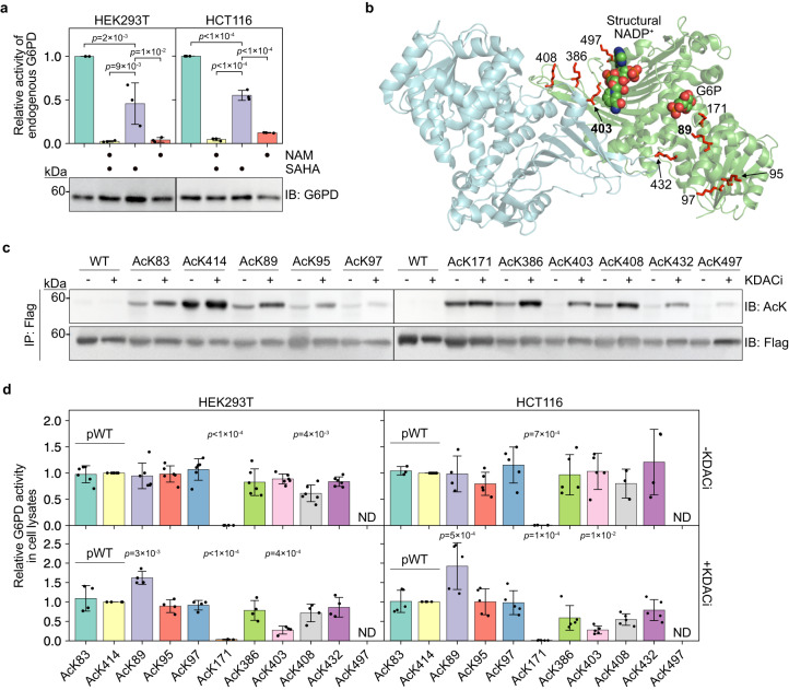
Lysine acetylation has been discovered in thousands of non-histone human proteins, including most metabolic enzymes. Deciphering the functions of acetylation is key to understanding how metabolic cues mediate metabolic enzyme regulation and cellular signaling. Glucose-6-phosphate dehydrogenase (G6PD), the rate-limiting enzyme in the pentose phosphate pathway, is acetylated on multiple lysine residues. Using site-specifically acetylated G6PD, we show that acetylation can activate (AcK89) and inhibit (AcK403) G6PD. Acetylation-dependent inactivation is explained by structural studies showing distortion of the dimeric structure and active site of G6PD. We provide evidence for acetylation-dependent K95/97 ubiquitylation of G6PD and Y503 phosphorylation, as well as interaction with p53 and induction of early apoptotic events. Notably, we found that the acetylation of a single lysine residue coordinates diverse acetylation-dependent processes. Our data provide an example of the complex roles of acetylation as a posttranslational modification that orchestrates the regulation of enzymatic activity, posttranslational modifications, and apoptotic signaling.


Conflict of interest statement

The authors declare no competing interests.

Figures

Fig. 1
Fig. 1. Site-specific acetylation modulates the catalytic activity of G6PD in cultured mammalian cells.
a Inhibition of KDACs downregulates G6PD activity. Bars represent the relative Vmax of endogenous G6PD measured in cleared lysates of HEK293T and HCT116 cells cultured in the presence or absence of KDACi. Data were analyzed using one-way ANOVA followed by Tukey’s post hoc test and are presented as mean values ± SD; n = 3 biologically independent samples. b Crystal structure of dimeric human G6PD (PDB ID: 6E08). Putative lysine acetylation sites selected for this study are presented in sticks model. G6P (predicted position) and structural NADP+ are displayed as spheres. The main focus of this study is on residues K89 and K403, which are highlighted in bold. c Acetylation of specific lysine residues is dynamic and differentially regulated by KDACs. Immunoblots show the acetylation level of immunopurified acetylated G6PD-Flag variants, expressed in HEK293T cells cultured in the presence (+) or absence (−) of KDACi. d Site-specific acetylation modulates G6PD activity. Bars represent the relative Vmax of exogenous full-length G6PD measured in cell lysate. Indicated acetylated G6PD variants were expressed in HEK293T (left) and HCT116 (right) cells cultured in the absence (top) or presence (bottom) of KDACi. G6PD activity was normalized to immunoblot intensities of expressed G6PD and displayed relative to AcK414 (pWT G6PD). Data were analyzed using one-way ANOVA followed by Tukey’s post hoc test and are presented as mean values ± SD; n = 4 or 6 (HEK293T), 3, 4, or 5 (HCT116) biologically independent samples. ND, not determined. Source data are provided as a Source Data file.
Fig. 2
Fig. 2. Lysine 403 acetylation inhibits and destabilizes G6PD in vitro and in cells.
a Purified site-specifically acetylated full-length G6PD expressed in E. coli. b Site-specific acetylation significantly affects the catalytic activity of recombinant G6PD. Bars represent the mean apparent kinetic parameters (obtained by fitting the data to the Michaelis–Menten equation) ± fitting error; n = 8 (Vmax), or 4 (KMapp) independent experiments (Supplementary Fig. 4a, b). Data were analyzed using one-way ANOVA followed by Tukey’s post hoc test. * p < 1 × 10−4. c Acetylation of K403 destabilizes the dimeric structure of G6PD. Indicated purified G6PD variants were cross-linked, resolved by SDS-PAGE, and visualized by immunoblotting using an antibody against the C-terminal 6×His tag. Bars represent the percentage of dimeric G6PD out of the total G6PD population [monomeric (Mr: ~59 KDa), and dimeric (Mr: ~120 KDa)]. Data were analyzed using one-way ANOVA followed by Tukey’s post hoc test and are presented as mean values ± SD; n = 3 independent experiments. d Lysine 403 acetylation reduces the thermodynamic stability of G6PD. Bars represent mean T1/2 values ± SD, measured at increasing concentrations of NADP+ (Supplementary Fig. 5); n = 3 (WT, AcK89, AcK386), or 4 (AcK403, AcK408, AcK432, AcK497). Data were analyzed using one-way ANOVA followed by Tukey’s post hoc test. e Lysine 403 acetylation increases the in vitro aggregation propensity of G6PD. Curves represent the accumulation of G6PD aggregates as a function of time and indicated concentration of NADP+. Aggregation was monitored by measuring the increase in scattered 340 nm light. Data are the mean ± SD, n = 3 independent experiments. f Lysine 403 acetylation increases the proteolytic degradation rate of G6PD in vitro. Western blots show the amount of indicated G6PD variants following chymotrypsin (Chy) digestion as a function of time (top) or as a function of chymotrypsin concentration (bottom). g Lysine 403 acetylation destabilizes G6PD in cells. Curves represent the relative amounts of G6PD variants in HCT116 cells as a function of time after CHX treatment. Data are the mean ± SD, n = 3 (WT, AcK403) or 5 (pWT, AcK89) biologically independent samples. ND not determined. Source data are provided as a Source Data file.
Fig. 3
Fig. 3. Lysine 89 acetylation promotes partial unwinding of helix αb’ and ubiquitylation of residues K95/97.
a Lysine 89 acetylation promotes ubiquitylation of G6PD. Representative Western blots showing the ubiquitylation level of immunoprecipitated Flag-tagged G6PD variants, co-expressed with HA-tagged ubiquitin in HEK293T cells treated with 5 μM MG132 for 16 h. b Mutation of lysine 95 and 97 to arginine does not affect G6PD activity. Bars represent the relative Vmax of pWT, AcK89, and indicated Lys-to-Arg G6PD mutants, measured in lysates of cells incubated in the absence of KDACi, and displayed relative to pWT G6PD. Data are the mean ± SD, n = 3 biologically independent samples. c AcK89 G6PD is ubquitylated on residues K95/97. Representative Western blots showing the ubiquitylation level of immunoprecipitated Flag-tagged G6PD variants, co-expressed with HA-tagged ubiquitin in HEK293T cells treated with 5 μM MG132 for 16 h. d Lysine 89 acetylation has minimal effect on the three-dimensional structure of monomeric G6PD. Superposition of AcK89 G6PD (green) and non-acetylated G6PD (cyan, PDB ID: 6E08). Structural NADP+ and K89 are shown in sticks model. e Lysine 89 acetylation promotes local conformational changes. Left: zoom in on K89 and nearby helices, showing the position of AcK89 (green) relative to the position of K89 (cyan, PDB ID: 6E08). Right: electron density map around residue AcK89. The 2Fo–Fc map was contoured at 1 σ. f Lysine 89 acetylation promotes partial unwinding of helix αb'. Local conformational changes around AcK89 with the partial unwinding of helix αb' (green), relative to the structure of non-acetylated G6PD (cyan, PDB ID: 6E08). Source data are provided as a Source Data file.
Fig. 4
Fig. 4. Lysine 403 acetylation is regulated by Sirt1 and Sirt2.
a The effect of acetylation on G6PD activity is maintained following the inhibition of endogenous KDACs. Bars represent relative Vmax of acetylated G6PD variants expressed in HEK293T cells cultured in the presence or absence of indicated KDACi. The activity was measured in cleared total cell lysates, corrected to G6PD expression level, and normalized to the activity of pWT expressed without KDACi treatment. Data were analyzed using one-way ANOVA followed by Tukey’s post hoc test and are presented as mean values ± SD; n = 4 (AcK171), 5 or 6 (AcK403), 6 (AcK89), or 9 (pWT) biologically independent samples. b Lysine 403 acetylation is stabilized upon inhibition of endogenous NAD+-dependent deacetylases. Representative Western blots showing the acetylation level of immunoprecipitated AcK89 and AcK403 G6PD, expressed in HEK293T cells cultured in the presence or absence of indicated KDACi. c The inhibitory effect of K403 acetylation is alleviated by NAD+-dependent deacetylases in vitro. Bars represent the relative Vmax of recombinant G6PD incubated with cleared lysates of HEK293T cells pretreated with indicated KDACi [mean ± SD; n = 4 (AcK89), or 5 (AcK403) independent experiments]. Western blots show the amount of G6PD (anti 6×His) and acetylation level following incubation with cleared cell lysates. d Lysine 403-acetylated G6PD is a substrate of Sirt1 and Sirt2. Western blots show the acetylation level of recombinant WT, AcK89, and AcK403 G6PD, following incubation with immunoprecipitated Sirt1, Sirt2, or HDAC6, in the presence or absence of indicated KDACi. e Sirt1 and Sirt2 reactivate AcK403 G6PD. Bars represent the relative Vmax of recombinant WT and AcK403 G6PD, following incubation with deacetylases as described in d. Data are the mean ± SD; n = 3 (Sirt1 treatment), or 7 (Sirt2, HDAC6 treatment) independent experiments. f Sirt1 and Sirt2 deacetylate AcK403 in a time-dependent manner. Representative Western blots showing K403 acetylation level as a function of incubation time with immunoprecipitated Sirt1, Sirt2, or HDAC6. Source data are provided as a Source Data file.
Fig. 5
Fig. 5. Lysine 403 acetylation promotes structural changes at the dimer interface and the active site.
a Dimeric structure of AcK403 G6PD. The expected position of catalytic NADP+ (light blue), structural NADP+ (light orange), and G6P (light purple) are presented as transparent spheres, based on superposition of AcK403 G6PD structure with structures of WT and mutant G6PD (PDB ID: 6VA7, 6E08, and 2BHL),,. Acetylated lysine residue 403 is highlighted in red. b Lysine 403 acetylation affects the dimeric structure of G6PD. Space-filling model of dimeric AcK403 G6PD (left) and WT G6PD (center, PDB ID: 6E08), with residue Glu93 highlighted in red. Right: The orientation of the lower monomers (darker shades) relative to the upper monomers (lighter shades) is compared by superimposing the upper monomers of each dimeric structure. The distance between Cα atoms of Glu93 residues is shown. c Lysine 403 acetylation is accompanied by distortion of βN and unwinding of αl. Superposition of AcK403 G6PD (green) and WT G6PD (cyan, PDB ID: 6E08), with a close-up view of the structural NADP+ binding site. Lysine 403 is presented in sticks model. d Close-up view of helices αf and αa' in AcK403 G6PD (green) and WT G6PD (cyan, PDB ID: 6E08). Backbone hydrogen bonds between residues V5–L7 and Y428–K432 that form a β-sheet-like structure are shown in gray. Residues L16, L20, and L442 that form a hydrophobic core between helices αa' and αm are rendered in sticks model. Also shown is residue K205, which rotates by ~180° following K403 acetylation and unfolding of helix αf'. e Lysine 403 acetylation promotes long-range conformational changes in the active site. Close-up view of the G6P binding site in AcK403 G6PD (green) and WT G6PD (cyan, PDB ID: 2BHL). The position of the substrate G6P in WT G6PD is shown, together with polar interactions with residues H201, Y202, and K205 (gray).
Fig. 6
Fig. 6. p53 modulates Fyn-dependent phosphorylation as a function of K403 acetylation.
a Y401 and Y503 are major Fyn phosphorylation sites in pWT and AcK403 G6PD, respectively. Representative Western blots showing tyrosine phosphorylation of immunoprecipitated G6PD co-expressed with Fyn. Data were analyzed using one-way ANOVA followed by Tukey’s post hoc test and presented as mean values ± SD; n = 3 biologically independent samples. b G6PD is phosphorylated by Fyn in vitro. Bars represent the relative phosphorylation level of recombinant G6PD, following in vitro incubation with Fyn. Data were analyzed using one-way ANOVA followed by Tukey’s post hoc test and are presented as mean values ± SD; n = 5 independent experiments. c Acetylation of G6PD K403 promotes the interaction with p53. Top: Co-IP using HA-tagged p53. Bottom: Reciprocal binding using 6×His-tagged G6PD. d AcK403 G6PD stabilizes p53 in cells. Bars represent the relative immunoblot intensities of endogenous p53, 120 min post-CHX treatment (Supplementary Fig. 10b). Data were analyzed using one-way ANOVA followed by Tukey’s post hoc test and are presented as mean values ± SD; n = 3 (pWT, AcK89, AcK171), or 4 (GFP, AcK403) biologically independent samples. e, f G6PD K403 acetylation-dependent interaction with p53' regulates Fyn-mediated G6PD phosphorylation at substoichiometric amounts of p53'. Representative Western blot (e) and quantification (f) of G6PD and p53' phosphorylation levels following in vitro incubation with active Fyn at indicated p53':G6PD molar ratios. Data were analyzed using one-way ANOVA followed by Tukey’s post hoc test and are presented as mean values ± SD; n = 6 or 7 independent experiments (Supplementary Fig. 10e). g p53' impairs Fyn-dependent G6PD phosphorylation at stoichiometric p53':G6PD ratio. Representative Western blot showing p53' and G6PD phosphorylation levels, following incubation with active Fyn. h G6PD K403 acetylation results in increased Bax levels and decreased Bcl2 levels in HEK293T cells. i G6PD K403 acetylation correlates with increased cleaved caspase-3 levels in HEK293T cells. j Annexin V/DAPI double staining of HEK293T cells expressing pWT or AcK403. Data were analyzed using two-sided t-test and are presented as mean values ± SD; n = 3 biologically independent samples. ns not significant, ND not detected. Source data are provided as a Source Data file.
Fig. 7
Fig. 7. Suggested model for the effect of K89 and K403 acetylation on activity and cellular functions of G6PD.
Left: Acetylation of K89 enhances the catalytic activity of G6PD and promotes its ubiquitylation on residues K95/K97. Sirt1 and Sirt2 can potentially deacetylate AcK89. Right: K403 is acetylated by KAT9 (Wang et al.) and CBP (this work) and deacetylated by Sirt2 (Wang et al. and this work) and Sirt1 (this work). Acetylation of K403 inhibits G6PD activity and promotes the interaction with p53, leading to the stabilization of p53 and induction of pro-apoptotic signaling. K403 acetylation also promotes Fyn-dependent phosphorylation of Y503, which can be partially inhibited by the interaction between K403-acetylated G6PD and p53. Y503 phosphorylation has no effect on K403 deacetylation by Sirt1 and Sirt2. Created with BioRender.com.

References

    1. Lin R, et al. Acetylation stabilizes ATP-citrate lyase to promote lipid biosynthesis and tumor growth. Mol. Cell. 2013;51:506–518. - PMC - PubMed
    1. Jiang W, et al. Acetylation regulates gluconeogenesis by promoting PEPCK1 degradation via recruiting the UBR5 ubiquitin ligase. Mol. Cell. 2011;43:33–44. - PMC - PubMed
    1. De Boor S, et al. Small GTP-binding protein Ran is regulated by posttranslational lysine acetylation. Proc. Natl. Acad. Sci. USA. 2015;112:E3679–E3688. - PMC - PubMed
    1. Mu A, et al. Regulation of INF2-mediated actin polymerization through site-specific lysine acetylation of actin itself. Proc. Natl. Acad. Sci. USA. 2020;117:439–447. - PMC - PubMed
    1. Wang Q, et al. Acetylation of metabolic enzymes coordinates carbon source utilization and metabolic flux. Science. 2010;327:1004–1007. - PMC - PubMed

Publication types