. 2023 Dec;37(12):2448-2456.
doi: 10.1038/s41375-023-02039-z. Epub 2023 Oct 5.

An "off-the-shelf" CD2 universal CAR-T therapy for T-cell malignancies

Affiliations

An "off-the-shelf" CD2 universal CAR-T therapy for T-cell malignancies

Jingyu Xiang et al. Leukemia. 2023 Dec.

Abstract

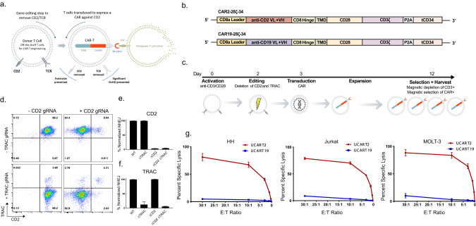
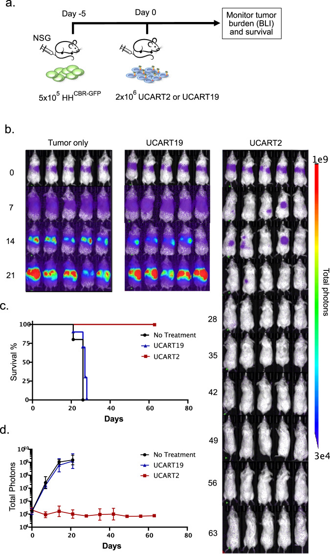
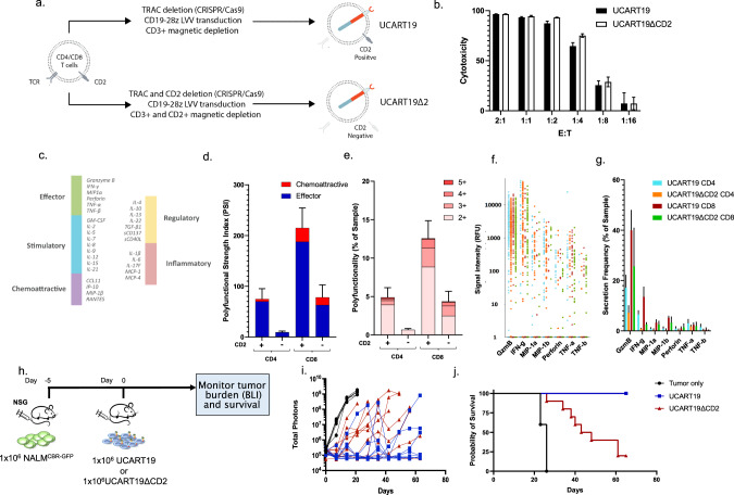
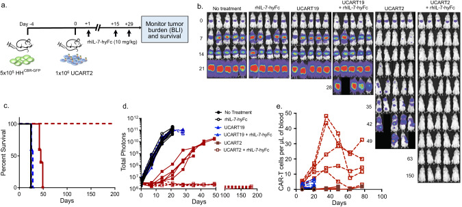
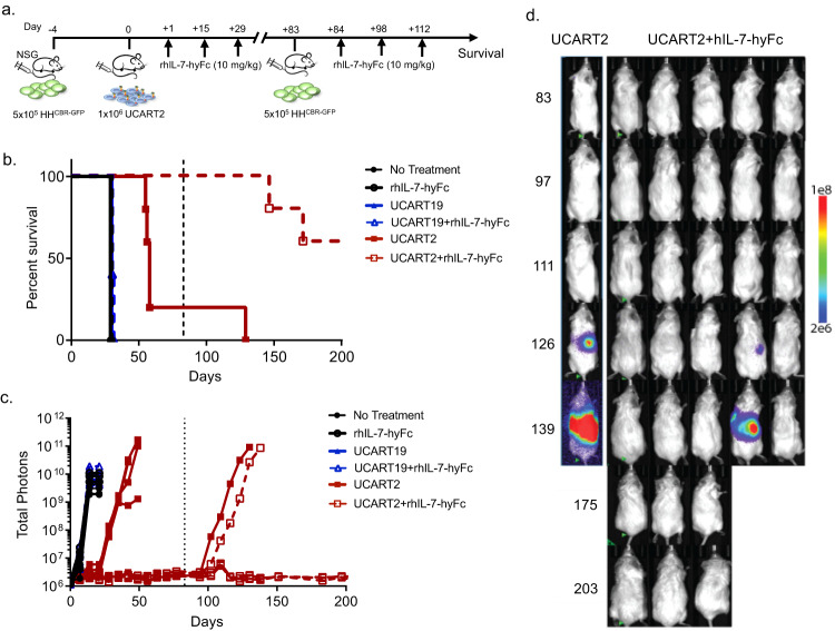
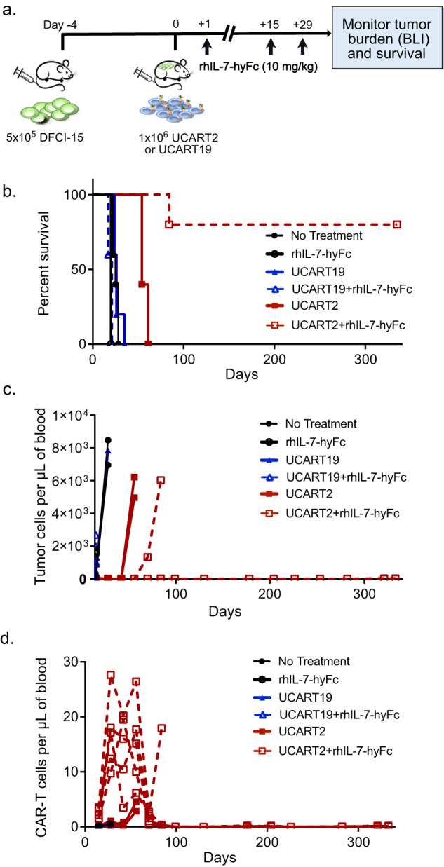
T-cell malignancies are associated with frequent relapse and high morbidity, which is partly due to the lack of effective or targeted treatment options. To broaden the use of CAR-T cells in pan T-cell malignancies, we developed an allogeneic "universal" CD2-targeting CAR-T cell (UCART2), in which the CD2 antigen is deleted to prevent fratricide, and the T-cell receptor is removed to prevent GvHD. UCART2 demonstrated efficacy against T-ALL and CTCL and prolonged the survival of tumor-engrafted NSG mice in vivo. To evaluate the impact of CD2 on CAR-T function, we generated CD19 CAR-T cells (UCART19) with or without CD2 deletion, single-cell secretome analysis revealed that CD2 deletion in UCART19 reduced frequencies of the effector cytokines (Granzyme-B and IFN-γ). We also observed that UCART19ΔCD2 had reduced anti-tumor efficacy compared to UCART19 in a CD19+NALM6 xenograft model. Of note is that the reduced efficacy resulting from CD2 deletion was reversed when combined with rhIL-7-hyFc, a long-acting recombinant human interleukin-7. Treatment with rhIL-7-hyFc prolonged UCART2 persistence and increased survival in both the tumor re-challenge model and primary patient T-ALL model in vivo. Together, these data suggest that allogeneic fratricide-resistant UCART2, in combination with rhIL-7-hyFc, could be a suitable approach for treating T-cell malignancies.


Conflict of interest statement

MYK, KWS, JO, MLC, and JFD are creators/inventors of a patent on the use of IL-7 to enhance CAR T cell function. MLC is currently employed by and has equity ownership in Wugen. JFD receives research funding from Amphivena Therapeutics, NeoImmuneTech, Macrogenics, Incyte, Bioline Rx, Wugen; has equity ownership in Magenta Therapeutics, Wugen; consults for Incyte, RiverVest Venture Partners, hC Bioscience, Inc.; and is a board member for RiverVest Venture Partners, Magenta Therapeutics. AJC is currently employed by BlueSphere Bio and has equity ownership in Wugen. GR and RT are currently employed by Integrated DNA Technologies. BHL is currently employed by NeoImmuneTech, Inc. The remaining authors declare no competing interests.

Figures

Fig. 1
Fig. 1. UCART2 kills T-ALL and CTCL in vitro.
a Schematic of UCART2 design. b Schematic of CAR2-28ζ-34 and CAR19-28ζ-34 CAR constructs. c Timeline of UCART2 production with CRISPR/Cas9 gene editing. T cells were cultured in Xcyte media supplemented with 50 U/mL IL-2 and 10 ng/ml IL-15 and activated with anti-CD3/CD28 beads (bead to cell ratio 3:1) for two days followed by T-cell transduction of either CD2 or CD19 CAR construct. Transduced T cells were expanded for 9 days, followed by CD3+ depletion and hCD34 enrichment. Efficiencies of multiplex CRISPR/Cas9 gene editing of CD2 and TRAC were assessed by d. flow cytometry (CD2 vs. TRAC), or e, f. targeted deep sequencing of CD2 or TRAC. % NHEJ was determined as a percentage of sequencing reads with indels relative to WT cells. g In vitro killing assay. UCART2 or UCART19 cells were cultured with 51Cr-labeled HH, Jurkat, or Molt-3 cells at various E: T ratios for 4 h. Specific lysis was calculated based on released 51Cr in the culture medium.
Fig. 2
Fig. 2. In vivo efficacy of UCART2 in a xenogeneic model of CTCL.
a Schema of the xenogeneic mouse model of CTCL. NSG mice were injected with 5 × 105 HHCBR-GFP cells on day -5, then infused with 2 × 106 UCART2 or UCART19 on day 0. b, c tumor burden was assessed with BLI weekly (n = 5 per group). d Kaplan–Meier survival curve of mice treated UCART19, UCART2, or untreated control. Median survival: untreated mice (26 days), UCART19 treated mice (27 days), UCART2 treated (no death at the end of the experiment on day 65, p < = 0.0001).
Fig. 3
Fig. 3. CD2 deletion affects the efficacy of UCART19.
a Schema of the generation of UCART19 (TRAC-CD2+) and UCART19ΔCD2 (TRAC-CD2-). b in vitro killing efficacy of UCART19 and U CART19ΔCD2. UCART19 and UCART19ΔCD2 were cultured with Nalm6 CBRGFP CD19+ targets at various E: T ratios for 16 h and target-specific killing was measured by luciferase activity. c Single-cell cytokine analysis using the IsoCode assay. UCART19 and UCART19ΔCD2 were incubated with B-cell lymphoma cell line Ramos at an E: T 1:2 for 20 h prior to loading purified CAR-T populations (CD19 depleted and CD34-affinity purified) onto the IsoCode chip. d Polyfunctionality and e Polyfunctional strength index (PSI) of the CD4 or CD8 subpopulation from UCART19 (CD2 +) or UCART19ΔCD2 (CD2-). f The signal strength of key cytokines driving polyfunctionality (GMZB, IFN-γ, MIP-1α, MIP-1β, Perforin, TNF-α, and TNF-β) was not affected by CD2 deletion. g The frequency of cells secreting Granzyme (Gzmb), IFN-γ, MIP1α, MIP1-β, Perforin, TNF-α, and TNF-β in the CD4 or CD8 subpopulation from UCART19 or UCART19ΔCD2. h Schema of the xenogeneic mouse model of CD19 + B cell acute lymphoblastic leukemia: 1 × 106 NALM6CBR-GFP cells were I.V. inoculated into NSG mice on day -5, followed by infusion of 1 × 106 UCART19 or UCART19ΔCD2 on day 0 (n = 10 per group). i Tumor burden as determined by BLI. j Kaplan–Meier survival curve. Median survival: untreated mice, 26 days, mice treated with UCART19 ΔCD2, 45.5 days, all mice treated with UCART19 were alive at day +65.
Fig. 4
Fig. 4. In vivo efficacy of UCART2 in combination with rhIL-7-hyFc.
a Schema of the xenogeneic mouse model of CTCL. NSG mice were injected with 5 × 105 HHCBR-GFP cells on day -4, then infused with 1 × 106 UCART2 or UCART19 on day 0. 10 mg/kg of rhIL-7-hyFc was delivered subcutaneously on day 1, day 15, and day 29. b tumor burden was measured weekly by BLI. c Kaplan–Meier survival curve of mice treated with UCART19, UCART2, UCART19+rhIL-7-hyFc, and UCART2+rhIL-7-hyFc. Median survival: untreated mice, mice treated with NT-17 alone, and mice treated with UCART19 were 25 days, mice treated with UCART19 + NT-17 was 26.8 days, mice treated with UCART2 alone was 45.4 days, mice treated with UCART2+rhIL-7-hyFc were all alive at 200 days when the experiment was ended. d Tumor burden as determined by BLI. e Flow cytometry analysis was performed bi-weekly to assess circulating CAR-T cells present per µl of peripheral blood. CAR-T population determined by hCD45+, 7aad-, hCD34+ cells. P values < 0.05 considered significant, *p ≤ 0.05, **p ≤ 0.01, ***p ≤ 0.001, ****p ≤ 0.0001. d BLI images normalized to a color gradient scale.
Fig. 5
Fig. 5. rhIL-7-hyFc prolongs UCART2 persistence in vivo and overcomes tumor re-challenge.
a Schema of the CTCL re-challenge mouse model. NSG mice were injected with 5 × 105 HHCBR-GFP cells on day -4, then infused with 1 × 106 UCART2 or UCART19 on day 0. 10 mg/kg of rhIL-7-hyFc was delivered subcutaneously on day +1, day +15, and day +29. Surviving mice were re-challenged with 5 × 105 HHCBR-GFP cells on day +83. 10 mg/kg of rhIL-7-hyFc was delivered subcutaneously on days +84, +98, and 112. b Kaplan–Meier survival curve. Mice receiving UCART. UCART19 Vs. UCART2, Median survival 30 days Vs. 58 days p = 0.0027. UCART19+rhIL-7-hyFc Vs. UCART2+rhIL-7-hyFc p = 0.0023 30 days Vs. 200+ days. c Tumor burden as determined by BLI imaging. d Normalized BLI images.
Fig. 6
Fig. 6. UCART2, in combination with rhIL-7-hyFc, kills primary patient T-ALL in vivo.
a Schema of the patient-derived T-ALL xenograft model. NSG mice were injected with 5 × 105 DCFI-15 cells on day -12, then infused with 1 × 106 UCART2 or UCART19 on day 0. 10 mg/kg of rhIL-7-hyFc was delivered subcutaneously on day +1, day +15, and day +29. b Kaplan–Meier survival curve Median survival: untreated mice 26 days, rhIL-7-hyFc alone - 20 days, UCART19 26 days, UCART19+rhIL-7-hyFc - 21 days, UCART2 - 54 days, mice treated with UCART2+rhIL-7-hyFc 300+ days. UCART19 Vs. UCART2 p = 0.0019, UCART19+rhIL-7-hyFc Vs. UCART2+rhIL-7-hyFc, p = 0.0031. c Flow cytometry analysis was performed by-weekly to assess circulating tumor cells present per µl in peripheral blood. Tumor population was determined as hCD45 +, 7-AAD-, hCD34-, hCD2+ cells. d. Flow cytometry analysis was performed bi-weekly to assess circulating CAR-T cells present per µl in peripheral blood. CAR-T population determined by hCD45 +, 7-AAD-, hCD34 +, hCD2- cells.

References

    1. Vadillo E, Dorantes-Acosta E, Pelayo R, Schnoor M. T cell acute lymphoblastic leukemia (T-ALL): New insights into the cellular origins and infiltration mechanisms common and unique among hematologic malignancies. Blood Rev. 2018;32:36–51. doi: 10.1016/j.blre.2017.08.006. - DOI - PubMed
    1. Alcantara M, Tesio M, June CH, Houot R. CAR T-cells for T-cell malignancies: challenges in distinguishing between therapeutic, normal, and neoplastic T-cells. Leukemia. 2018;32:2307–15. doi: 10.1038/s41375-018-0285-8. - DOI - PMC - PubMed
    1. Cooper ML, Choi J, Staser K, Ritchey JK, Devenport JM, Eckardt K, et al. An “off-the-shelf” fratricide-resistant CAR-T for the treatment of T cell hematologic malignancies. Leukemia. 2018;32:1970–83. doi: 10.1038/s41375-018-0065-5. - DOI - PMC - PubMed
    1. Gomes-Silva D, Srinivasan M, Sharma S, Lee CM, Wagner DL, Davis TH, et al. CD7-edited T cells expressing a CD7-specific CAR for the therapy of T-cell malignancies. Blood. 2017;130:285–96. doi: 10.1182/blood-2017-01-761320. - DOI - PMC - PubMed
    1. Png YT, Vinanica N, Kamiya T, Shimasaki N, Coustan-Smith E, Campana D. Blockade of CD7 expression in T cells for effective chimeric antigen receptor targeting of T-cell malignancies. Blood Adv. 2017;1:2348–60. doi: 10.1182/bloodadvances.2017009928. - DOI - PMC - PubMed

Publication types