. 2023 Oct 5;13(1):16763.
doi: 10.1038/s41598-023-43711-9.

Design of hypoxia responsive CRISPR-Cas9 for target gene regulation

Affiliations

Design of hypoxia responsive CRISPR-Cas9 for target gene regulation

Yan An et al. Sci Rep. .

Abstract

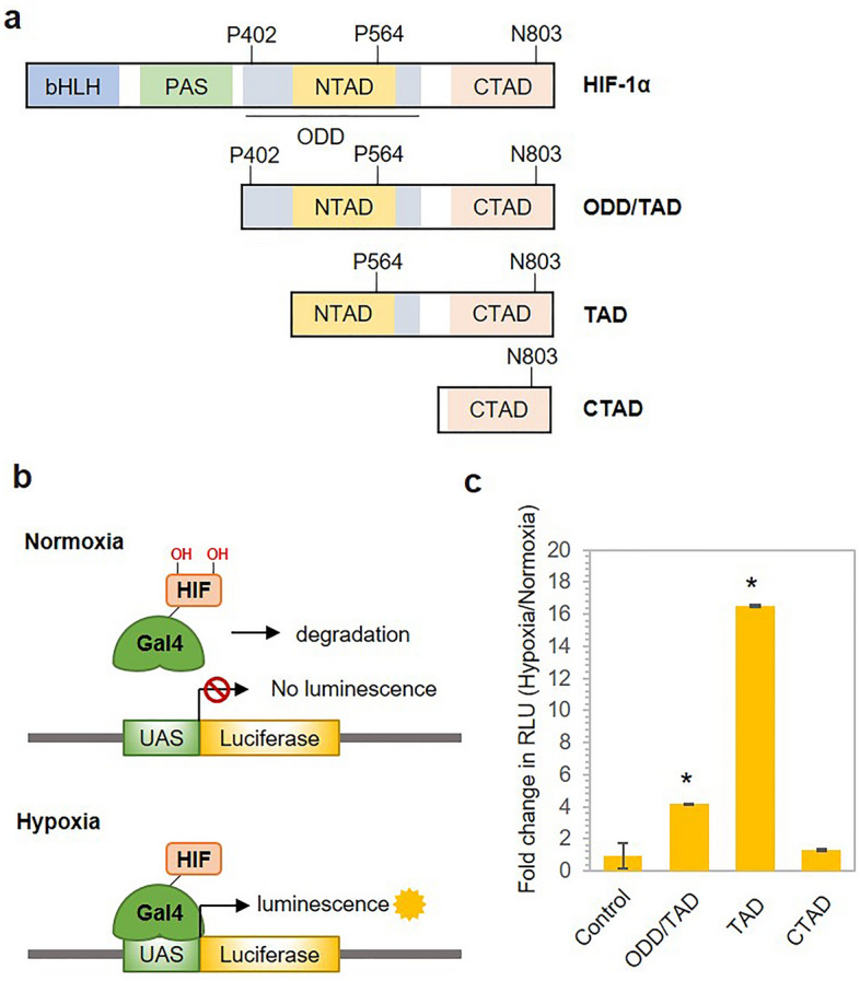
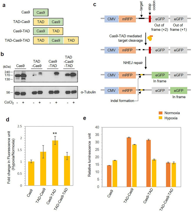
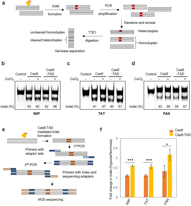
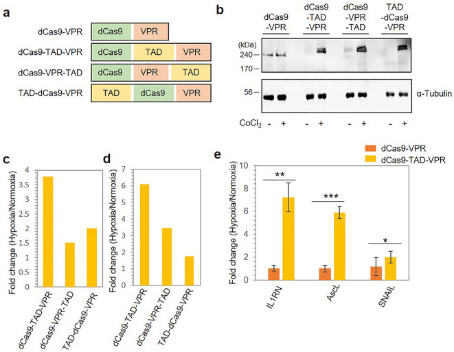
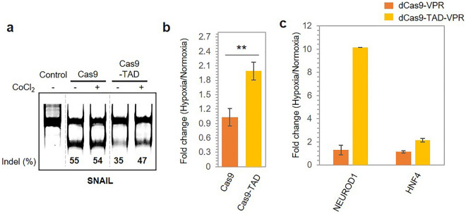
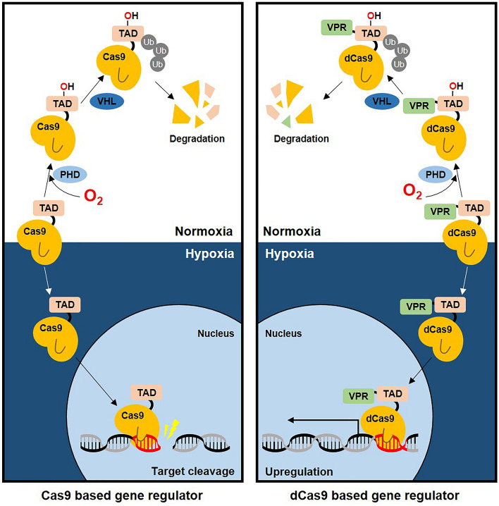
The CRISPR-Cas9 system is a widely used gene-editing tool, offering unprecedented opportunities for treating various diseases. Controlling Cas9/dCas9 activity at specific location and time to avoid undesirable effects is very important. Here, we report a conditionally active CRISPR-Cas9 system that regulates target gene expression upon sensing cellular environmental change. We conjugated the oxygen-sensing transcription activation domain (TAD) of hypoxia-inducing factor (HIF-1α) with the Cas9/dCas9 protein. The Cas9-TAD conjugate significantly increased endogenous target gene cleavage under hypoxic conditions compared with that under normoxic conditions, whereas the dCas9-TAD conjugate upregulated endogenous gene transcription. Furthermore, the conjugate system effectively downregulated the expression of SNAIL, an essential gene in cancer metastasis, and upregulated the expression of the tumour-related genes HNF4 and NEUROD1 under hypoxic conditions. Since hypoxia is closely associated with cancer, the hypoxia-dependent Cas9/dCas9 system is a novel addition to the molecular tool kit that functions in response to cellular signals and has potential application for gene therapeutics.


Conflict of interest statement

The authors declare no competing interests.

Figures

Figure 1
Figure 1
Selection of the hypoxia-sensing domain of HIF-1α. (a) Schematic diagram showing the domain distribution of HIF-1α. Residues marked in the diagram represent the key residues involved in sensing hypoxia. (b) Graphical representation showing the principle of the luciferase reporter assay. (c) Bar graph showing the hypoxia-sensing efficiency of the truncated variants of HIF-1α as examined with a luciferase reporter assay. The Y-axis shows the fold change in the relative luminescence units (RLUs) under hypoxic conditions normalized to those obtained under normoxic conditions. The error bars represent the standard deviation (SD) of the results from three technical replicates. P values were calculated using two-tailed t test. *P < 0.05, **P < 0.01 and ***P < 0.001 vs. the control group.
Figure 2
Figure 2
Construction of a hypoxia-dependent Cas9 regulator by conjugation with TAD. (a) Schematic presentation of the conjugate proteins created by the fusion of the TAD and Cas9. The diagrams shows the placement of the TAD with respect to Cas9 in each of the three variants constructed. (b) Western blotting results showing the protein expression of the conjugate proteins under normoxic and hypoxic conditions. The protein expression was investigated using anti-Cas9 antibodies 12 h after transfection. The lower lane shows the protein level of α-Tubulin. (c) Schematic representation of the mRFP-eGFP reporter assay used to investigate the hypoxia-sensitive target cleavage activity of the conjugate proteins. (d) Bar graph showing the quantified results from the mRFP-eGFP reporter assay. The fluorescence intensity was quantified using ImageJ software and is expressed as the fold change in fluorescence normalized to the fluorescence observed under normoxic conditions. The error bars represent the standard deviation (SD) of the results from three technical replicates. P values were calculated using two-tailed t test. *P < 0.05, **P < 0.01 and ***P < 0.001 vs. the Cas9 group. (e) Hypoxia-dependent Cas9-mediated cleavage of the target luciferase gene was assessed with a luciferase reporter assay. The marked value indicates the luciferase activity observed under hypoxic conditions normalized to that observed under normoxic conditions. The error bars represent the standard deviation (SD) of two technical replicates.
Figure 3
Figure 3
Hypoxia-dependent regulation of endogenous gene expression by Cas9-TAD. (a) Schematic representation of the T7 endonuclease assay. Cas9-TAD activity on the endogenous genes (b) INIP, (c) TAT, and (d) FAN as shown by the T7 endonuclease assay. The gel images are shown. Different parts of the same gel are grouped together to place Cas9 and Ca9-TAD conditions next to each other. Original gel image can be found in Supplementary data (Supplementary Fig. S10a–c). Representative gel image of T7 endonuclease-treated PCR products amplified from the target sites. (−) and (+) represent the presence and absence of cobalt chloride, respectively. The intensity of the bands from the T7 endonuclease assay was quantified using ImageJ software, and the percentage of indels was calculated as the ratio of the intensity of the cleaved fragments to the sum of the intensities of the cleaved and uncleaved products and is shown below each lane. (e) Schematic presentation of the steps involved in NGS sequencing. (f) Cas9-TAD activity on the endogenous genes INIP, TAT and FAN as shown by NGS sequencing. The graph shows the fold change in indel formation. The plotted values indicate the average of the results, and the error bars represent the standard deviation of the results from three technical replicates. P values were calculated using two-tailed t test. *P < 0.05, **P < 0.01 and ***P < 0.001 vs. the Cas9 group.
Figure 4
Figure 4
Construction of a hypoxia-dependent regulator by conjugation of dCas9 with TAD. (a) Schematic presentation of the conjugate proteins created by the fusion of TAD, dCas9 and VPR. (b) Western blotting results showing the protein expression of the conjugate proteins under normoxic and hypoxic conditions. The protein expression was investigated using anti-Cas9 antibodies 12 h after transfection. (c) The luciferase gene located on a plasmid under a GAL4 promoter was targeted using sgRNA, and the luciferase activity is presented as RLUs. The fold change in RLUs is shown after normalization to the RLUs observed under normoxic conditions. (d) Hypoxia-dependent transcriptional activation of the target luciferase gene expressed from a genomic locus. (e) dCas9-TAD-VPR activity on the endogenous genes IL1RN, AscL and SNAIL as shown by qPCR. The fold change in the mRNA level is plotted on the y-axis. The standard deviation was calculated from the results of three replicates. P values were calculated using two-tailed t test. *P < 0.05, **P < 0.01 and ***P < 0.001 vs. the dCas9-VPR group.
Figure 5
Figure 5
Hypoxia-dependent modulation of cancer target gene expression by Cas9/dCas9 gene regulators. (a) Cas9-TAD mediated the downregulation of SNAIL expression, as shown by the T7 endonuclease assay. Different parts of the same gel are grouped together to place Cas9 and Ca9-TAD conditions next to each other. Original gel image is shown in supplementary data (Supplementary Fig. S10d). (b) Cas9-TAD activity on the endogenous gene SNAIL as shown by NGS sequencing. The error bars represent the standard deviation of the results from three technical replicates. P values were calculated using two-tailed t test. *P < 0.05, **P < 0.01 and ***P < 0.001 vs. the Cas9 group. (c) dCas9-VPR-TAD mediated the upregulation of the expression of the endogenous genes NEUROD 1 and HNF4 as shown by an qPCR assay. The standard deviation was calculated from the results of two replicates.
Figure 6
Figure 6
Working diagram of the function of hypoxia-dependent Cas9/dCas9 regulators. Graphical representation of the proposed working model of the function of hypoxia-dependent Cas9/dCas9 gene regulators.

Similar articles

Cited by

References

    1. Li H, et al. Applications of genome editing technology in the targeted therapy of human diseases: Mechanisms, advances and prospects. Signal Transduct. Target. Ther. 2020;5:1. doi: 10.1038/s41392-019-0089-y. - DOI - PMC - PubMed
    1. Doerflinger M, Forsyth W, Ebert G, Pellegrini M, Herold MJ. CRISPR/Cas9—The ultimate weapon to battle infectious diseases? Cell. Microbiol. 2017;19:e12693. doi: 10.1111/cmi.12693. - DOI - PubMed
    1. Chen Y, et al. Genetic engineering of bacteriophages against infectious diseases. Front. Microbiol. 2019;10:1. doi: 10.3389/fmicb.2019.00954. - DOI - PMC - PubMed
    1. Cai L, Fisher AL, Huang H, Xie Z. CRISPR-mediated genome editing and human diseases. Genes Dis. 2016;3:244–251. doi: 10.1016/j.gendis.2016.07.003. - DOI - PMC - PubMed
    1. Liu W, Li L, Jiang J, Wu M, Lin P. Applications and challenges of CRISPR-Cas gene-editing to disease treatment in clinics. Precis. Clin. Med. 2021;4:179–191. doi: 10.1093/pcmedi/pbab014. - DOI - PMC - PubMed

Publication types

Substances