. 2023 Oct 5;6(1):1010.
doi: 10.1038/s42003-023-05354-4.

Muscimol inhibits plasma membrane rupture and ninjurin-1 oligomerization during pyroptosis

Affiliations

Muscimol inhibits plasma membrane rupture and ninjurin-1 oligomerization during pyroptosis

Andreas B den Hartigh et al. Commun Biol. .

Erratum in

Abstract

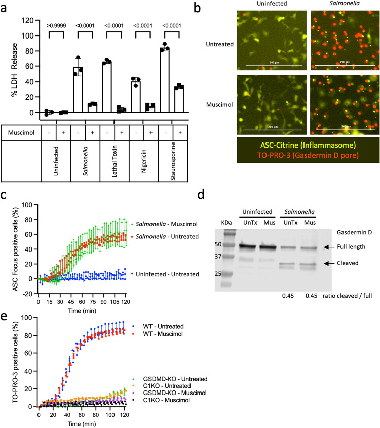
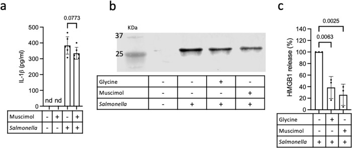
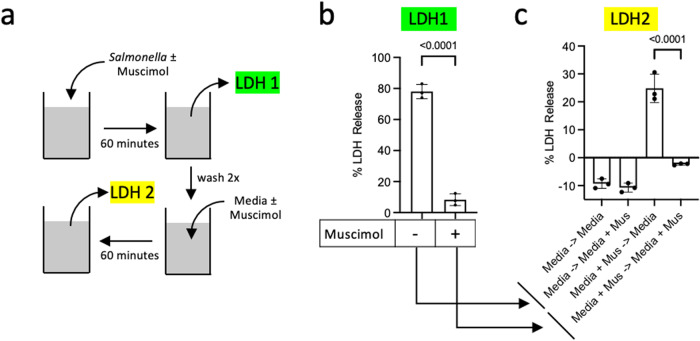
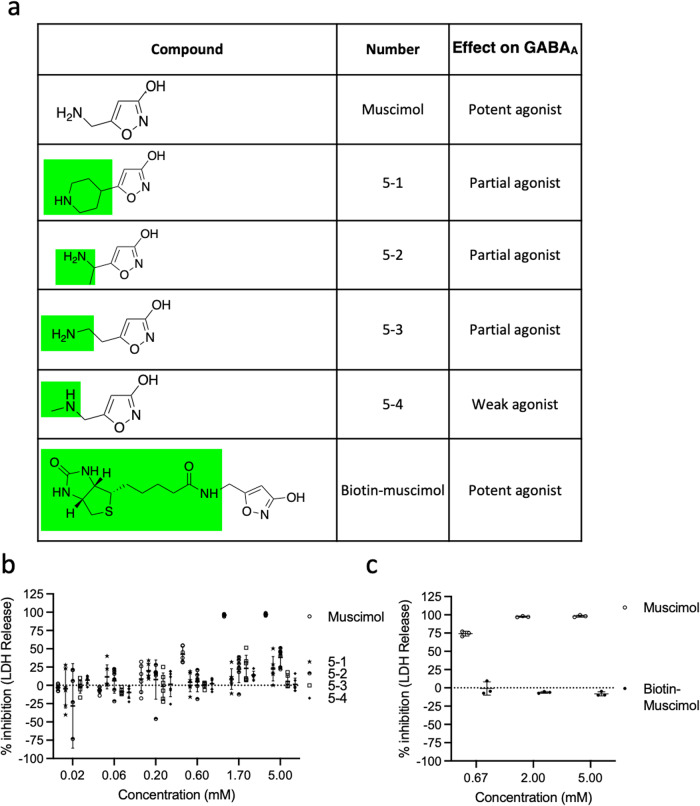
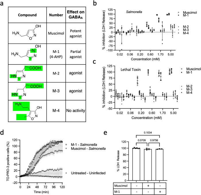
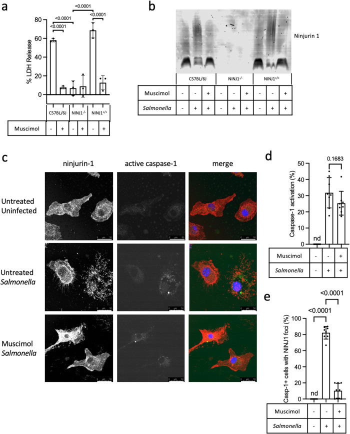
Pyroptosis is a cell death process that causes inflammation and contributes to numerous diseases. Pyroptosis is mediated by caspase-1 family proteases that cleave the pore-forming protein gasdermin D, causing plasma membrane rupture and release of pathogenic cellular contents. We previously identified muscimol as a small molecule that prevents plasma membrane rupture during pyroptosis via an unidentified mechanism. Here, we show that muscimol has reversible activity to prevent cellular lysis without affecting earlier pyroptotic events. Although muscimol is a well-characterized agonist for neuronal GABAA receptors, muscimol protection is not altered by GABAA receptor antagonists or recapitulated by other GABAA agonists, suggesting that muscimol acts via a novel mechanism. We find that muscimol blocks oligomerization of ninjurin-1, which is required for plasma membrane rupture downstream of gasdermin D pore formation. Our structure-activity relationship studies reveal distinct molecular determinants defining inhibition of pyroptotic lysis compared to GABAA binding. In addition, we demonstrate that muscimol reduces lethality during LPS-induced septic shock. Together, these findings demonstrate that ninjurin-1-mediated plasma membrane rupture can be pharmacologically modulated and pave the way toward identification of therapeutic strategies for pathologic conditions associated with pyroptosis.


Conflict of interest statement

The authors declare no competing interests.

Figures

Fig. 1
Fig. 1. Muscimol prevents pyroptotic lysis without affecting the formation of ASC inflammasome foci or membrane pores.
a Bone marrow-derived macrophages (BMMs) were treated with Salmonella, Lethal Toxin, Nigericin, or Staurosporine in the presence of 1 mM muscimol as indicated. LDH released during cell lysis was measured. b Localization of the inflammasome adapter ASC (yellow) and uptake of the small membrane-impermeant nuclear dye TO-PRO-3 (red) were assessed in ASC-Citrine-expressing BMMs infected with Salmonella for 90 min. c ASC foci formation was quantified. d Cleavage of gasdermin D was determined by Western blot in untreated (UnTx) and muscimol (Mus) treated cells infected with Salmonella or uninfected controls. The ratio of cleaved/uncleaved gasdermin D was determined. e TO-PRO-3 uptake was assessed in wild type, caspase−1/11−/−, or gasdermin D−/− BMMs infected with Salmonella in the presence or absence of muscimol. The percentage of cells with TO-PRO-3+ nuclei was quantified. Representative data in a, c, and e (mean ± SD, n  =  3) from two or three independent experiments are shown. Images representative of 5 taken per condition in three independent experiments are shown in b; scale bars represent 200 mm. Western blot representative of two independent experiments is shown in d. Statistics: 2-way ANOVA + Tukey’s multiple comparisons.
Fig. 2
Fig. 2. Muscimol prevents lytic release of inflammatory mediator HMGB1.
a BMMs were infected with Salmonella or treated with PBS in the presence of muscimol as indicated and IL-1β secretion was measured by ELISA. Representative data (mean ± SD, n  =  6) from three independent experiments are shown. b, c BMM were infected with Salmonella or treated with PBS in the presence of glycine or muscimol as indicated. Release of HMGB1 was determined by western blot (b) and quantified (c). Representative blot is shown in b and quantification in c is mean ± SD of n = 3 independent experiments. M: MW marker in KDa. Statistics: One-way ANOVA + Tukey’s multiple comparisons. nd is none detected.
Fig. 3
Fig. 3. Muscimol protects mice against LPS-induced lethality.
a C57BL/6J mice were injected with 3 mg/kg muscimol, or PBS, followed by 10 mg/kg LPS. Mice were observed for clinical signs of illness and euthanized when overt signs of sepsis were observed. b Sera from mice injected with PBS, LPS alone, or muscimol + LPS were analyzed for the level of blood urea nitrogen (BUN). Data are combined values from three independent experiments, n = 23 mice per condition for LPS and muscimol + LPS, n = 5 uninfected mice. Statistics: (a) Mantel-Cox Log-rank test, (b) one-way ANOVA + Tukey’s multiple comparisons.
Fig. 4
Fig. 4. Inhibition of cellular lysis by muscimol is reversible.
a Schematic representation of the experiment. b BMM were infected with Salmonella for 60 min in a medium containing muscimol (as indicated) and cell lysis was measured by LDH release (LDH 1). c New medium with or without muscimol was added and LDH released during the next 60 min was measured (LDH 2). Statistics: t-test (b) or one-way ANOVA + Tukey’s multiple comparisons (c). nd is none detected. Representative data in b and c (mean ± SD, n  =  3) from two independent experiments are shown.
Fig. 5
Fig. 5. Modification of muscimol at positions 3 and 4 abolishes the inhibitory capacity of muscimol.
a Structures of muscimol (with numbers indicating positions of modification sites) and analogs modified at positions 3 and 4 of the ring structure. The effect on GABAA is based on previously published findings. b BMM were infected with Salmonella in the presence of indicated concentrations of muscimol or muscimol analogs. LDH released during cell lysis was measured and used to calculate the inhibitory percentage. Combined data from two independent experiments (mean ± SD, n = 3 per experiment) are shown.
Fig. 6
Fig. 6. Modification of muscimol at position 5 abolishes the inhibitory capacity of muscimol.
a Structures of muscimol analogs modified at position 5 of the ring structure that maintains activity at the GABAA receptor. b, c BMM were infected with Salmonella in the presence of indicated concentrations of muscimol or muscimol analogs. LDH released during cell lysis was measured and used to calculate the inhibitory percentage. Combined data from two independent experiments (mean ± SD, n = 3 per experiment) are shown.
Fig. 7
Fig. 7. A muscimol analog with modifications at multiple positions retains inhibitory potential.
a Structures of muscimol analogs modified at multiple positions of the ring structure. b, c BMM were infected with Salmonella or exposed to Lethal Toxin in the presence of indicated concentrations of muscimol or muscimol analogs. LDH released during cell lysis was measured and used to calculate the inhibitory percentage. d TO-PRO-3 uptake was assessed in BMM infected with Salmonella in the presence of 1 mM muscimol or analog M1. e Total cell lysis was assessed in the presence of muscimol or analog M-1 to determine if the LDH assay was affected by the addition of muscimol or muscimol analogs. Data in b, c, and d are from two independent experiments (mean ± SD, n = 3 per experiment). Data in e are representative (mean ± SD, n = 3) from two independent experiments.
Fig. 8
Fig. 8. Ninjurin-1 oligomerization is blocked by muscimol.
BMM from wild type C57BL/6 J (WT), ninjurin-1−/− (NINJ1−/−), and ninjurin+/+ (NINJ1+/+) littermate controls were infected with Salmonella in the presence of muscimol, as indicated. a LDH released during cellular lysis was measured at 90 min post infection. b BN-PAGE Western blot for ninjurin-1 oligomerization in uninfected, Salmonella-infected, and Salmonella-infected muscimol-treated cells. Representative blot from two independent experiments. c Images representative of 8 taken per condition in 2 independent experiments of NINJ1+/+BMM infected with Salmonella for 60 min in the presence or absence of muscimol. Ninjurin-1 oligomerization was visualized as punctate staining with an antibody specific for the N-terminal domain of ninjurin-1 (red). Caspase-1 activation was detected using FAM-YVAD-FMK (green). TO-PRO-3 staining of permeabilized cells (blue) was used to identify nuclei. The percentage of cells containing active caspase-1 was quantified (d). The percentage of caspase-1 positive cells containing ninjurin-1 puncta was then quantified (e). Statistics: (a, d, e) one-way ANOVA + Tukey’s multiple comparisons. ns is not significant. nd is none detected. Data in a, d, and e is from representative experiments (mean ± SD, n = 3 (a) or n = 8 (d and e)) from two independent experiments. Scale bars in c represent 50 µm.

References

    1. Jorgensen I, Rayamajhi M, Miao EA. Programmed cell death as a defence against infection. Nat. Rev. Immunol. 2017;17:151–164. - PMC - PubMed
    1. Jia C, et al. Role of pyroptosis in cardiovascular diseases. Int. Immunopharmacol. 2019;67:311–318. - PubMed
    1. Zhaolin Z, Guohua L, Shiyuan W, Zuo W. Role of pyroptosis in cardiovascular disease. Cell Prolif. 2019;52:e12563. - PMC - PubMed
    1. Barrington J, Lemarchand E, Allan SM. A brain in flame; do inflammasomes and pyroptosis influence stroke pathology? Brain Pathol. 2017;27:205–212. - PMC - PubMed
    1. Chen S, et al. Gasdermin family: a promising therapeutic target for stroke. Transl. Stroke Res. 2018;9:555–563. - PubMed

Publication types

MeSH terms

LinkOut - more resources