Review
. 2023 Oct 5;118(1):42.
doi: 10.1007/s00395-023-01009-x.

Mitophagy for cardioprotection

Affiliations
Review

Mitophagy for cardioprotection

Allen Sam Titus et al. Basic Res Cardiol. .

Abstract

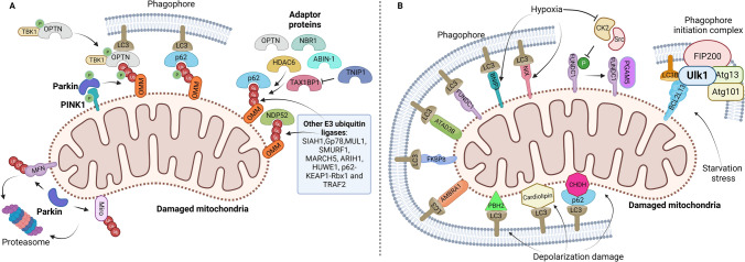
Mitochondrial function is maintained by several strictly coordinated mechanisms, collectively termed mitochondrial quality control mechanisms, including fusion and fission, degradation, and biogenesis. As the primary source of energy in cardiomyocytes, mitochondria are the central organelle for maintaining cardiac function. Since adult cardiomyocytes in humans rarely divide, the number of dysfunctional mitochondria cannot easily be diluted through cell division. Thus, efficient degradation of dysfunctional mitochondria is crucial to maintaining cellular function. Mitophagy, a mitochondria specific form of autophagy, is a major mechanism by which damaged or unnecessary mitochondria are targeted and eliminated. Mitophagy is active in cardiomyocytes at baseline and in response to stress, and plays an essential role in maintaining the quality of mitochondria in cardiomyocytes. Mitophagy is mediated through multiple mechanisms in the heart, and each of these mechanisms can partially compensate for the loss of another mechanism. However, insufficient levels of mitophagy eventually lead to mitochondrial dysfunction and the development of heart failure. In this review, we discuss the molecular mechanisms of mitophagy in the heart and the role of mitophagy in cardiac pathophysiology, with the focus on recent findings in the field.

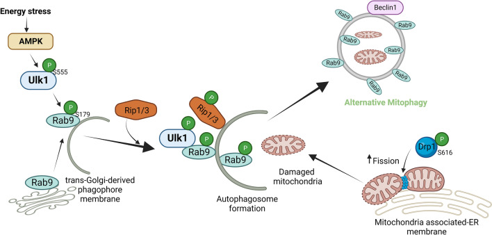
Keywords: Alternative mitophagy; Drp1; Mitochondrial quality control; Mitophagy.


Conflict of interest statement

The authors declare no coflict of interests.

Figures

Fig. 1
Fig. 1
Mechanisms of mitophagy. A Ubiquitin-dependent mitophagy. In the PINK1–Parkin pathway of mitophagy, PINK1 is stabilized on the OMM following stress, which then stimulates Parkin recruitment. Several components of the outer membrane are ubiquitinated by Parkin. Following this, PINK1 phosphorylates poly-Ub chains, acting as an "eat me" signal for the autophagic machinery. Phosphorylated poly-Ub chains on mitochondrial proteins are recognized by adaptor proteins (p62, OPTN, TAX1BP1, NBR1, NDP52, HDAC6, and ABIN-1 or TNIP1), which then bind to LC3 to start the formation of autophagosomes. TNIP1 has been shown to inhibit TAX1BP1 by competition and downregulate mitophagy. By phosphorylating OPTN, TBK1 increases the protein's ability to bind to Ub chains. A feed-forward mechanism promoting mitochondrial clearance is established by the OPTN-TBK1 complex. Prior to mitophagy, alternative E3 ubiquitin ligases that target OMM proteins include Gp78, SMURF1, MUL1, SIAH1, ARIH1, MARCH5, HUWE1, p62-Keap1-Rbx1, and TRAF2. B Receptor-mediated mitophagy. Mitophagy receptors BNIP3, NIX, FKBP8, BCL2L13, AMBRA1, ATAD3, and FUNDC1 localize to the OMM and interact with LC3 directly to mediate mitochondrial elimination. BCL2L13 has also been shown to activate mitophagy by recruiting the Ulk1/FIP200/Atg13/Atg101 initiation complex and LC3B during starvation stress. Following mitochondrial depolarization, PHB2 and cardiolipin are externalized to the OMM and interact with LC3. Choline dehydrogenase (CHDH) accumulates in the OMM of depolarized mitochondria and interacts with p62 and LC3. FUNDC1 phosphorylation status is influenced by PGAM5 phosphatase, CK2 and Src kinases, all of which control mitochondrial dynamics during hypoxia. The figure was created with Biorender.com
Fig. 2
Fig. 2
Mechanism of alternative mitophagy. Energy stress activates AMPK-mediated phosphorylation of Ulk1 at Ser555. The activated Ulk1 phosphorylates Rab9 at Ser179 and initiates phagophore formation utilizing trans-Golgi-derived membrane. Activated Ulk1-Rab9 recruits Rip1/3 kinase and phosphorylates Drp1 at Ser616, which activates mitochondrial fission to segregate damaged mitochondria and promote alternative mitophagy at the mitochondrial-associated ER membrane. The figure was created with Biorender.com
Fig. 3
Fig. 3
Mitophagy in ischemia–reperfusion injury. During myocardial reperfusion following ischemic injury, increased ROS production causes mPTP opening, thereby leading to cardiomyocyte apoptosis and necrosis. In this condition, accumulated CO2 and bicarbonate inhibit mitophagy and promote myocardial injury. ZIP7 and TRPML1 upregulation during I/R inhibits mitophagy thereby contributing to increased reperfusion injury. Upregulated Mst1 and CK2α or downregulated PLK1 inhibit FUNDC1-mediated mitophagy, resulting in exacerbated cardiac injury. Mst1 may inhibit mitophagy through Beclin 1 phosphorylation and inhibition of autophagosome formation. Drp1-mediated and PGAM5-PINK1-mediated mitophagy protects the heart against I/R injury. MORN4 promotes ROCK2-mediated phosphorylation and activation of MFN2 leading to increased mitochondrial dynamics and mitophagy to enhance cardioprotection during I/R injury. Inhibition of overactivation of mitophagy during I/R injury is cardioprotective in certain conditions. M2AChR signaling inhibits excessive mitophagy. Notch1 inhibits PTEN-PINK1-Parkin signaling-mediated mitophagy. YTHDF2 inhibits BNIP3 mRNA expression and downregulates mitophagy. The figure was created with Biorender.com
Fig. 4
Fig. 4
Mitophagy in pressure overload-induced hypertrophy. Transient activation of conventional autophagy during pressure overload occurs one day after TAC, whereas activation of mitophagy occurs thereafter, peaking at around 3 and 7 days after TAC. In particular, mitophagy activation after inactivation of conventional autophagy occurs through an Ulk1-Rab9-dependent alternative mitophagy mechanism. Both autophagy and mitophagy contribute to cardioprotection under pressure overload stress. Pressure overload causes mitochondrial dysfunction and inhibits the action of DNase II in the lysosome, leading to non-degradation of mitochondrial DNA. The accumulated mitochondrial DNA provokes inflammation through TLR9-dependent signaling and leads to heart failure. Drp1-mediated mitophagy is critical for the protection of the heart during pressure overload. TAT-Beclin 1 can activate both general and alternative mitophagy. Parkin has also been shown to enhance autophagy during pressure overload. The figure was created with Biorender.com
Fig. 5
Fig. 5
Mitophagy in diabetic cardiomyopathy. A Accumulated glucose elevates mitochondrial superoxide levels in type 1 diabetes and leads to myocardial cell death. While general autophagy is inhibited by high glucose levels, a compensatory mechanism simultaneously activates alternative autophagy and mitophagy and is cardioprotective. B In a mouse model of type 2 diabetes, mitophagy is stimulated in the heart through multiple mechanisms in a time-dependent manner. During the acute phase of HFD consumption, mitophagy is activated by Atg7- and Parkin-dependent mechanisms. Drp1 is also involved in mitophagy during the acute phase of HFD consumption by inhibiting Bcl-2/Bcl-xL interaction with Beclin 1, which allows activation of Beclin 1. In the chronic phase of HFD consumption, Drp1 is phosphorylated at Ser616 through unknown mechanisms and localized to ER–mitochondria-associated membrane, where it activates Rab9-mediated alternative mitophagy. These mechanisms may be compensatory for the downregulation of conventional mitophagy. Despite activation of conventional mitophagy in the acute phase and alternative mitophagy during the chronic phase, the level of mitophagy appears to be insufficient, and thus mitochondrial dysfunction in the heart develops during the chronic phase of type 2 diabetes. The figure was created with Biorender.com

References

    1. Abeliovich H, Zarei M, Rigbolt KT, Youle RJ, Dengjel J. Involvement of mitochondrial dynamics in the segregation of mitochondrial matrix proteins during stationary phase mitophagy. Nat Commun. 2013;4:2789. doi: 10.1038/ncomms3789. - DOI - PMC - PubMed
    1. Acin-Perez R, Lechuga-Vieco AV, Del Mar MM, Nieto-Arellano R, Torroja C, Sánchez-Cabo F, Jiménez C, González-Guerra A, Carrascoso I, Benincá C, Quiros PM, López-Otín C, Castellano JM, Ruíz-Cabello J, Jiménez-Borreguero LJ, Enríquez JA. Ablation of the stress protease OMA1 protects against heart failure in mice. Sci Transl Med. 2018;10:eaan4935. doi: 10.1126/scitranslmed.aan4935. - DOI - PubMed
    1. Ahn J, Kim J. Nutritional status and cardiac autophagy. Diabetes Metab J. 2013;37:30–35. doi: 10.4093/dmj.2013.37.1.30. - DOI - PMC - PubMed
    1. Ambivero CT, Cilenti L, Main S, Zervos AS. Mulan E3 ubiquitin ligase interacts with multiple E2 conjugating enzymes and participates in mitophagy by recruiting GABARAP. Cell Signal. 2014;26:2921–2929. doi: 10.1016/j.cellsig.2014.09.004. - DOI - PubMed
    1. Anand R, Wai T, Baker MJ, Kladt N, Schauss AC, Rugarli E, Langer T. The i-AAA protease YME1L and OMA1 cleave OPA1 to balance mitochondrial fusion and fission. J Cell Biol. 2014;204:919–929. doi: 10.1083/jcb.201308006. - DOI - PMC - PubMed

Publication types