. 2023 Oct 5;21(1):275.
doi: 10.1186/s12964-023-01275-1.

The Golgi stacking protein GRASP55 is targeted by the natural compound prodigiosin

Affiliations

The Golgi stacking protein GRASP55 is targeted by the natural compound prodigiosin

Lena Berning et al. Cell Commun Signal. .

Abstract

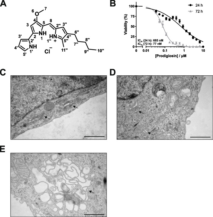
Background: The bacterial secondary metabolite prodigiosin has been shown to exert anticancer, antimalarial, antibacterial and immunomodulatory properties. With regard to cancer, it has been reported to affect cancer cells but not non-malignant cells, rendering prodigiosin a promising lead compound for anticancer drug discovery. However, a direct protein target has not yet been experimentally identified.

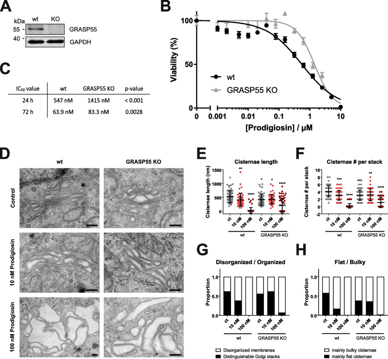
Methods: We used mass spectrometry-based thermal proteome profiling in order to identify target proteins of prodigiosin. For target validation, we employed a genetic knockout approach and electron microscopy.

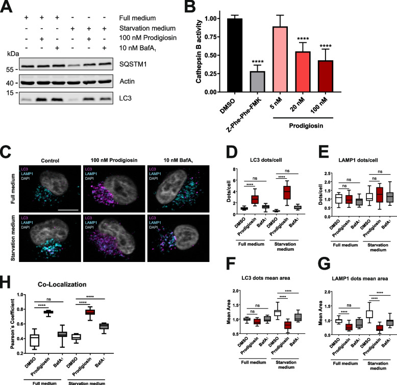
Results: We identified the Golgi stacking protein GRASP55 as target protein of prodigiosin. We show that prodigiosin treatment severely affects Golgi morphology and functionality, and that prodigiosin-dependent cytotoxicity is partially reduced in GRASP55 knockout cells. We also found that prodigiosin treatment results in decreased cathepsin activity and overall blocks autophagic flux, whereas co-localization of the autophagosomal marker LC3 and the lysosomal marker LAMP1 is clearly promoted. Finally, we observed that autophagosomes accumulate at GRASP55-positive structures, pointing towards an involvement of an altered Golgi function in the autophagy-inhibitory effect of this natural compound.

Conclusion: Taken together, we propose that prodigiosin affects autophagy and Golgi apparatus integrity in an interlinked mode of action involving the regulation of organelle alkalization and the Golgi stacking protein GRASP55. Video Abstract.

Keywords: Autophagy; Golgi apparatus; Natural compound; Prodigiosin; Target identification.


Conflict of interest statement

The authors declare no competing interests.

Figures

Fig. 1
Fig. 1
Prodigiosin exhibits cytotoxic properties in HeLa wt cells and alters Golgi apparatus morphology. A Chemical structure of prodigiosin. B HeLa wt cells were treated with different concentrations of prodigiosin for 24 h or 72 h. After treatment, cell viability was measured using a thiazolyl blue (MTT) assay. Results are shown as the mean ± SEM of three independent experiments performed in triplicates for each treatment. HeLa wt cells were treated with C DMSO, D 10 nM or E 100 nM prodigiosin for 24 h and effects on cell morphology were investigated by transmission electron microscopy. Arrows indicate changes in Golgi apparatus morphology after prodigiosin treatment. Representative electron micrographs are shown. Scale bar: 1 µm
Fig. 2
Fig. 2
Pre-treatment with prodigiosin impairs Golgi apparatus reassembly after BFA treatment. HeLa wt cells were seeded on cover slips. On the next day, cells were treated with DMSO, 10 nM prodigiosin or 100 nM prodigiosin for 24 h. Cells were washed once and treated with 5 μg/mL brefeldin A (BFA) or DMSO (ct) for 2 h. BFA was washed out with DPBS 4 times and cells were incubated in fresh growth medium to wash out (W/O) BFA for 0/15/30/45/60/120 min. After treatment, cover slips were prepared for microscopy. A Representative sections are depicted. Scale bar: 10 µm. B-E The relative number per cell and mean area of TGN46 and B4GALT1 positive structures of 15 representative images from three biological replicates for each treatment were quantified using ImageJ 1.53c
Fig. 3
Fig. 3
Thermal proteome profiling (TPP) for the identification of prodigiosin targets and prodigiosin-affected proteins: GRASP55 is thermally stabilized by treatment with prodigiosin. A RTSA analysis plot of the statistical significance vs. the extent of the effect of prodigiosin on protein intensity collated over the different temperatures. Differential protein intensities may result from prodigiosin-mediated thermal protein stability alternation and/or a change in protein abundance (caused by, e.g., differential protein expression, degradation or secretion), where these two effects cannot be distinguished in the current plot. Significant proteins given by the RTSA software are colored in green or red for positive or negative RTSA distance score, respectively. B Volcano-like plot of the statistical significance vs. the extent of prodigiosin-mediated thermal protein stabilization (mean ΔTm > 0) or destabilization (mean ΔTm < 0). The color code refers to significant proteins by RTSA (see panel A). GRASP55 is the RTSA significant protein with the highest statistical significance among the stabilized proteins and, thus, a highly promising prodigiosin target protein candidate. C Volcano-like plot of the statistical significance vs. the extent of prodigiosin-mediated change in protein abundance (higher or lower for mean log2 ratio > 0 or < 0, respectively) calculated from protein intensities at 36.5 °C. The color code refers to significant proteins by RTSA (see panel A). D Melting curves (solid lines) of GRASP55 from prodigiosin or DMSO treated cells (RTSA software output of TPP-TR analysis). Datapoints and whiskers represent the arithmetic mean ± SD of three replicates. Datapoints for temperatures showing significant intensity differences are dash-boxed, next to which the collation ratio and (uncorrected) p-value are given. The thermal stabilization of GRASP55 by prodigiosin (with a mean melting point difference of 2.1 °C) is indicated by dashed lines and a measure for statistical significance is provided (-lg(p), same as y-axis of panel B). E Immunoblotting for GRASP55 protein quantification from the non-denatured protein fractions of prodigiosin or DMSO treated HeLa cells (CETSA). F Melting curves of GRASP55 from quantitative immunoblotting (CETSA, see panel E) using the same RTSA analysis and representation as for TPP-TR (see panel D). The thermal stabilization of GRASP55 by prodigiosin was confirmed
Fig. 4
Fig. 4
GRASP55 is stabilized at low nanomolar prodigiosin concentrations. For thermal proteome profiling compound concentration range (TPP-CCR) experiments, HeLa wt cells were treated with ten different concentrations of prodigiosin for 6 h, harvested, and cell suspensions were exposed to a short (3 min) constant temperature treatment at 50 °C (or 37 °C to test for abundance effects). Cells were lysed and the non-denatured protein fraction was recovered after centrifugation followed by quantitative MS analysis as described for TPP-TR, resulting in dose response characteristics for prodigiosin-affected proteins. A Plot of the dose response curve fitting parameters pseudo-R2 (proteins with a pseudo-R2 value of > 0.8 were considered to have prodigiosin dose–response characteristics) vs. pEC50 (the negative decadic logarithm of the half-maximal effective concentration) for proteins exhibiting increasing intensities at increasing prodigiosin concentrations (positive dose response). The data point diameter encodes the pseudo-R2 (50 °C – 37 °C) difference (high for solely (de)stabilized proteins, low for solely abundance affected proteins). Proteins with pEC50 > 8 (EC50 < 10 nM) are labeled. GRASP55 is the protein affected (stabilized) at the lowest prodigiosin concentrations among the proteins with positive dose response. B Same representation as for panel A but for proteins with negative dose response. Lysosome associated proteins (UniProt annotated keyword KW-0458) are underlined. C Dose response characteristics and fitting results for GRASP55 at 50 °C. Data points and whiskers represent the arithmetic mean ± SD of three replicates and the fitted dose response curve is shown in red. D Same representation as in panel C but for 37 °C, with the absence of a dose–response effect (irrelevant pseudo-R2 <  < 0.8) represented by a thin dotted fit curve (in red)
Fig. 5
Fig. 5
Knockout of GRASP55 impairs prodigiosin cytotoxicity and alters prodigiosin effects on the Golgi apparatus. A GRASP55 was knocked out in HeLa wt cells and KO was verified by western blot. B HeLa wt and HeLa GRASP55 KO cells were treated with different concentrations of prodigiosin for 24 h. After treatment, cell viability was measured using a thiazolylblue (MTT) assay. Results are shown as the mean ± SEM of three independent experiments performed in triplicates for each treatment. C IC50 values and statistical analysis for MTT assays in HeLa wt and HeLa GRASP55 KO cells after 24 and 72 h treatment with prodigiosin. Please note that the MTT assay for HeLa wt cells was independent from the one shown in Fig. 1B; accordingly, IC50 values slightly differ. (D-H) HeLa wt and HeLa GRASP55 KO cells were treated with DMSO, 10 nM or 100 nM prodigiosin for 24 h and effects on Golgi apparatus structure were investigated by transmission electron microscopy. D Representative electron micrographs are shown. Scale bar: 200 nm. E Cisternae length and F cisternae number per stack were quantified. G Golgi stacks were categorized into organized (stacked structures with three or more cisternae) and disorganized (multiple unstacked cisternae and vesicles). H Stacks were classified as containing mainly flat or mainly bulky/swollen cisternae. At least 50 Golgis per treatment were quantified after blinding and randomization. Bars represent the means ± SD. p values were determined by ordinary one-way ANOVA with Dunnett´s post hoc test. * p < 0.05; ** p < 0.01; *** p < 0.001; **** p < 0.0001
Fig. 6
Fig. 6
GRASP55 and LC3 co-localize upon treatment with prodigiosin. HeLa wt cells were seeded on cover slips. On the next day, cells were treated with different concentrations of prodigiosin, 10 nM bafilomycin A1 (BafA1) or DMSO for 6 h. After treatment, cover slips were prepared for microscopy. A Representative sections are depicted. Scale bar: 10 µm. B-E The relative number per cell and mean area of GRASP55 and LC3 positive structures and F the co-localization (Pearson´s coefficient) after Costes thresholding of GRASP55 and LC3 of 15 representative images from three biological replicates for each treatment were quantified using ImageJ 1.53c. p values were determined by ordinary one-way ANOVA with Dunnett´s post hoc test. * p < 0.05; ** p < 0.01; *** p < 0.001; **** p < 0.0001; ns, non-significant
Fig. 7
Fig. 7
Prodigiosin blocks autophagy and inhibits cathepsin activity. A HeLa wt cells were treated with DMSO, 100 nM prodigiosin or 10 nM bafilomycin A1 (BafA1) in DMEM or EBSS. After 6 h, the cells were lysed and cellular lysates were immunoblotted for the indicated proteins. One representative immunoblot of three independent experiments is shown. LC3: light chain 3; SQSTM1: sequestosome 1. B HeLa wt cells were treated with different concentrations of prodigiosin or DMSO. After 24 h, the cells were lysed and a cathepsin B assay was performed according to the manufacturer´s instructions. 20 µM Z-Phe-Phe-FMK was used as inhibitor control. The fluorescence of duplicates for each treatment of three independent experiments was measured and the mean of the DMSO control was set as 100%. Bars represent the means + SD. C HeLa wt cells were seeded on cover slips. On the next day, cells were treated with DMSO, 100 nM prodigiosin or 10 nM BafA1 in DMEM or EBSS for 2 h. After treatment, cover slips were prepared for microscopy. Representative sections are depicted. Scale bar: 10 µm. D-G The relative number per cell and mean area of LC3- and LAMP1-positive structures and H the co-localization (Pearson´s coefficient) after Costes thresholding of LC3 and LAMP1 of 15 representative images from three biological replicates for each treatment were quantified using ImageJ 1.53c. p values were determined by ordinary one-way ANOVA with Dunnett´s post hoc test. **** p < 0.0001; ns, non-significant

Similar articles

Cited by

References

    1. Ahat E, Bui S, Zhang J, da Veiga Leprevost F, Sharkey L, Reid W, Nesvizhskii AI, Paulson HL, Wang Y. GRASP55 regulates the unconventional secretion and aggregation of mutant huntingtin. J Biol Chem. 2022;298:102219. - PMC - PubMed
    1. Ahat E, Li J, Wang Y. New Insights Into the Golgi Stacking Proteins. Front Cell Dev Biol. 2019;7:131. - PMC - PubMed
    1. Anderson RG, Pathak RK. Vesicles and cisternae in the trans Golgi apparatus of human fibroblasts are acidic compartments. Cell. 1985;40:635–643. - PubMed
    1. Barr FA, Puype M, Vandekerckhove J, Warren G. GRASP65, a protein involved in the stacking of Golgi cisternae. Cell. 1997;91:253–262. - PubMed
    1. Bekier ME, 2nd, Wang L, Li J, Huang H, Tang D, Zhang X, Wang Y. Knockout of the Golgi stacking proteins GRASP55 and GRASP65 impairs Golgi structure and function. Mol Biol Cell. 2017;28:2833–2842. - PMC - PubMed

Publication types