

A Systematic Review of the Role of Magnetic Resonance Imaging in the Diagnosis and Detection of Neurovascular Conflict in Patients With Trigeminal Neuralgia
- PMID: 37799230
- PMCID: PMC10547583
- DOI: 10.7759/cureus.44614
A Systematic Review of the Role of Magnetic Resonance Imaging in the Diagnosis and Detection of Neurovascular Conflict in Patients With Trigeminal Neuralgia
Abstract
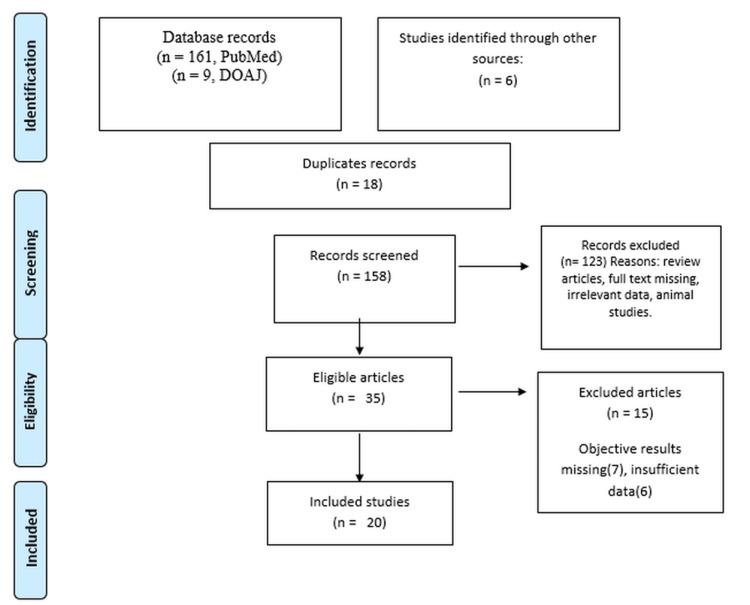
Trigeminal neuralgia (TN) is a debilitating disorder causing severe, episodic, unilateral stabbing facial pain disturbing enough to disrupt the activities of daily life. Classic TN is caused due to compression injury of the trigeminal nerve at the cistern segment caused by either an artery or a vein, referred to as neurovascular contact or conflict (NVC). Magnetic resonance imaging (MRI) has been the standard tool for the diagnosis of NVC. This study aimed to determine the incidence of NVC in TN, as identified by MRI, assess the various MRI grading patterns among patients with TN, and identify the vessels primarily involved in NVC. A systematic search of studies that used MRI for the diagnosis of TN in reference to NVC was conducted on DOAJ and PubMed/PubMed Central. Data were extracted and entered into a Microsoft Excel spreadsheet. The outcomes measured were the incidence of NVC as shown in MRI, vessels involved in NVC, and MRI grading patterns. We identified and selected 20 studies that fulfilled inclusion/exclusion criteria. In total, 1,436 patients were enrolled in all included studies. The type of MRI used was 1.5 T or 3 T MRI. The mean age of the patients varied from 49 to 63 years, with an equivalent male-to-female ratio. NVC was seen in 1,276 cases out of 1,436 cases (88.85%) of TN on the ipsilateral side, as shown by MRI. The vessels involved were arteries in 80-90% of the cases, followed by veins. Among the arteries, the superior cerebellar artery was the most common artery (80-90% of cases). The grades of NVC as assessed by MRI included grades I, II, and III with varied proportions in different studies. NVC is a common problem in TN, wherein there is compression at the nerve root entry zone, and it shows a strong predilection for the elderly population. MRI seems to be a novel imaging diagnostic investigation to identify NVC associated with TN. Moreover, NVC grading must be done with MRI so that it may help the surgeon in stratifying the patient's treatment.
Keywords: magnetic resonance imaging; microvascular decompression; neurovascular contact; root entry zone; trigeminal neuralgia.
Copyright © 2023, Bora et al.
Conflict of interest statement
The authors have declared that no competing interests exist.
Figures
References
-
- The role of magnetic resonance imaging in the evaluation of trigeminal neuralgia. Rangaswamy VK, Srinivas MR, Basavalingu D, Nagaraj BR. https://ijars.net/article_abstract.aspx?issn=0973-709x&year=2016&month=A... Int J Anat Radiol Surg. 2016;5:24–29.
-
- A study to evaluate neurovascular conflict of trigeminal nerve in trigeminal neuralgia patients with the help of 1.5 T MR imaging. Anwar HA, Krishna MR, Sadiq S, Kumar RR, Venkatarathnam V, Saikiran G. Egypt J Radiol Nucl Med. 2022;53:66.
-
- Trigeminal neuralgia: pathology and pathogenesis. Love S, Coakham HB. Brain. 2001;124:2347–2360. - PubMed
-
- Magnetic resonance imaging contribution for diagnosing symptomatic neurovascular contact in classical trigeminal neuralgia: a blinded case-control study and meta-analysis. Antonini G, Di Pasquale A, Cruccu G, et al. Pain. 2014;155:1464–1471. - PubMed
Publication types
LinkOut - more resources
Full Text Sources