. 2023 Sep 19:11:1219739.
doi: 10.3389/fcell.2023.1219739. eCollection 2023.

Self-assembled adipose-derived mesenchymal stem cells as an extracellular matrix component- and growth factor-enriched filler

Affiliations

Self-assembled adipose-derived mesenchymal stem cells as an extracellular matrix component- and growth factor-enriched filler

Choa Park et al. Front Cell Dev Biol. .

Abstract

The clinical application of mesenchymal stem cells (MSCs) is attracting attention due to their excellent safety, convenient acquisition, multipotency, and trophic activity. The clinical effectiveness of transplanted MSCs is well-known in regenerative and immunomodulatory medicine, but there is a demand for their improved viability and regenerative function after transplantation. In this study, we isolated MSCs from adipose tissue from three human donors and generated uniformly sized MSC spheroids (∼100 µm in diameter) called microblocks (MiBs) for dermal reconstitution. The viability and MSC marker expression of MSCs in MiBs were similar to those of monolayer MSCs. Compared with monolayer MSCs, MiBs produced more extracellular matrix (ECM) components, including type I collagen, fibronectin, and hyaluronic acid, and growth factors such as vascular endothelial growth factor and hepatocyte growth factor. Subcutaneously injected MiBs showed skin volume retaining capacity in mice. These results indicate that MiBs could be applied as regenerative medicine for skin conditions such as atrophic scar by having high ECM and bioactive factor expression.

Keywords: atrophic scar in addition; extracellular matrix; growth factor; mesenchymal stem cell; microblock; regenerative medicine; spheroid.


Conflict of interest statement

The authors declare that the research was conducted in the absence of any commercial or financial relationships that could be construed as a potential conflict of interest.

Figures

FIGURE 1
FIGURE 1
Isolation and characterization of ADMSCs from donors I and II. (A) Representative microscopic image of ADMSCs in monolayer culture. Cells were isolated from donor I. Scale bar, 200 μm. (B) The cumulative population doubling level (CPDL) and (C) population doubling time (PDT) of ADMSCs were measured at each passage (n = 6). (D) Expression of positive markers (CD29, CD44, CD73, CD90, and CD105) and negative markers (CD31, CD45, CD79a, CD117, and HLA-DR) of ADMSCs were analyzed using flow cytometry. (E) The differentiation potential of ADMSCs into adipocytes (b), osteocytes (c), and chondrocytes (d) was compared with that of undifferentiated cells (a) by Oil Red O, Alizarin Red S, and Alcian Blue staining, respectively. Scale bar, 200 μm. (F) Gene expression of differentiation markers was analyzed by RT-PCR. Total RNA was isolated from undifferentiated ADMSCs (a), and differentiated adipocytes (b), osteocytes (c), and chondrocytes (d).
FIGURE 2
FIGURE 2
Generation of MiBs using Aggrewell™400 plates. (A) Images of uniformly sized MiBs grown inside microwells for 1, 2, 3, and 7 days were taken at ×40 magnification. MiBs were generated using ADMSCs isolated from donor I. Scale bar, 200 μm. (B) The diameters of MiBs cultured for 2 days in AggreWell™400 plates were measured using ImageJ software. MiBs were generated using MSCs in passages 3, 5, and 7. Over 130 MiBs were analyzed per passage to determine their diameter. Data represent the mean MiB diameter ± SD. (C) The viability of MiBs was assessed using the MTT assay kit. MiBs were prepared at passages 3, 5, and 7, and their viability was compared to monolayer MSCs (day 0) (n = 4). (D) Analysis of live (green) and dead (red) cells in MiBs at day 2 was performed using a LIVE/DEAD kit. Scale bar, 200 μm. (E) SEM analysis showed small aggregates around MiBs at day 2 (2,000×) (left panel, Scale bar, 50 μm). Higher magnification showed the formation of small membrane vesicles on MiB surfaces (30,000×) (right panel, Scale bar, 5 μm). (F) MiBs were cultured in AggreWell™400 plates for 2 days, and expression of MSC surface markers was confirmed using flow cytometry. Positive (CD29, CD44, CD73, CD90, and CD105) and negative (CD31, CD45, CD79a, CD117, and HLA-DR) markers of human MSCs were analyzed. (G) Immunofluorescence staining showed CD73, CD90, and CD105 expression in MiBs at day 2. DAPI was used to stain nuclei. Images were taken at ×400 magnification. Scale bar, 100 μm.
FIGURE 3
FIGURE 3
Enhanced ECM expression in MiBs on day 2. (A) Type I collagen expression was compared between MiBs and monolayer MSCs (SC). Type I collagen was measured by ELISA (n = 3). (B) Fibronectin (FN) expression was measured in monolayer MSCs and MiBs using ELISA (n = 3). (C) Immunofluorescence staining using anti-type I collagen antibody confirmed higher expression of type I collagen in MiBs than in monolayer MSCs. Scale bar, 100 μm. (D) Immunohistochemistry (brown) and Masson’s trichrome staining (blue) showed enrichment of collagen expression in MiBs. Scale bar, 50 μm (E) HA was increased in MiBs. Relative HA level was normalized by total protein level (n = 3). (F) GAG expression was measured by a GAG assay kit and normalized by DNA content (n = 3). Data represent the mean of fold change ± SD. Statistical analysis was conducted using independent samples t-test. * p < 0.05, ** p < 0.01, *** p < 0.001 vs. monolayer MSCs (SC).
FIGURE 4
FIGURE 4
Expression of growth factors in MiBs on day 2. Expression of FGF2 (A), PDGFA (B), VEGFA (C), and HGF (D) were analyzed by real-time PCR. Relative expression was compared with monolayer MSCs (SCs) (n = 3). Data represent the mean ± SD. Statistical analysis was conducted using independent samples t-test. **p < 0.01, ***p < 0.001 vs. monolayer MSCs (SC).
FIGURE 5
FIGURE 5
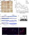
Sustained filling effect of subcutaneously injected MiBs. Low-dose (500 MiB) and high-dose (5,000 MiB) MiBs were subcutaneously injected into NSG mice. Saline (SA) and diluted HA (dHA) were used as controls. (A) Stereoscopic images of injected MiBs under the skin were taken after surgical removal of injected sites. The retained volumes were confirmed 8 and 12 weeks post-transplantation of MiBs. Scale bar, 200 μm. (B) The retention volume of MiBs, which were subcutaneously injected into NSG mice, was measured weekly for up to 12 weeks post-transplantation. From weeks 1 to 8, the volume was measured in 24 mice, and subsequently, 12 mice were sacrificed for histological examination. From weeks 9 to 12, the volume was measured in 12 mice. Data represent the mean ± SD. Statistical analysis was conducted using one-way ANOVA followed by Dunnett’s and Tukey’s Honestly Significant Difference tests. *p < 0.05, **p < 0.01, ***p < 0.001 vs. dHA. (C) Masson’s trichrome-stained images of skin tissue sections from different experimental groups. Scale bar, 1 mm. (D) Human Alu-sx and mouse c-mos sequences were detected in skin tissue from the MiB-injected group by PCR. (E) Transplanted cells are shown in merged images. Skin tissue sections were stained by STEM 101 (red). DAPI (blue) was used for nuclear staining. The white arrows indicate representative STEM 101-positive cells. Scale bar, 500 μm in the left image and 20 μm in the right image.

Similar articles

References

    1. Ahn J. M., Mao J. J. (2010). Adipose tissue engineering from adult human stem cells: A new concept in biosurgery. Facial Plast. Surg. 26 (5), 413–420. 10.1055/s-0030-1265022 - DOI - PubMed
    1. Bhang S. H., Cho S.-W., La W.-G., Lee T.-J., Yang H. S., Sun A.-Y., et al. (2011). Angiogenesis in ischemic tissue produced by spheroid grafting of human adipose-derived stromal cells. Biomaterials 32 (11), 2734–2747. 10.1016/j.biomaterials.2010.12.035 - DOI - PubMed
    1. Borena B. M., Martens A., Broeckx S. Y., Meyer E., Chiers K., Duchateau L., et al. (2015). Regenerative skin wound healing in mammals: State-of-the-art on growth factor and stem cell based treatments. Cell Physiol. Biochem. 36 (1), 1–23. 10.1159/000374049 - DOI - PubMed
    1. Cesarz Z., Tamama K. (2016). Spheroid culture of mesenchymal stem cells. Stem Cells Int. 2016, 9176357. 10.1155/2016/9176357 - DOI - PMC - PubMed
    1. Cheng L.-Y., Sun X.-M., Tang M.-Y., Jin R., Cui W.-G., Zhang Y.-G. (2016). An update review on recent skin fillers. Plast. Aesthet. Res. 3, 92–99. 10.20517/2347-9264.2015.124 - DOI

LinkOut - more resources