. 2023 Oct 3;7(10):e957.
doi: 10.1097/HS9.0000000000000957. eCollection 2023 Oct.

Clinical Implications and Dynamics of Clonal Hematopoiesis in Anti-CD19 CAR T-cell Treated Patients

Affiliations

Clinical Implications and Dynamics of Clonal Hematopoiesis in Anti-CD19 CAR T-cell Treated Patients

Victoria Panagiota et al. Hemasphere. .

Abstract

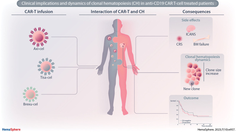
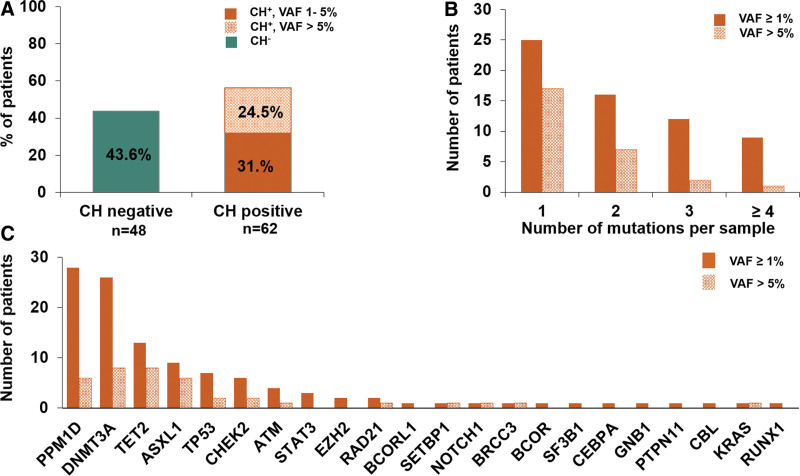
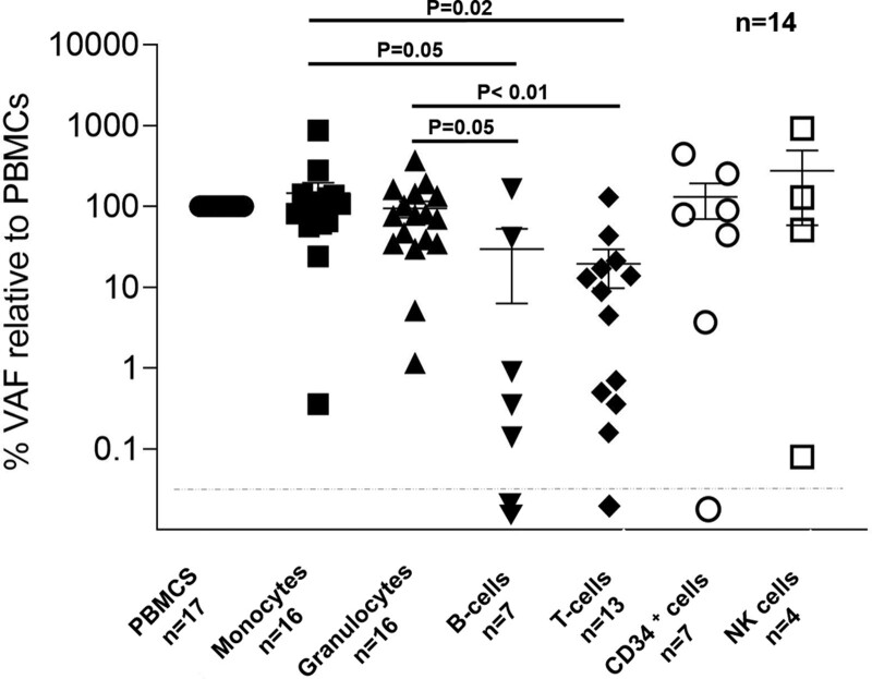
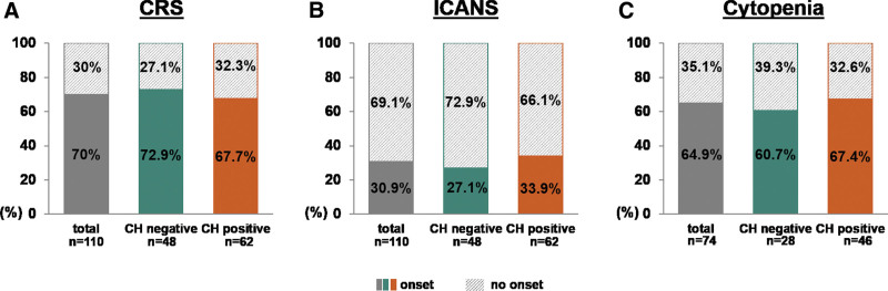
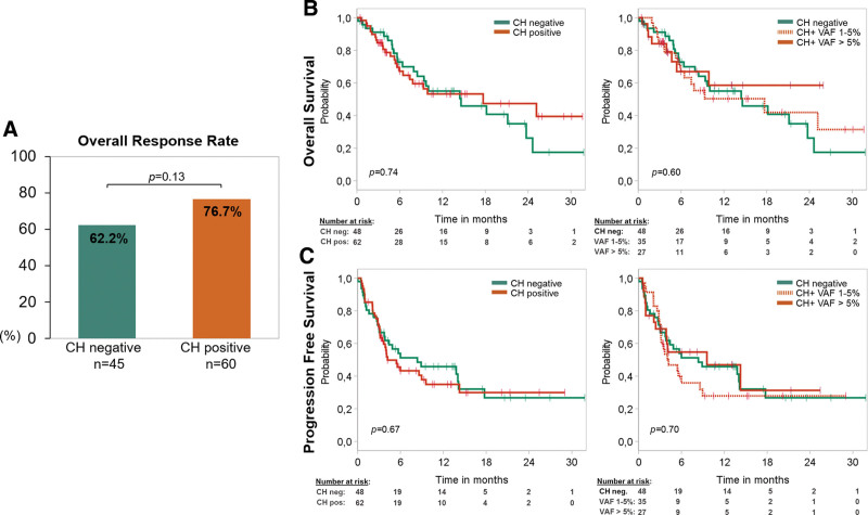
Recent evidence revealed important interactions between clonal hematopoiesis (CH) and cellular therapies established for the treatment of hematologic malignancies. The impact of CH on safety, efficacy, and outcome of chimeric antigen receptor (CAR) T-cell therapy is currently under investigation. We analyzed 110 patients with relapsed/refractory B-cell non-Hodgkin lymphoma (n = 105) or acute lymphoblastic leukemia (ALL) (n = 5), treated with Axicabtagene-Ciloleucel (39%), Tisagenlecleucel (51%), or Brexucabtagene autoleucel (10%). Using error-corrected targeted sequencing, a high CH prevalence of 56.4% (variant allele frequency [VAF] ≥1%) at the time of CAR T-cell infusion was detected. The most frequently mutated gene was PPM1D followed by DNMT3A, TET2, ASXL1, and TP53. Variant allele frequencies were significantly lower in B and T cells compared with monocytes and granulocytes. CH did not increase the risk of CAR T-related toxicities. The incidences of cytokine release syndrome and immune effector-cell-associated neurotoxicity syndrome were similar between CHpos and CHneg patients, regardless of clone size, age, or CAR T product. Prolonged cytopenias were not associated with CH. Best overall response rates (ORRs) were numerically but not significantly higher in CHpos patients (ORR 76.7% versus 62.2%; P = 0.13). Furthermore, CH status did not predict progression-free survival or overall survival. Lastly, sequential analysis showed a modest VAF increase of 1.3% and acquisition of novel mutations within 100 days postinfusion. CH was frequent in large B-cell lymphoma/ALL patients receiving CAR T-cells but did not affect toxicity nor treatment response or outcome.


Conflict of interest statement

MH reports fees for advisory or consultancy services from Abbvie, Agios, BMS, Daiichi Sankyo, Eurocept, Glycostem, Janssen, Jazz Pharmaceuticals, Kura Oncology, Novartis, Pfizer, PinotBio, Roche, Takeda, and Tolremo. FD reports personal fees from Gilead, Incyte, Roche, Novartis, AbbVie, Astra Zeneca outside the submitted work. JFK reports personal fees from Gilead and Janssen outside the submitted work. GW reports honoraria from Gilead, Novartis, Clinigen, and Janssen outside the submitted work. OP has no conflicts of interest directly related to this work. OP has received honoraria or travel support from Gilead, Jazz, MSD, Novartis, Pfizer, and Therakos. He has received research support from Incyte and Priothera. He is member of advisory boards to Equillium Bio, Jazz, Gilead, Novartis, MSD, Omeros, Priothera, Sanofi, Shionogi, and SOBI. All the other authors have no conflicts of interest to disclose.

Figures

None
Graphical abstract
Figure 1.
Figure 1.
CH at the time of treatment is very common among patients receiving CAR T-cell therapy. (A) Frequency of CH across the entire cohort (n = 110) as measured by the VAF using a cutoff of 1%. (B) Distribution of the number of mutations found in patients of total cohort. Quantity of mutations stratified by the size of the clone considering low VAF %1–5% and high VAF >5%. (C) Number of mutated patients (y-axis) according to gene information (x-axis) at differing VAF groups (VAF ≥1% and VAF ≥5%). CAR = chimeric antigen receptor; CH = clonal hematopoiesis; VAF = variant allele frequency.
Figure 2.
Figure 2.
Cellular distribution of clonal hematopoiesis. VAFs of 18 mutations were studied in PBMNCs and flow-sorted peripheral blood cell fractions (monocytes, granulocytes, B cells, T cells, CD34+ cells,and NK cells). PBMNCs = peripheral blood mononuclear cells; VAF = variant allele frequency.
Figure 3.
Figure 3.
CH and CAR T-therapy toxicity. (A, B) Histogram plots showing prevalence of CRS/ICANS (onset: filled, no onset: hatched) according to the absence (n = 48, green) or presence (n = 62, orange) of clonal hematopoiesis across the total cohort (n = 110, gray). (C) Histogram plots illustrating prevalence of cytopenias at day 100 after CAR T-cell therapy as defined by leukopenia (total WBC count <3000/µL) and/or thrombocytopenia (platelet count <100,000/µL) and/or anemia (hemoglobin <10 g/dL) in patients with available information (n = 74) according to the presence (n = 46) or absence (n = 28) of CH with a VAF cutoff 1%. CAR = chimeric antigen receptor; CH = clonal hematopoiesis; CRS = cytokine release syndrome; ÍCANS = immune effector-cell-associated neurotoxicity syndrome; VAF = variant allele frequency.
Figure 4.
Figure 4.
CH and survival after treatment with CAR T- therapy. (A) Diagram demonstrating the best ORR during the first 180 days after CAR T-cell treatment stratified by absence (green, n = 45) or presence (orange, n = 60) of CH with a VAF cutoff 1%, respectively. P-value was calculated with Fisher exact test. Patients with lack of follow-up data or death before progress/response were excluded (n = 5). (B) Kaplan-Meier curves showing overall survival of 110 patients undergoing CAR T-cell therapy stratified by absence (green, n = 48) or presence (orange, n = 62) of CH with a VAF cutoff 1% and stratified by the size of the clone considering low VAF 1%–5% (orange dashed) and high VAF >5% (orange). (C) Kaplan-Meier curves showing progression-free survival of 110 patients undergoing CAR T-cell therapy stratified by absence (green, n = 48) or presence (orange, n = 62) of CH with a VAF cutoff 1% and stratified by the size of the clone considering low VAF 1%–5% (orange dashed) and high VAF >5% (orange). P-value was calculated for (B) and (C) with log-rank test. CAR = chimeric antigen receptor; CH = clonal hematopoiesis; ORR = overall response rate; VAF = variant allele frequency.
Figure 5.
Figure 5.
Clonal dynamics after CAR T-cell therapy. (A–D) VAFs at the time of CAR T-cell treatment and individual time points of sampling and corresponding mutational and clinical data. The gray dashed line depicts our VAF detection limit. CAR = chimeric antigen receptor; VAF = variant allele frequency.

References

    1. Abelson S, Collord G, Ng SWK, et al. Prediction of acute myeloid leukaemia risk in healthy individuals. Nature. 2018;559:400–404. - PMC - PubMed
    1. Acuna-Hidalgo R, Sengul H, Steehouwer M, et al. Ultra-sensitive sequencing identifies high prevalence of clonal hematopoiesis-associated mutations throughout adult life. Am J Hum Genet. 2017;101:50–64. - PMC - PubMed
    1. Arends CM, Weiss M, Christen F, et al. Clonal hematopoiesis in patients with anti-neutrophil cytoplasmic antibody-associated vasculitis. Haematologica. 2020;105:e264–e267. - PMC - PubMed
    1. Buscarlet M, Provost S, Zada YF, et al. DNMT3A and TET2 dominate clonal hematopoiesis and demonstrate benign phenotypes and different genetic predispositions. Blood. 2017;130:753–762. - PubMed
    1. Bolton KL, Ptashkin RN, Gao T, et al. Cancer therapy shapes the fitness landscape of clonal hematopoiesis. Nat Genet. 2020;52:1219–1226. - PMC - PubMed

LinkOut - more resources