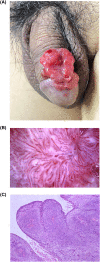
'Seaweed appearance' in squamous cell carcinoma of the penis: A new dermoscopic finding
- PMID: 37799367
- PMCID: PMC10549818
- DOI: 10.1002/ski2.275
'Seaweed appearance' in squamous cell carcinoma of the penis: A new dermoscopic finding
Abstract
Squamous cell carcinoma of the penis is an uncommon cancer. Vascular feature on dermoscopy is common in all forms of invasive squamous cell carcinoma, and the presence of the specific vascular features is often used to aid diagnosis. Here, we reported a new dermoscopic finding-seaweed-like vascular pattern in squamous cell carcinoma of the penis.
© 2023 The Authors. Skin Health and Disease published by John Wiley & Sons Ltd on behalf of British Association of Dermatologists.
Conflict of interest statement
None declared.
Figures

References
-
- Renaud‐Vilmer C, Cavelier‐Balloy B, Verola O, Morel P, Servant JM, Desgrandchamps F, et al. Analysis of alterations adjacent to invasive squamous cell carcinoma of the penis and their relationship with associated carcinoma. J Am Acad Dermatol. 2010;62(2):284–290. 10.1016/j.jaad.2009.06.087 - DOI - PubMed
LinkOut - more resources
Full Text Sources
