Review
. 2023 Oct;13(10):4025-4059.
doi: 10.1016/j.apsb.2023.06.015. Epub 2023 Jun 30.

Antibody-drug conjugates: Recent advances in payloads

Affiliations
Review

Antibody-drug conjugates: Recent advances in payloads

Zhijia Wang et al. Acta Pharm Sin B. 2023 Oct.

Abstract

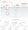
Antibody‒drug conjugates (ADCs), which combine the advantages of monoclonal antibodies with precise targeting and payloads with efficient killing, show great clinical therapeutic value. The ADCs' payloads play a key role in determining the efficacy of ADC drugs and thus have attracted great attention in the field. An ideal ADC payload should possess sufficient toxicity, low immunogenicity, high stability, and modifiable functional groups. Common ADC payloads include tubulin inhibitors and DNA damaging agents, with tubulin inhibitors accounting for more than half of the ADC drugs in clinical development. However, due to clinical limitations of traditional ADC payloads, such as inadequate efficacy and the development of acquired drug resistance, novel highly efficient payloads with diverse targets and reduced side effects are being developed. This perspective summarizes the recent research advances of traditional and novel ADC payloads with main focuses on the structure-activity relationship studies, co-crystal structures, and designing strategies, and further discusses the future research directions of ADC payloads. This review also aims to provide valuable references and future directions for the development of novel ADC payloads that will have high efficacy, low toxicity, adequate stability, and abilities to overcome drug resistance.

Keywords: Antibody‒drug conjugates; DNA damaging agents; Dual payloads; PROTACs; RNA targeting agents; Tubulin inhibitors.


Conflict of interest statement

The authors have no conflicts of interest to declare.

Figures

Image 1
Graphical abstract
Figure 1
Figure 1
Key structures and mechanisms of action of ADCs. (A) The general mechanism of action of ADCs; (B) The mechanism of DNA inhibitors as ADC payloads; (C) The mechanism of Splicing inhibitors as ADC payloads; (D) The mechanism of tubulin inhibitors as ADC payloads; (E) The mechanism of PROTAC molecules as ADC payloads; (F) The mechanism of Bcl-xL inhibitors and proteasome inhibitors as ADC payloads; (G) The mechanism of NAMPT inhibitors as ADC payloads; (H) The mechanism of NIR-PIT ADC.
Figure 2
Figure 2
Milestones in the development of ADCs payloads.
Figure 3
Figure 3
Design and SAR analysis of maytansinoids. (A) Chemical structures of maytansinoids 15; (B) Binding mode of maytansine (1) in complex with tubulin (PDB code 4TV8); (C) Binding mode of compound 5 in complex with tubulin (PDB code 5SBA).
Figure 4a
Figure 4-1
Design and SAR analysis of auristatin analogs. (A) Chemical structures of auristatin analogs 710, 2124; (B) Binding mode of compound 10 in complex with tubulin (PDB code 4X1I).
Figure 4b
Figure 4-2
Chemical structures of auristatin analogs 1120.
Figure 5
Figure 5
Design and SAR analysis of eribulin analogs. (A) Chemical structures of eribulin analogs 2527; (B) Binding mode of eribulin 26 in complex with tubulin (PDB code 5JH7).
Figure 6
Figure 6
Design and SAR analysis of tubulysin analogs. (A) Chemical structures of tubulysin analogs 2834; (B) Binding mode of compound 30 in complex with tubulin (PDB code 6Y4N).
Figure 7
Figure 7
Design and SAR analysis of cryptophycin analogs. (A) Chemical structures of cryptophycin analogs 3540; (B) Binding mode of cryptophycin-52 (36) in complex with tubulin (PDB code 7M20).
Figure 8
Figure 8
Design and SAR analysis of EG5 inhibitors. (A) Chemical structures of EG5 inhibitors 4145; (B) Binding mode of ispinesib (41) in complex with EG5 (PDB code 4AP0).
Figure 9
Figure 9
Design and SAR analysis of enediyne. (A) Chemical structures of enediyne 4652; (B) Binding mode of calicheamicin γI1 (46) in complex with DNA (PDB code 2PIK).
Figure 10
Figure 10
Design and SAR analysis of Topoisomerase I inhibitors. (A) Binding mode of Topotecan (53) in complex with TOPO I-DNA (PDB code 1K4T); (B) Chemical structures of Topoisomerase I inhibitors 5360.
Figure 11
Figure 11
Design and SAR analysis of PBD analogs 6172.
Figure 12
Figure 12
Design and SAR analysis of duocarmycins analogs. (A) Chemical structures of duocarmycins analogs 7380; (B) Binding mode of Duocarmycins A (73) in complex with DNA (PDB code 1DSM).
Figure 13
Figure 13
Design and SAR analysis of thailanstatin analogs 8187.
Figure 14
Figure 14
Design and SAR analysis of amatoxins. (A) Chemical structures of amatoxins 88, 89; (B) Binding mode of α-Amanitin (88) in complex with RNA (PDB code 3CQZ).
Figure 15
Figure 15
Design and SAR analysis of TLR agonists 9092.
Figure 16
Figure 16
Design and SAR analysis of STING agonists. (A) Chemical structures of STING agonists 9396; (B) Binding mode of CDN (93) in complex with STING (PDB code 6A05).
Figure 17
Figure 17
Design and SAR analysis of glucocorticoid receptor modulators (GRMs). (A) Chemical structures of GRMs 97102; (B) Binding mode of compound 97 in complex with GR (PDB code 4UDC); (C) Binding mode of compound 98 in complex with GR (PDB code 4UDD).
Figure 18
Figure 18
Design and SAR analysis of Bcl-xL inhibitors. (A) Chemical structures of Bcl-xL inhibitors 103, 104; (B) Binding mode of ABT-737 (103) in complex with Bcl-xL (PDB code 2YXJ).
Figure 19
Figure 19
Design and SAR analysis of NAMPT inhibitors. (A) Chemical structures of NAMPT inhibitors 105113; (B) Binding mode of A-1293201 (106) in complex with NAMPT (PDB code 5U2M).
Figure 20
Figure 20
Design of carmaphycins analogs 114122.
Figure 21
Figure 21
Chemical structures of PROTAC molecules 123127.
Figure 22
Figure 22
Design and analysis of IRDye700DX (128). (A) Chemical structures of IRDye700DX (128); (B) The mechanism of NIR-PIT ADC.
Figure 23
Figure 23
The mode pattern of dual-payloads drug ADC (The red and green shapes mean different payloads).
Figure 24
Figure 24
Key structures and mechanisms of action of PDCs. (A) The general mechanism of action of PDCs; (B) Chemical structures of the common PDC payloads.

References

    1. Weingart S.N., Zhang L., Sweeney M., Hassett M. Chemotherapy medication errors. Lancet Oncol. 2018;19:e191–e199. - PubMed
    1. Zraik I.M., Hess-Busch Y. Management of chemotherapy side effects and their long-term sequelae. Urologe. 2021;60:862–871. - PubMed
    1. Ponziani S., Di Vittorio G., Pitari G., Cimini A.M., Ardini M., Gentile R., et al. Antibody‒drug conjugates: the new frontier of chemotherapy. Int J Mol Sci. 2020;21:5510. - PMC - PubMed
    1. McKertish C.M., Kayser V. Advances and limitations of antibody drug conjugates for cancer. Biomedicines. 2021;9:872. - PMC - PubMed
    1. Chau C.H., Steeg P.S., Figg W.D. Antibody‒drug conjugates for cancer. Lancet. 2019;394:793–804. - PubMed

LinkOut - more resources