. 2021 Sep 3;11(17):10974-10987.
doi: 10.1021/acscatal.1c03088. Epub 2021 Aug 18.

Structural Determinants of Substrate Recognition and Catalysis by Heparan Sulfate Sulfotransferases

Affiliations

Structural Determinants of Substrate Recognition and Catalysis by Heparan Sulfate Sulfotransferases

Tarsis Ferreira Gesteira et al. ACS Catal. .

Abstract

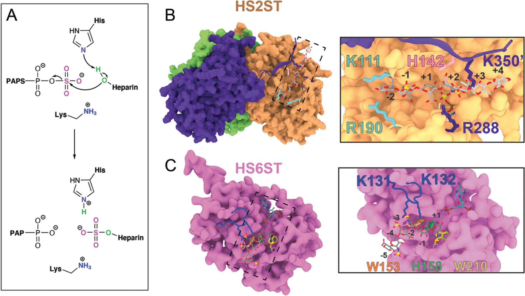
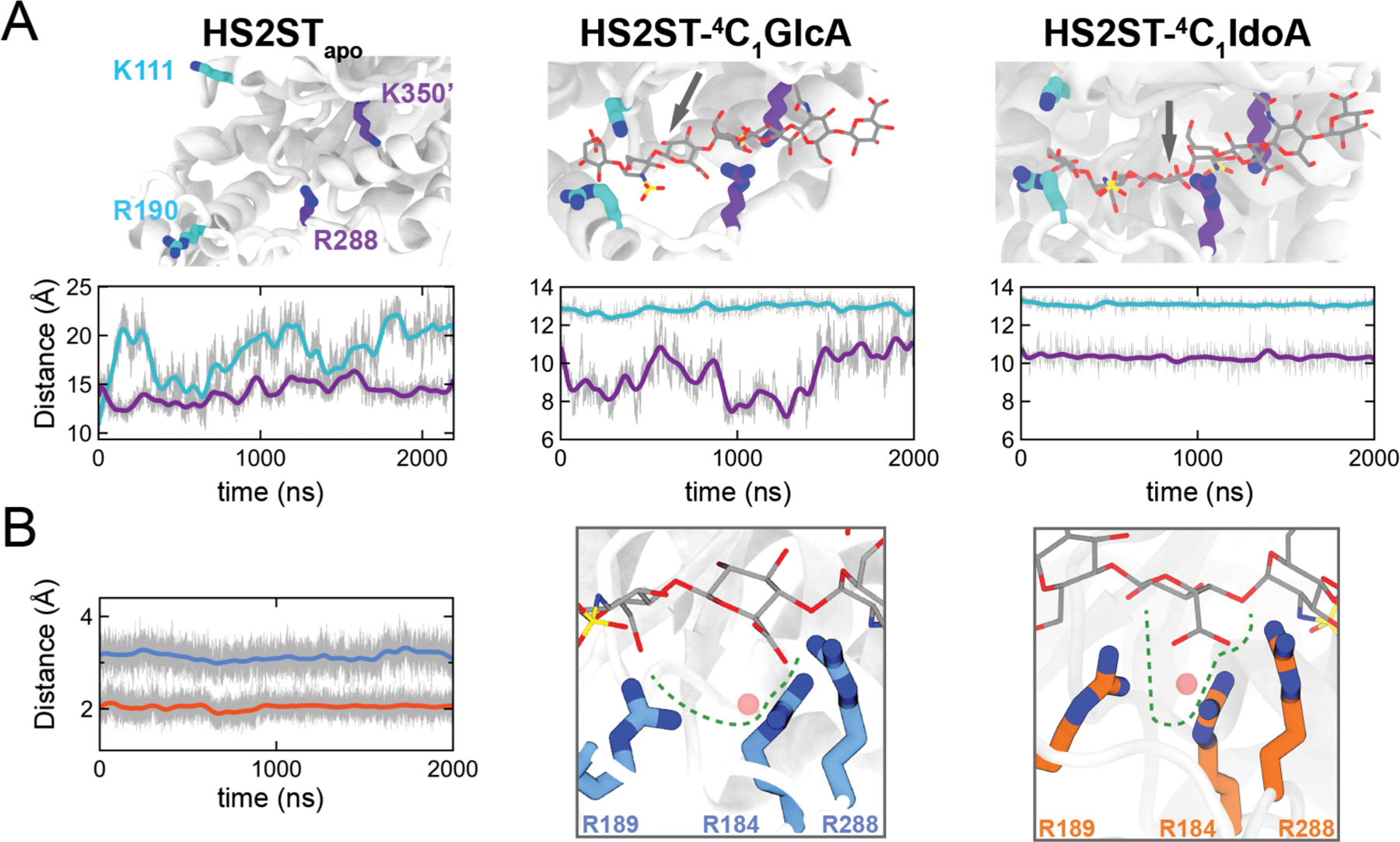
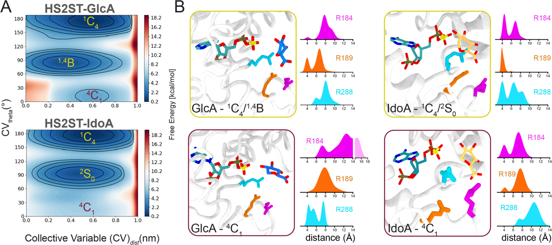
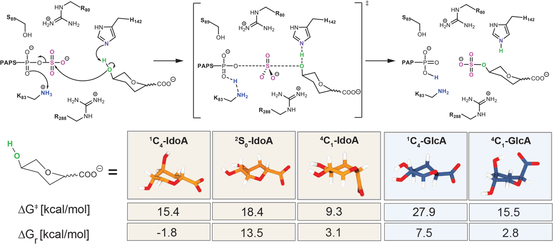
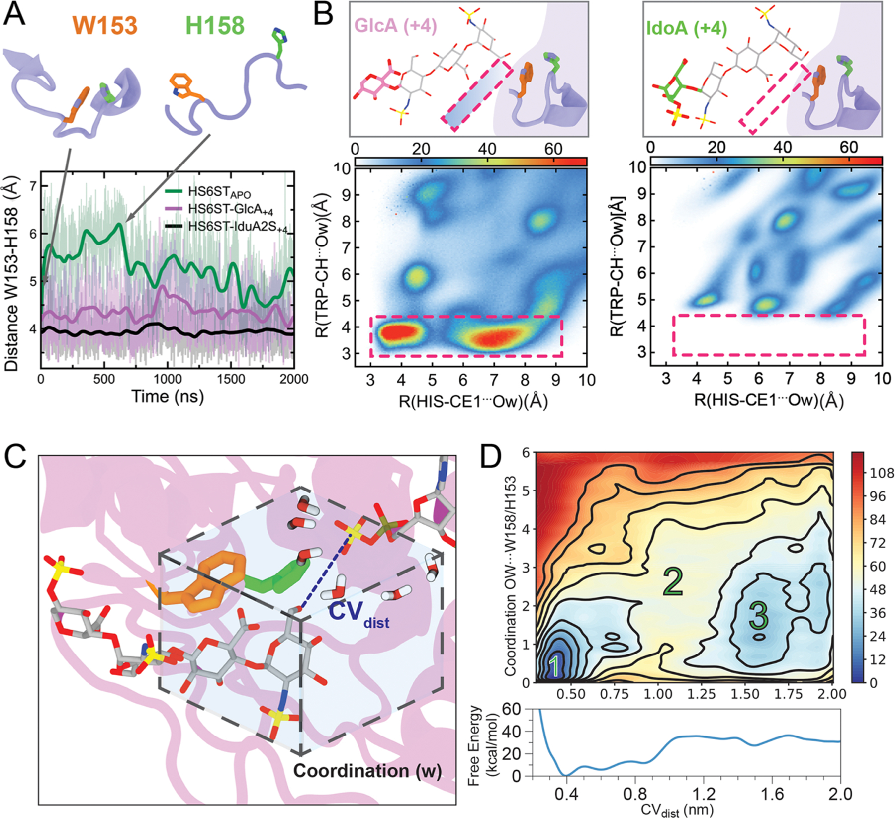
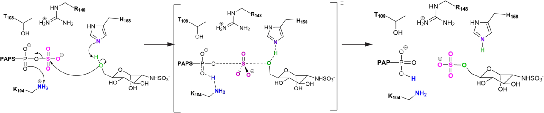
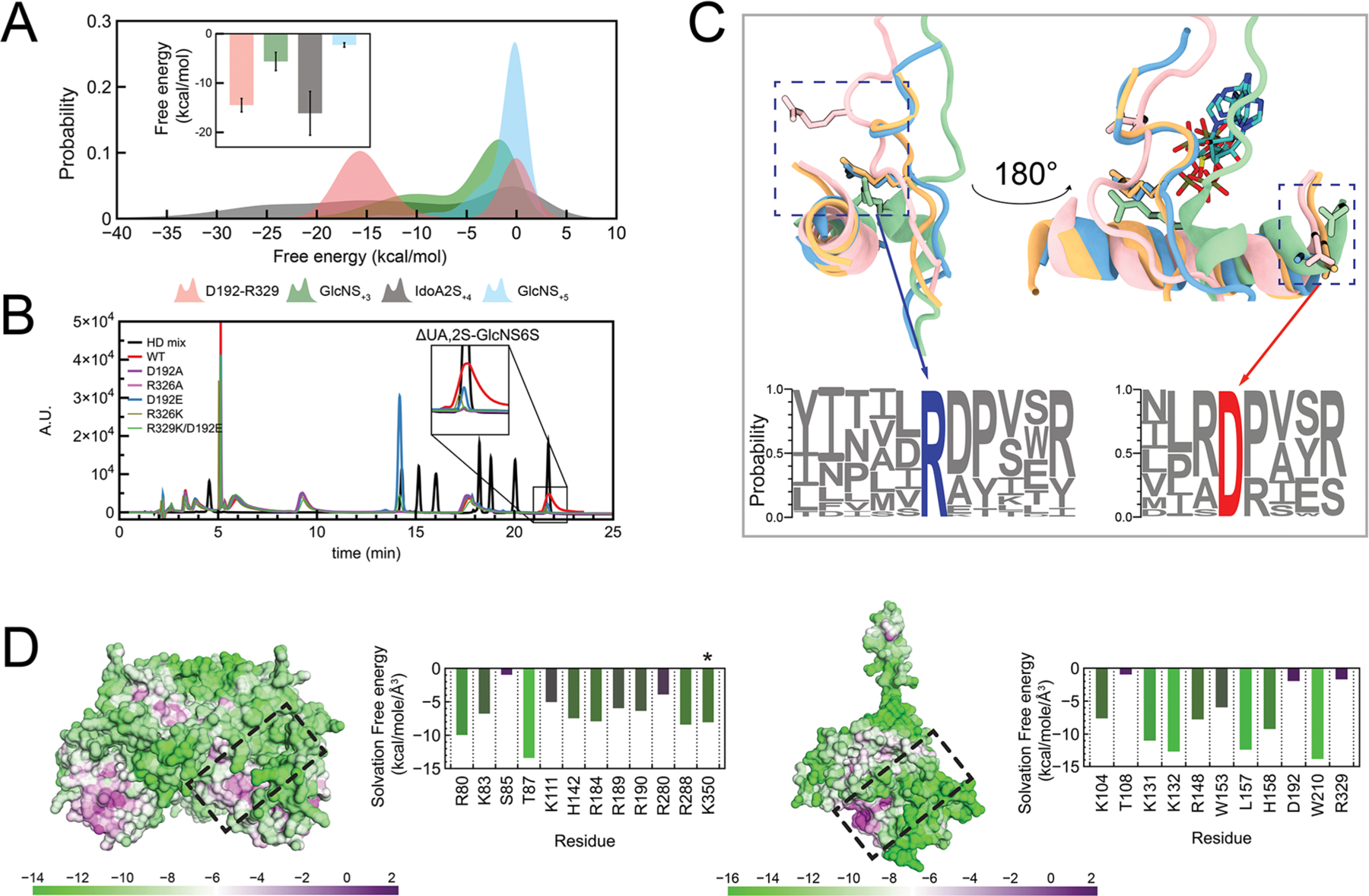
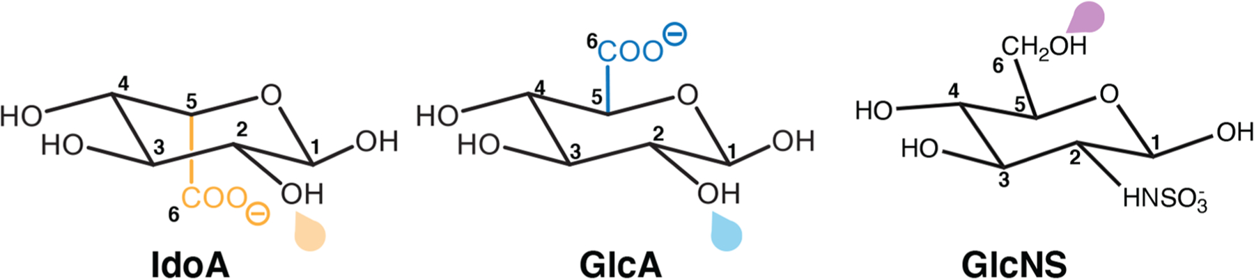
Heparan sulfate (HS) and heparin contain imprinted "sulfation codes", which dictate their diverse physiological and pathological functions. A group of orchestrated biosynthetic enzymes cooperate in polymerizing and modifying HS chains. The biotechnological development of enzymes that can recreate this sulfation pattern on synthetic heparin is challenging, primarily due to the paucity of quantitative data for sulfotransferase enzymes. Herein, we identified critical structural characteristics that determine substrate specificity and shed light on the catalytic mechanism of sugar sulfation of two HS sulfotransferases, 2-O-sulfotransferase (HS2ST) and 6-O-sulfotransferase (HS6ST). Two sets of molecular clamps in HS2ST recognize appropriate substrates; these clamps flank the acceptor binding site on opposite sides. The hexuronic epimers, and not their puckers, have a critical influence on HS2ST selectivity. In contrast, HS6ST recognizes a broader range of substrates. This promiscuity is granted by a conserved tryptophan residue, W210, that positions the acceptor within the active site for catalysis by means of strong electrostatic interactions. Lysines K131 and K132 act in concert with a second tryptophan, W153, shedding water molecules from within the active site, thus providing HS6ST with a binding preference toward 2-O-sulfated substrates. QM/MM calculations provided valuable mechanistic insights into the catalytic process, identifying that the sulfation of both HS2ST and HS6ST follows a SN2-like mechanism. When they are taken together, our findings reveal the molecular basis of how these enzymes recognize different substrates and catalyze sugar sulfation, enabling the generation of enzymes that could create specific heparin epitopes.

Keywords: HS2ST; HS6ST; active-site solvation; glycosaminoglycan biosynthesis; heparin; sugar ring puckering; sulfation; sulfotransferase.


Conflict of interest statement

The authors declare no competing financial interest.

Figures

Figure 1.
Figure 1.
Representation of HS2ST and HS6ST. (A) General proposed catalytic mechanism exerted by HS2ST and HS6ST. (B) Surface representation of the HS2ST trimer. Each monomer is represented with a unique color. The insert highlights the glycan clamps (K111/R190 in cyan and R288/K350′ in purple) and the H142 catalytic base. (C) Surface representation of the HS6ST monomer. The insert highlights W153 and W210 near the active site, catalytic H153, and the loop containing the K131/K132 pair. The numbers indicate the position of the saccharide units in relation to the acceptor unit.
Figure 2.
Figure 2.
Uronic acid epimers binding to HS2ST. (A) Clamp pairs center of mass distances during simulations with the apoprotein, HS2ST:4C1-GlcA or HS2ST:4C1-IdoA. (B) Center of mass (pink sphere) distance of arginine triad R184/R189/R288 to the C6 atom of either IdoA (in orange) or GlcA (in blue).
Figure 3.
Figure 3.
Metadynamics of the HS2ST acceptor sugars. (A) Conformational FES (θ vs distance from PAPS) obtained for HS2ST:GlcA and HS2ST:IdoA. Each contour line of the diagram corresponds to 5 kcal/mol. (B) Distribution of key interactions in terms of the distances from the acceptor hexosamine to the arginines composing the fork in the 1C4/4C1 states (yellow and purple outlines, respectively). The distances of the uronic acceptor heavy atoms to the guanidine groups of R184, R189, and R288 were calculated.
Figure 4.
Figure 4.
Schematic representation of the critical points computed at the ONIOM[M06–2x/6–31+G*:ff14SB] level of theory for HS2ST catalysis on 4C1-, 2S0-, and 1C4-IdoA and 4C1- and 1C4-GlcA saccharide units. The activation (ΔG) and reaction (ΔGr) Gibbs free energies are reported.
Figure 5.
Figure 5.
HS6ST substrate at position +4 influences HS6ST motility and active site hydration. (A) Distances between W153 and H158 during HS6ST simulations. (B) 2D-radial distribution functions (2D-RDF) function for the two closest atoms from W153 and H158 for both HS6ST:IdoA2S+4 and HS6ST:GlcA+4 simulations. Pink dashed lines outline the two water clusters observed in HS6ST:GlcA+4 simulations. Atoms are labeled according to AMBER force field nomenclature. (C) Diagrams of coordination parameters defined for free energy calculations of active site hydration and acceptor coupling. The dashed box displays the coordination of waters surrounding the active site collective variable parameter (w). The substrate is shown as gray sticks. (D) Projection of the FES onto CVdist and CVw, as well as onto CVdist alone (bottom). Numbers indicate the most relevant metastable states 1 and 3, separated by a high-energy barrier, 2. Contours are drawn every 12 kcal/mol.
Figure 6.
Figure 6.
Schematic representation of the critical points computed at the ONIOM[M06–2x/6–31+G*:ff14SB] level of theory for HS6ST catalysis.
Figure 7.
Figure 7.
Primer on the shared topologies of heparin sulfotransferases. (A) Probability distribution of calculated pairwise energies during molecular dynamics simulations for (I) the substrate IdoA2S-GlcNS and (II) the D192-R329 salt bridge during HS6ST apoprotein simulations. The inset shows calculated averages. (B) SAX-HPLC analysis of total disaccharide composition after the action of R329/D192 mutants incubated with IdoA2S-GlcNS substrates. The insert highlights the formation of IdoA2S-GlcNS6S moieties. HD mix (black line), heparin disaccharide mix (Iduron, UK). (C) A salt bridge is conserved among glycan sulfotransferases, and its conformation change is triggered by substrate binding. Superposition of HSSTs: light blue, HSNST; pink, HS2ST; orange, HS3ST; green, HS6ST. The bottom logo plot highlights ARG (blue) and ASP (red) conservation among all HSSTs known to date. (D) Calculated solvation free energies on the surface of the binding interfaces of HS2ST (left) and HS6ST (right) as a measure of their hydrophobicity. Tables show the per-residue average hydrophobicity for each apoprotein enzyme. Table colors range from more hydrophobic (purple) to more hydrophilic (green). The asterisk denotes C-terminal “prime” residues.
Scheme 1.
Scheme 1.. Schematic Representation of IdoA, GlcA, and GlcNS Monosaccharidesa
aThe C5 centers of the epimers IdoA and GlcA are shown in orange and blue, respectively. Drops (orange/blue for HS2ST and purple for HS6ST) point toward the sulfation sites.

References

    1. Nader HB; Dietrich CP; Buonassisi V; Colburn P Heparin sequences in the heparan sulfate chains of an endothelial cell proteoglycan. Proc. Natl. Acad. Sci. U. S. A 1987, 84 (11), 3565–9. - PMC - PubMed
    1. Gesteira TF; Coulson-Thomas VJ; Ogata FT; Farias EH; Cavalheiro RP; de Lima MA; Cunha GL; Nakayasu ES; Almeida IC; Toma L; Nader HB A novel approach for the characterisation of proteoglycans and biosynthetic enzymes in a snail model. Biochim. Biophys. Acta, Proteins Proteomics 2011, 1814 (12), 1862–9. - PubMed
    1. Attreed M; Saied-Santiago K; Bulow HE Conservation of anatomically restricted glycosaminoglycan structures in divergent nematode species. Glycobiology 2016, 26 (8), 862–870. - PMC - PubMed
    1. Heparin Market Size, Share & Trends Analysis Report By Type (Low Molecular Weight, Ultra-low Molecule Weight) Grand View Research. https://www.grandviewresearch.com/industry-analysis/heparin-market; accessed December 15, 2019.
    1. Gallagher JT; Walker A Molecular distinctions between heparan sulphate and heparin. Analysis of sulphation patterns indicates that heparan sulphate and heparin are separate families of N-sulphated polysaccharides. Biochem. J 1985, 230 (3), 665–74. - PMC - PubMed