

Albumin-based nanosystem for dual-modality imaging-guided chem-phototherapy against immune-cold triple-negative breast cancer
- PMID: 37799708
- PMCID: PMC10548782
- DOI: 10.1093/rb/rbad073
Albumin-based nanosystem for dual-modality imaging-guided chem-phototherapy against immune-cold triple-negative breast cancer
Abstract
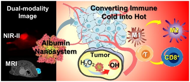
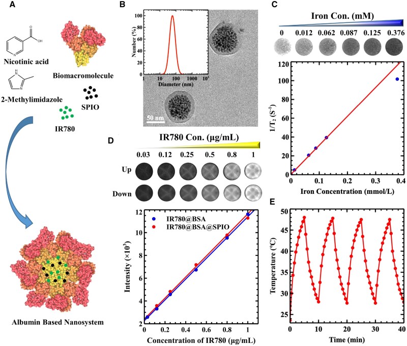
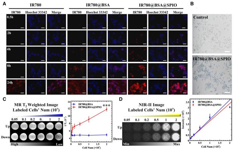
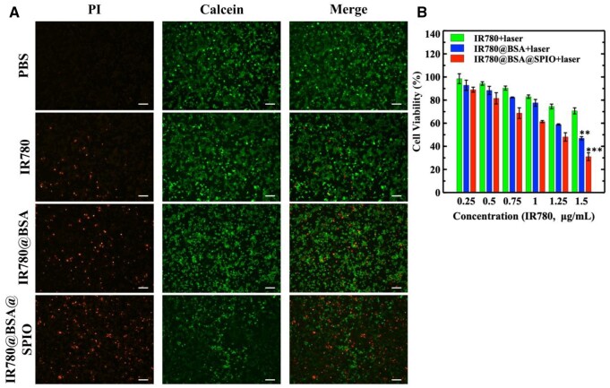
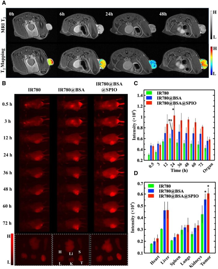
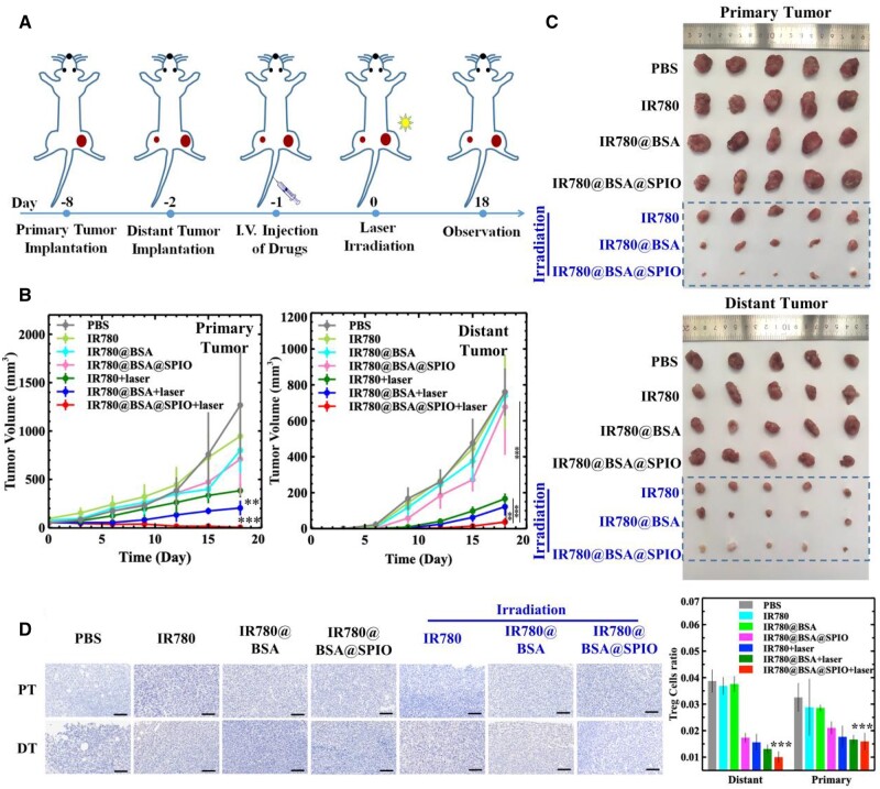
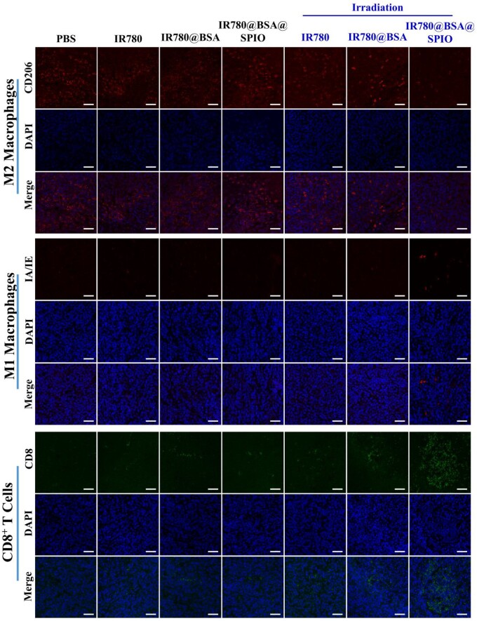
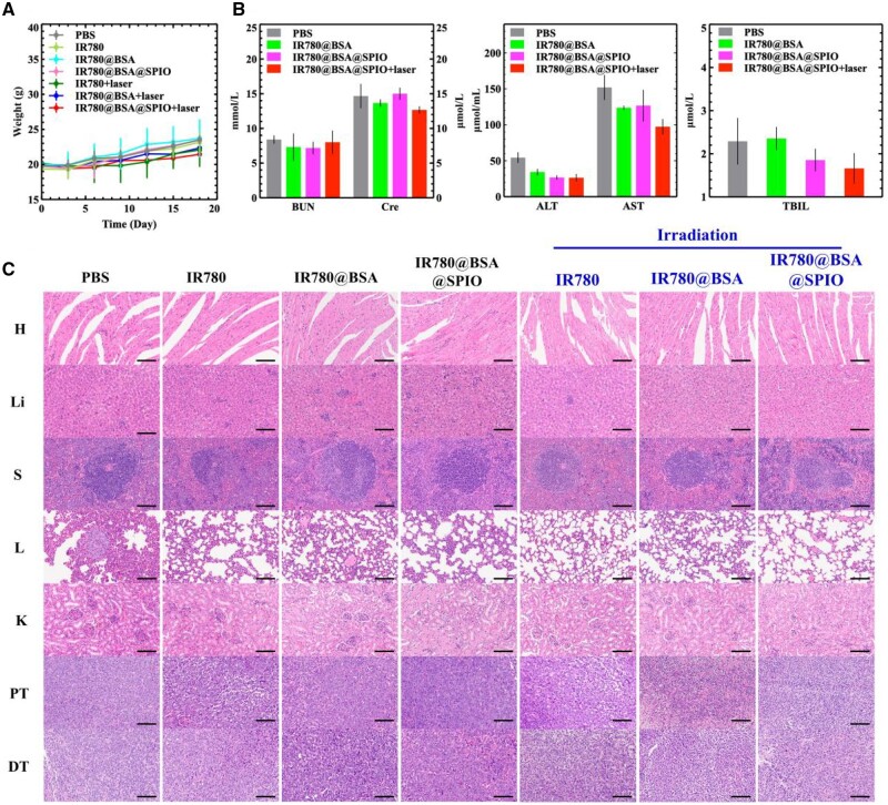
Triple-negative breast cancer is a highly aggressive and metastatic tumor; diagnosing it in the early stages is still difficult, and the prognosis for conventional radio-chemotherapy and immunotreatment is not promising due to cancer's immunosuppressive microenvironment. The utilization of protein-based nanosystem has proven to be effective in delivering agents with limited adverse effects, yet the combination of diagnosis and treatment remains a difficult challenge. This research took advantage of natural albumin and organic molecules to construct a self-assemble core-shell nanostructure combining with superparamagnetic iron oxide nanocrystals and heptamethine cyanine dye IR780 through non-covalent interactions. This nanocomposite successfully decreased the transverse relaxation time of the magnetic resonance hydrogen nucleus, resulting in outstanding T2 imaging, as well as emitting near-infrared II fluorescence, thereby the resulting dual-modality imaging tool was applied to improve diagnostic competency. It is noteworthy that the nanocomposites exhibited impressive enzyme-like catalytic and photothermal capabilities, resulting in a successful activation of the immune system to efficiently suppress distant metastatic lesions in vivo. Consequently, this nano-drug-based therapy could be an advantageous asset in reinforcing the immune system and hindering the growth and reappearance of the immune-cold breast cancer.
Keywords: albumin; fluorescence near-infrared II imaging; magnetic resonance imaging; nanozyme; phototherapy.
© The Author(s) 2023. Published by Oxford University Press.
Figures
Similar articles
-
pH-Sensitive nanotheranostics for dual-modality imaging guided nanoenzyme catalysis therapy and phototherapy.J Mater Chem B. 2020 Jun 10;8(22):4859-4869. doi: 10.1039/c9tb02731a. J Mater Chem B. 2020. PMID: 32100793
-
In situ Injection of pH- and Temperature-Sensitive Nanomaterials Increases Chemo-Photothermal Efficacy by Alleviating the Tumor Immunosuppressive Microenvironment.Int J Nanomedicine. 2022 Jun 16;17:2661-2678. doi: 10.2147/IJN.S367121. eCollection 2022. Int J Nanomedicine. 2022. Retraction in: Int J Nanomedicine. 2023 Mar 09;18:1193-1194. doi: 10.2147/IJN.S411463. PMID: 35733417 Free PMC article. Retracted.
-
Core-Shell-Structured Covalent-Organic Framework as a Nanoagent for Single-Laser-Induced Phototherapy.ACS Appl Mater Interfaces. 2021 Apr 21;13(15):17243-17254. doi: 10.1021/acsami.1c01125. Epub 2021 Apr 7. ACS Appl Mater Interfaces. 2021. PMID: 33825447
-
Thermo- and pH-dual responsive polymeric micelles with upper critical solution temperature behavior for photoacoustic imaging-guided synergistic chemo-photothermal therapy against subcutaneous and metastatic breast tumors.Theranostics. 2018 Jul 16;8(15):4097-4115. doi: 10.7150/thno.26195. eCollection 2018. Theranostics. 2018. PMID: 30128039 Free PMC article.
-
Recent progress on near-infrared fluorescence heptamethine cyanine dye-based molecules and nanoparticles for tumor imaging and treatment.Wiley Interdiscip Rev Nanomed Nanobiotechnol. 2023 Sep-Oct;15(5):e1910. doi: 10.1002/wnan.1910. Epub 2023 Jun 12. Wiley Interdiscip Rev Nanomed Nanobiotechnol. 2023. PMID: 37305979 Review.
Cited by
-
In vivo dynamic visualization and evaluation of collagen degradation utilizing NIR-II fluorescence imaging in mice models.Regen Biomater. 2025 Apr 11;12:rbaf025. doi: 10.1093/rb/rbaf025. eCollection 2025. Regen Biomater. 2025. PMID: 40405872 Free PMC article.
-
Ferroptosis boosting system based on a sonodynamic therapy cascade-augmented strategy for triple-negative breast cancer therapy.Regen Biomater. 2025 May 20;12:rbaf042. doi: 10.1093/rb/rbaf042. eCollection 2025. Regen Biomater. 2025. PMID: 40567832 Free PMC article.
-
Cancer cell response to extrinsic and intrinsic mechanical cue: opportunities for tumor apoptosis strategies.Regen Biomater. 2024 Feb 20;11:rbae016. doi: 10.1093/rb/rbae016. eCollection 2024. Regen Biomater. 2024. PMID: 38476678 Free PMC article. Review.
-
Nanomaterial-assisted pancreatic cancer theranostics.Regen Biomater. 2025 Jun 11;12:rbaf054. doi: 10.1093/rb/rbaf054. eCollection 2025. Regen Biomater. 2025. PMID: 40585530 Free PMC article. Review.
-
Facile construction of manganese-based contrast agent with high T 1 relaxivity for magnetic resonance imaging via flash technology-based self-assembly.Regen Biomater. 2025 Mar 11;12:rbaf009. doi: 10.1093/rb/rbaf009. eCollection 2025. Regen Biomater. 2025. PMID: 40270577 Free PMC article.
References
-
- Hyuna S, Jacques F, Rebecca LS, Mathieu L, Isabelle S, Ahmedin J, Freddie B.. Global cancer statistics 2020: GLOBOCAN estimates of incidence and mortality worldwide for 36 cancers in 185 countries. CA Cancer J Clin 2021;71:209–49. - PubMed
-
- Dharambir K, Deeksha P, Riya S, Vivek KG, Neelam G, Deepika K, Atef Z, Shubham K, Assaye B.. Global increase in breast cancer incidence: risk factors and preventive measures. BioMed Res Int 2022;10:1–16. - PubMed
-
- Fernández JÁ, Ozores PP, López VC, Mosquera AC, López RI.. Breast cancer. Medicine (Spain) 2021;13:1506–17.
-
- William H, Xinkai Z, Antonia P, Ngan C, Stephanie LF, Anne H, Neil W, David E, Arash N.. Role of clinical and imaging risk factors in predicting breast cancer diagnosis among BI-RADS 4 cases. Clin Breast Cancer 2019;19:e142–51. - PubMed
LinkOut - more resources
Full Text Sources
