

Anatomic evaluation of the posterior superior alveolar artery using cone-beam computed tomography: A systematic review and meta-analysis
- PMID: 37799745
- PMCID: PMC10548151
- DOI: 10.5624/isd.20230009
Anatomic evaluation of the posterior superior alveolar artery using cone-beam computed tomography: A systematic review and meta-analysis
Abstract
Purpose: This systematic review examined the detection of the posterior superior alveolar artery, along with various anatomic characteristics, on cone-beam computed tomography images.
Materials and methods: Studies were identified electronically through the Web of Science, MEDLINE, Scopus, and Embase databases. The quality of the included studies was evaluated using a 5-item binary scale. The detection rate, location, and classified diameter of the posterior superior alveolar artery were estimated as prevalence values. The diameter of this artery, as well as the distances from the artery to the alveolar crest and sinus floor, were estimated as means with associated 95% confidence intervals.
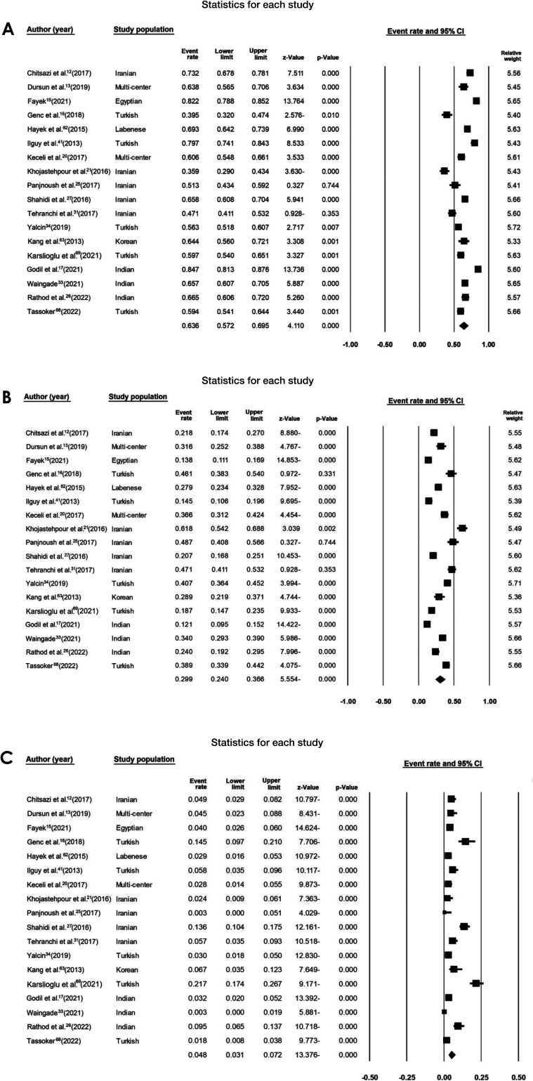
Results: Thirty-seven studies were enrolled, with 34 of these included in the meta-analysis. The mean detection rate was 79% (range: 72%-84%), and the mean diameter was 1.06±0.05 mm (range: 0.96-1.16 mm). The posterior superior alveolar artery was located intraosseously in 64% of cases. The mean distance of the artery from the alveolar crest was 16.71±0.49 mm (range: 15.75-17.68 mm), while the mean distance from the artery to the sinus floor was 8.85±0.4 mm (range: 8.05-9.64 mm).
Conclusion: According to the findings of this meta-analysis regarding various anatomic characteristics of the posterior superior alveolar artery, severe hemorrhage after damage to this artery during sinus augmentation procedures is not a substantial clinical problem.
Keywords: Cone-Beam Computed Tomography; Hemorrhage; Maxillary Artery; Sinus Floor Augmentation.
Copyright © 2023 by Korean Academy of Oral and Maxillofacial Radiology.
Conflict of interest statement
Conflict of Interest: None
Figures


References
-
- Bedeloğlu E, Yalçın M. Evaluation of the posterior superior alveolar artery prior to sinus floor elevation via lateral window technique: a cone-beam computed tomography study. J Adv Oral Res. 2020;11:215–223.
-
- Jensen OT, Shulman LB, Block MS, Iacono VJ. Report of the sinus consensus conference of 1996. Int J Oral Maxillofac Implants. 1998;13 Suppl:11–45. - PubMed
-
- Zijderveld SA, van den Bergh JP, Schulten EA, ten Bruggenkate CM. Anatomical and surgical findings and complications in 100 consecutive maxillary sinus floor elevation procedures. J Oral Maxillofac Surg. 2008;66:1426–1438. - PubMed
-
- Testori T, Wallace SS, Del Fabbro M, Taschieri S, Trisi P, Capelli M, et al. Repair of large sinus membrane perforations using stabilized collagen barrier membranes: surgical techniques with histologic and radiographic evidence of success. Int J Periodontics Restorative Dent. 2008;28:9–17. - PubMed
-
- Testori T, Rosano G, Taschieri S, Del Fabbro M. Ligation of an unusually large vessel during maxillary sinus floor augmentation. A case report. Eur J Oral Implantol. 2010;3:255–258. - PubMed
