

The Diagnostic Performance of Linked Color Imaging Compared to White Light Imaging in Endoscopic Diagnosis of Helicobacter pylori Infection: A Systematic Review and Meta-Analysis
- PMID: 37800315
- PMCID: PMC11096912
- DOI: 10.5009/gnl230244
The Diagnostic Performance of Linked Color Imaging Compared to White Light Imaging in Endoscopic Diagnosis of Helicobacter pylori Infection: A Systematic Review and Meta-Analysis
Abstract
Background/aims: Recognizing Helicobacter pylori infection during endoscopy is important because it can lead to the performance of confirmatory testing. Linked color imaging (LCI) is an image enhancement technique that can improve the detection of gastrointestinal lesions. The purpose of this study was to compare LCI to conventional white light imaging (WLI) in the endoscopic diagnosis of H. pylori infection.
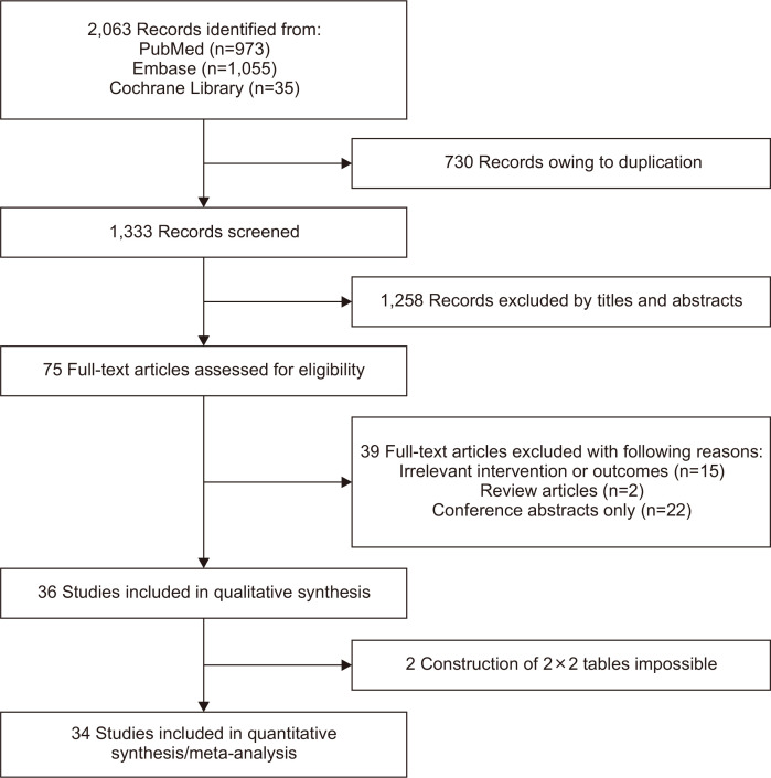
Methods: We conducted a comprehensive literature search using PubMed, Embase, and the Cochrane Library. All studies evaluating the diagnostic performance of LCI or WLI in the endoscopic diagnosis of H. pylori were eligible. Studies on magnifying endoscopy, chromoendoscopy, and artificial intelligence were excluded.
Results: Thirty-four studies were included in this meta-analysis, of which 32 reported the performance of WLI and eight reported the performance of LCI in diagnosing H. pylori infection. The pooled sensitivity and specificity of WLI in the diagnosis of H. pylori infection were 0.528 (95% confidence interval [CI], 0.517 to 0.540) and 0.821 (95% CI, 0.811 to 0.830), respectively. The pooled sensitivity and specificity of LCI in the diagnosis of H. pylori were 0.816 (95% CI, 0.790 to 0.841) and 0.868 (95% CI, 0.850 to 0.884), respectively. The pooled diagnostic odds ratios of WLI and LCI were 15.447 (95% CI, 8.225 to 29.013) and 31.838 (95% CI, 15.576 to 65.078), respectively. The areas under the summary receiver operating characteristic curves of WLI and LCI were 0.870 and 0.911, respectively.
Conclusions: LCI showed higher sensitivity in the endoscopic diagnosis of H. pylori infection than standard WLI.
Keywords: Gastrointestinal endoscopy; Helicobacter pylori; Image enhancement; Sensitivity and specificity.
Conflict of interest statement
No potential conflict of interest relevant to this article was reported.
Figures


Similar articles
-
Non-invasive diagnostic tests for Helicobacter pylori infection.Cochrane Database Syst Rev. 2018 Mar 15;3(3):CD012080. doi: 10.1002/14651858.CD012080.pub2. Cochrane Database Syst Rev. 2018. PMID: 29543326 Free PMC article.
-
Sequential versus standard triple first-line therapy for Helicobacter pylori eradication.Cochrane Database Syst Rev. 2016 Jun 28;2016(6):CD009034. doi: 10.1002/14651858.CD009034.pub2. Cochrane Database Syst Rev. 2016. PMID: 27351542 Free PMC article.
-
Signs and symptoms to determine if a patient presenting in primary care or hospital outpatient settings has COVID-19.Cochrane Database Syst Rev. 2022 May 20;5(5):CD013665. doi: 10.1002/14651858.CD013665.pub3. Cochrane Database Syst Rev. 2022. PMID: 35593186 Free PMC article.
-
Simultaneous detection of Helicobacter pylori infection comparing between white light and image-enhanced endoscopy.BMC Gastroenterol. 2024 Jan 26;24(1):46. doi: 10.1186/s12876-024-03132-y. BMC Gastroenterol. 2024. PMID: 38273222 Free PMC article.
-
Association of Helicobacter pylori with migraine headaches and the effects of this infection and its eradication on the migraine characteristics in adults: A comprehensive systematic review and meta-analysis.Helicobacter. 2023 Oct;28(5):e13010. doi: 10.1111/hel.13010. Epub 2023 Aug 2. Helicobacter. 2023. PMID: 37529895
Cited by
-
Diagnostic Methods for Helicobacter pylori.Med Princ Pract. 2024;33(3):173-184. doi: 10.1159/000538349. Epub 2024 Mar 14. Med Princ Pract. 2024. PMID: 38484713 Free PMC article. Review.
References
Publication types
MeSH terms
LinkOut - more resources
Full Text Sources
Medical
