. 2023 Nov;52(5):109.
doi: 10.3892/ijmm.2023.5312. Epub 2023 Oct 6.

Tanshinone IIA confers protection against myocardial ischemia/reperfusion injury by inhibiting ferroptosis and apoptosis via VDAC1

Affiliations

Tanshinone IIA confers protection against myocardial ischemia/reperfusion injury by inhibiting ferroptosis and apoptosis via VDAC1

Tie Hu et al. Int J Mol Med. 2023 Nov.

Abstract

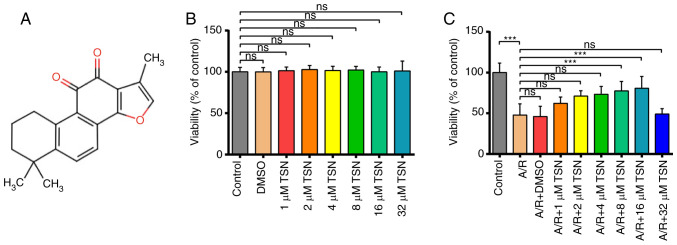
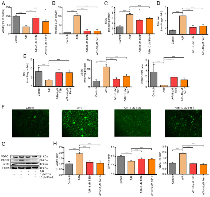
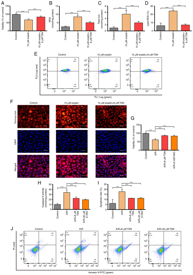
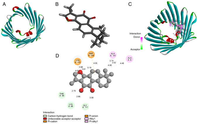
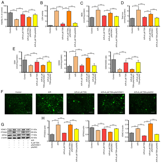
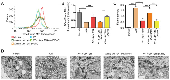
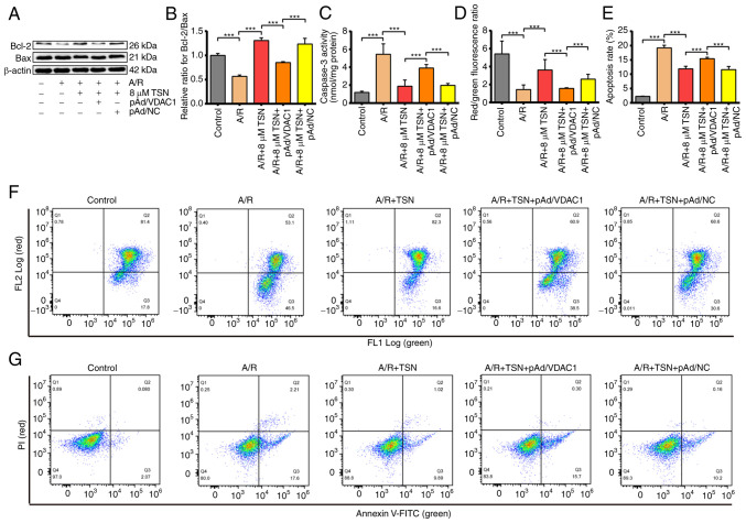
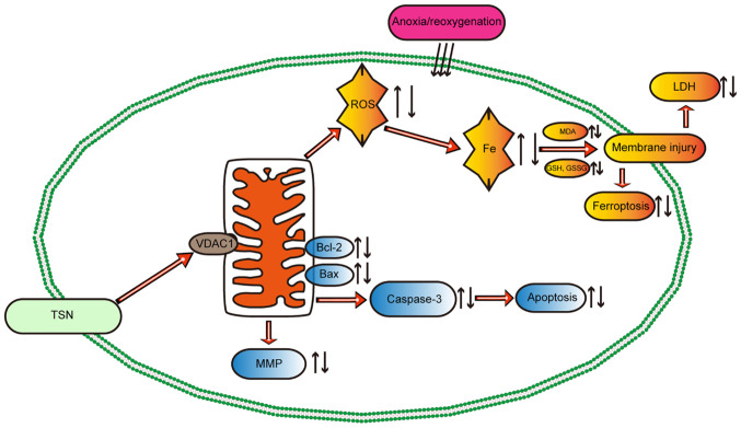
Tanshinone IIA (TSN) extracted from danshen (Salvia miltiorrhiza) could protect cardiomyocytes against myocardial ischemia/reperfusion injury (IRI), however the underlying molecular mechanisms of action remain unclear. The aim of the present study was to identify the protective effects of TSN and its mechanisms of action through in vitro studies. An anoxia/reoxygenation (A/R) injury model was established using H9c2 cells to simulate myocardial IRI in vitro. Before A/R, H9c2 cardiomyoblasts were pretreated with 8 µM TSN or 10 µM ferrostatin‑1 (Fer‑1) or erastin. The cell counting kit 8 (CCK‑8) and lactate dehydrogenase (LDH) assay kit were used to detect the cell viability and cytotoxicity. The levels of total iron, glutathione (GSH), glutathione disulfide (GSSG), malondialdehyde (MDA), ferrous iron, caspase‑3 activity, and reactive oxygen species (ROS) were assessed using commercial kit. The levels of mitochondrial membrane potential (MMP), lipid ROS, cell apoptosis, and mitochondrial permeability transition pore (mPTP) opening were detected by flow cytometry. Transmission electron microscopy (TEM) was used to observed the mitochondrial damage. Protein levels were detected by western blot analysis. The interaction between TSN and voltage‑dependent anion channel 1 (VDAC1) was evaluated by molecular docking simulation. The results showed that pretreatment with TSN and Fer‑1 significantly decreased cell viability, glutathione peroxidase 4 (GPX4) protein and GSH expression and GSH/GSSG ratio and inhibited upregulation of LDH activity, prostaglandin endoperoxide synthase 2 and VDAC1 protein expression, ROS levels, mitochondrial injury and GSSG induced by A/R. TSN also effectively inhibited the damaging effects of erastin treatment. Additionally, TSN increased MMP and Bcl‑2/Bax ratio, while decreasing levels of apoptotic cells, activating Caspase‑3 and closing the mPTP. These effects were blocked by VDAC1 overexpression and the results of molecular docking simulation studies revealed a direct interaction between TSN and VDAC1. In conclusion, TSN pretreatment effectively attenuated H9c2 cardiomyocyte damage in an A/R injury model and VDAC1‑mediated ferroptosis and apoptosis served a vital role in the protective effects of TSN.

Keywords: apoptosis; ferroptosis; ischemia/reperfusion injury; tanshinone IIA; voltage- dependent anion channel 1.


Conflict of interest statement

The authors declare that they have no competing interests.

Figures

Figure 1
Figure 1
TSN protects H9c2 cardiomyocytes against A/R injury. (A) Chemical structure of TSN. (B) CCK-8 detection of cell viability following TSN treatment. (C) CCK-8 detection of viability of A/R-induced cells following treatment with TSN. Data are expressed as the mean ± SD (n=3). ***P<0.05, TSN, Tanshinone IIA; A/R, Anoxia/reoxygenation; CCK, Cell Counting Kit; ns, not significant.
Figure 2
Figure 2
TSN alleviates ferroptosis of A/R-induced H9c2 cardiomyocytes via downregulation of VDAC1. (A) Cell Counting Kit-8 detection of viability in A/R-induced cells following TSN or Fer-1 pretreatment. (B) LDH, (C) MDA, (D) total iron, (E) GSH, GSSG, GSH/GSSG and (F) ROS were determined by quantitative kits in A/R-induced cells following TSN or Fer-1 treatment (magnification, ×200; scale bar, 50 μm). (G) Expression of (H) ferroptosis-related proteins and VDAC1 were detected by western blot analysis in A/R-induced cells following TSN or Fer-1 pretreatment. Data are expressed as the mean ± SD (n=3). ***P<0.05. TSN, Tanshinone IIA; A/R, Anoxia/reoxygenation; VDAC1, Voltage-dependent anion channel 1; Fer-1, ferrostatin-1; LDH, lactate dehydrogenase; MDA, malondialdehyde; GSH, Glutathione; GSSG, Glutathione disulfide; ROS, reactive oxygen species; PTGS2, Prostaglandin endoperoxide synthase 2; GPX, Glutathione peroxidase 4.
Figure 3
Figure 3
TSN attenuates ferroptosis of erastin-induced H9c2 cells and inhibits apoptosis of A/R-induced H9c2 cardiomyocytes. (A) CCK-8 detection of viability in H9c2 cells following erastin or TSN treatment. (B) MDA, (C) total iron, (D) lipid ROS were determined by quantitative kits in H9c2 cells after erastin or TSN treatment. Quantitative analysis for (E) the levels of intracellular lipid ROS. (F) Ferrous iron was measured by quantitative kits in H9c2 cells after erastin or TSN treatment (magnification, ×200; scale bar, 50 μm). (G) CCK-8 detected viability in A/R-induced cells after TSN or NAC pretreatment. (H) Caspase-3 activity was measured using a Caspase-3 quantitative kit in A/R-induced cells following TSN or Z-VAD treatment. (I) Apoptotic rate was (J) measured by Annexin V-FITC/PI detected by flow cytometry. Data are expressed as the mean ± SD (n=3). ***P<0.05. TSN, tanshinone IIA; A/R, Anoxia/reoxygenation; CCK, Cell Counting Kit; MDA, malondialdehyde; ROS, reactive oxygen species; NAC, N-acetylcysteine; Z-VAD, Z-Val-Ala-DL-Asp-fluoromethylketone.
Figure 4
Figure 4
TSN binds to VDAC1. (A) Chemical structure of TSN. (B) Molecular structure of VDAC1. (C) 3D and (D) 2D diagram of the interaction between TSN and VDAC1. TSN, Tanshinone IIA; VDAC1, Voltage-dependent anion channel 1.
Figure 5
Figure 5
TSN inhibits ferroptosis of A/R-induced H9c2 cardiomyocytes by downregulating VDAC1. (A) Cell Counting Kit-8 detection of viability in A/R-induced cells after TSN, pAd/VDAC1 and pAd/NC pretreatment. (B) LDH, (C) MDA, (D) total iron, (E) GSH, GSSG, GSH/GSSG and (F) ROS were determined by quantitative kits in A/R-induced cells following TSN, pAd/VDAC1 and pAd/NC treatment (magnification, x200; scale bar, 50 μm). (G) Expression of (H) ferroptosis-associated proteins and VDAC1 were detected by western blot analysis in A/R-induced cells after TSN, pAd/VDAC1 and pAd/NC pretreatment. Data are expressed as the mean ± SD (n=3). ***P<0.05. TSN, tanshinone IIA; A/R, anoxia/reoxygenation; VDAC1, voltage-dependent anion channel 1; NC, negative control; LDH, lactate dehydrogenase; MDA, malondialdehyde; GSH, Glutathione; GSSG, Glutathione disulfide; ROS, reactive oxygen species; PTGS2, Prostaglandin endoperoxide synthase 2; GPX4, Glutathione peroxidase 4.
Figure 6
Figure 6
TSN improves mitochondrial function and integrity in H9c2 cardiomyocytes exposed to A/R by downregulating VDAC1. (A) Fluorescent probe BBcellProbe M61 indicating mPTP opening was detected by flow cytometry with the FL1-A: B525-FITC channel. (B) mPTP flow cytometry. (C) Flameng score and (D) transmission electron microscopy of H9c2 cells (magnification, ×8,000; scale bar, 2 μm). Data are expressed as the mean ± SD (n=3). ***P<0.05. TSN, tanshinone IIA; A/R, Anoxia/reoxygenation; VDAC1, Voltage-dependent anion channel 1; mPTP, Mitochondrial permeability transition pore; NC, negative control.
Figure 7
Figure 7
TSN inhibits apoptosis of A/R-induced H9c2 cardiomyocytes by downregulating VDAC1. (A) Expression of (B) apoptosis-associated proteins was detected by western blot analysis in A/R-induced cells following TSN, pAd/VDAC1 and pAd/NC pretreatment. (C) Caspase-3 activity was measured using a Caspase-3 kit in A/R-induced cells after TSN, pAd/VDAC1 and pAd/NC treatment. (D) MMP and (E) apoptosis were detected by flow cytometry. (F) MMP levels detected by JC-1 in H9c2 cells indicated by the red/green fluorescence ratio. (G) Apoptotic rate measured by Annexin V-FITC/PI flow cytometry. Data are expressed as the mean ± SD (n=3). ***P<0.05. TSN, Tanshinone IIA; A/R, Anoxia/reoxygenation; VDAC1, Voltage-dependent anion channel 1; NC, negative control; MMP, mitochondrial membrane potential.
Figure 8
Figure 8
Potential mechanism of TSN in myocardial ischemia/reperfusion injury. TSN pretreatment upregulates the expression of VDAC1, thereby decreasing the accumulation of ROS and iron and abnormal lipid metabolism, maintaining mitochondrial function and protecting the myocardium against anoxia/reoxygenation-induced ferroptosis and apoptosis. TSN, tanshinone IIA; VDAC1, Voltage-dependent anion channel 1; ROS, reactive oxygen species; MDA, malondialdehyde; GSH, Glutathione; GSSG, Glutathione disulfide; LDH, lactate dehydrogenase; MMP, Mitochondrial membrane potential.

References

    1. Davidson SM, Ferdinandy P, Andreadou I, Bøtker HE, Heusch G, Ibáñez B, Ovize M, Schulz R, Yellon DM, Hausenloy DJ, et al. Multitarget strategies to reduce myocardial ischemia/reperfusion injury: JACC review topic of the week. J Am Coll Cardiol. 2019;73:89–99. doi: 10.1016/j.jacc.2018.09.086. - DOI - PubMed
    1. Hausenloy DJ, Bøtker HE, Ferdinandy P, Heusch G, Ng GA, Redington A, Garcia-Dorado D. Cardiac innervation in acute myocardial ischaemia/reperfusion injury and cardioprotection. Cardiovasc Res. 2019;115:1167–1177. doi: 10.1093/cvr/cvz053. - DOI - PMC - PubMed
    1. Lee JR, Park BW, Park JH, Lim S, Kwon SP, Hwang JW, Kim H, Park HJ, Kim BS. Local delivery of a senolytic drug in ischemia and reperfusion-injured heart attenuates cardiac remodeling and restores impaired cardiac function. Acta Biomater. 2021;135:520–533. doi: 10.1016/j.actbio.2021.08.028. - DOI - PubMed
    1. Gumpper-Fedus K, Park KH, Ma H, Zhou X, Bian Z, Krishnamurthy K, Sermersheim M, Zhou J, Tan T, Li L, et al. MG53 preserves mitochondrial integrity of cardiomyocytes during ischemia reperfusion-induced oxidative stress. Redox Biol. 2022;54:102357. doi: 10.1016/j.redox.2022.102357. - DOI - PMC - PubMed
    1. Ren D, Quan N, Fedorova J, Zhang J, He Z, Li J. Sestrin2 modulates cardiac inflammatory response through maintaining redox homeostasis during ischemia and reperfusion. Redox Biol. 2020;34:101556. doi: 10.1016/j.redox.2020.101556. - DOI - PMC - PubMed

MeSH terms