. 2023 Oct 31;89(10):e0080223.
doi: 10.1128/aem.00802-23. Epub 2023 Oct 6.

Unveiling the regulatory mechanisms of salicylate degradation gene cluster cehGHIR4 in Rhizobium sp. strain X9

Affiliations

Unveiling the regulatory mechanisms of salicylate degradation gene cluster cehGHIR4 in Rhizobium sp. strain X9

Zhijian Ke et al. Appl Environ Microbiol. .

Abstract

In a previous study, the novel gene cluster cehGHI was found to be involved in salicylate degradation through the CoA-mediated pathway in Rhizobium sp. strain X9 (Mol Microbiol 116:783-793, 2021). In this study, an IclR family transcriptional regulator CehR4 was identified. In contrast to other regulators involved in salicylate degradation, cehR4 forms one operon with the gentisyl-CoA thioesterase gene cehI, while cehG and cehH (encoding salicylyl-CoA ligase and salicylyl-CoA hydroxylase, respectively) form another operon. cehGH and cehIR4 are divergently transcribed, and their promoters overlap. The results of the electrophoretic mobility shift assay and DNase I footprinting showed that CehR4 binds to the 42-bp motif between genes cehH and cehI, thus regulating transcription of cehGH and cehIR4. The repeat sequences IR1 (5'-TTTATATAAA-3') and IR2 (5'-AATATAGAAA-3') in the motif are key sites for CehR4 binding. The arrangement of cehGH and cehIR4 and the conserved binding motif of CehR4 were also found in other bacterial genera. The results disclose the regulatory mechanism of salicylate degradation through the CoA pathway and expand knowledge about the systems controlled by IclR family transcriptional regulators.IMPORTANCEThe long-term residue of aromatic compounds in the environment has brought great threat to the environment and human health. Microbial degradation plays an important role in the elimination of aromatic compounds in the environment. Salicylate is a common intermediate metabolite in the degradation of various aromatic compounds. Recently, Rhizobium sp. strain X9, capable of degrading the pesticide carbaryl, was isolated from carbaryl-contaminated soil. Salicylate is the intermediate metabolite that appeared during the degradation of carbaryl, and a novel salicylate degradation pathway and the involved gene cluster cehGHIR4 have been identified. This study identified and characterized the IclR transcription regulator CehR4 that represses transcription of cehGHIR4 gene cluster. Additionally, the genetic arrangements of cehGH and cehIR4 and the binding sites of CehR4 were also found in other bacterial genera. This study provides insights into the biodegradation of salicylate and provides an application in the bioremediation of aromatic compound-contaminated environments.

Keywords: IclR-type transcriptional regulators; Rhizobium sp. strain X9; cehR4; salicylate degradation.


Conflict of interest statement

The authors declare no conflict of interest.

Figures

Fig 1
Fig 1
(A) The DNA fragments that are located at same positions in the genome are shown. The scale bar represents 1 kb. The amplification fragments for transcriptional unit evaluation are shown as lines under the cehGH and cehIR4. (B) PCR amplification of the cehGH and cehIR4 using DNA (D), total RNA (R), and cDNA (cD) as the templates. (C and D) TSS determination of the cehGH (C) and cehIR4 (D) by 5′-RACE. Red mark indicates the TSS. (E) Sequence elements in promoter region of cehIR4 and cehGH. The −35 box TTTCCT and the −10 box TTGCACAAT of PGH are shown in blue box, the −35 box TTCGTT and the −10 box TTCTATATT of PIR4 are shown in red box, and the two TSSs and start codon ATG are indicated with arrowheads. The CehR4 binding site in the promoter region is indicated by a black half-box. (F) Relative transcript abundance of cehG, cehH, cehI, and cehR4 in the wild strain X9 (WT), the cehR4 deletion mutant strain (MT), and the cehR4-complementary strain (MTC) in the presence of 0.21 mM glucose or salicylate. Transcriptional level of the 16S rRNA gene was used as an internal standard. The baseline condition is relative transcript abundance of cehG, cehH, cehI, and cehR4 in the wild strain X9 (WT) grown in glucose (relative transcript abundance was set to 1). The ΔCT represents the difference between transcription of the tested gene and 16S rRNA gene in the same treatment group, and ΔΔCT represents the difference between ΔCT (test group) and the ΔCT (strain X9 in the presence of 0.21 mM glucose) and the data in each column were calculated with the 2-ΔΔCT threshold cycle (CT) method using three biological repeats.
Fig 2
Fig 2
(A) Electrophoretic mobility shift assays on the binding of CehR4 to the cehGH promoter. Each lane contains 60 nM of DNA probe. The content of CehR4, increasing from left to right, is shown above the lanes. The control DNA was a 50-bp fragment that was amplified from the cehH gene. (B) Effects of increasing salicylate (0.01 to 0.2 mM) on the binding between 219.5 nM CehR4 and the DNA probe.
Fig 3
Fig 3
In vivo inducer identification. A β-galactosidase assay was performed with X9ΔcehG (pME6522-PGH) carrying the PGH-lacZ transcriptional fusion, grown in the presence (+X9ΔcehG [pME6522-PGH]) or absence (-X9ΔcehG [pME6522-PGH]) of 0.21 mM salicylate. β-Galactosidase activity was measured as described in Materials and Methods. Each value is the mean ± SD) of at least three cultures.
Fig 4
Fig 4
(A) DNase I footprinting analysis of the CehR4-binding site in the cehGH promoter. A total of 154 nM of 6-carboxyfluorescein-labeled DNA probe was incubated without CehR4 (red line) or with 145 nM CehR4 (blue line). The CehR4-protected region is shown in the dashed box, and the protected sequence is shown at the bottom. The small sequence in the protected region is shown in a black box. The blue arrow indicates transcription direction of cehGH and the red arrow indicates transcription direction of cehIR4. (B) Electrophoretic mobility shift assays on the binding of CehR4 to the 42-bp motif. The first two lanes are wild-type cehGH promoter DNA, which was used as the control, the next two lanes are 42 bp motif deleted DNA probes, the next two lanes contain promoter DNA of MT1, the next last two lanes contain promoter DNA of MT2, and finally two lanes contain promoter DNA of MT3. The nucleotide sequence 5′-TTTATATAAA-3′ in the CehR4-binding site was mutated to 5′-CGCGAGCACA-3′ (MT1), and the nucleotide sequence 5′-AATATAGAAA-3′ in the CehR4-binding site was mutated to 5′-ACGGACGGAG-3′ (MT2). The nucleotide sequence 5′-TTTATATAAA-3′ and 5′-AATATAGAAA-3′ in the CehR4-binding site was mutated to 5′-CGCGAGCACA-3′ and 5′-ACGGACGGAG-3′ (MT3), respectively.
Fig 5
Fig 5
β-Galactosidase assay was performed with X9 (pME6522-PIR) (A) and MT (pME6522-PIR) (B) carrying the PIR-lacZ transcriptional fusion, grown in the presence [+X9 (pME6522-PIR) or +MT (pME6522-PIR)] or absence [-X9 (pME6522-PIR) or −MT (pME6522-PIR)] of 0.21 mM salicylate. β-galactosidase activity was measured as described in Materials and Methods. Each value is the mean ± SD of at least three cultures.
Fig 6
Fig 6
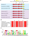
(A) Comparison of genetic organization of cehGH and cehIR4 from different strains. Arrows indicate the size and transcriptional direction of each gene. The red arrows indicate genes involved in salicylate degradation, and the depth of color represents the degree of CehR4 similarity. (B) Sequence alignment of CehR4-binding motif from diverse strains. The CehR4-binding site among strains Rhizobium sp. strain X9, Pseudaminobacter sp. DSM6986 (20), Agrobacterium tumefaciens W9/94 (21), Celeribacter indicus P73 (22), Pelagibaca abyssi JLT2014 (23), Ruegeria sp. PBVC088 (24), and Mameliella alba CGMCC 1.7290 (25). (C) The logo of the CehR4-binding motif in diverse strains was generated using software WebLogo (version 2.8.2). The conserved repeat sequences are indicated by red line.

Similar articles

References

    1. Ke ZJ, Zhu Q, Jiang WK, Zhou YD, Zhang ML, Jiang ML, Hong Q. 2021. Heterologous expression and exploration of the enzymatic properties of the carbaryl hydrolase CarH from a newly isolated carbaryl-degrading strain. Ecotoxicol Environ Saf 224:112666. doi:10.1016/j.ecoenv.2021.112666 - DOI - PubMed
    1. Trivedi VD, Jangir PK, Sharma R, Phale PS. 2016. Insights into functional and evolutionary analysis of carbaryl metabolic pathway from Pseudomonas sp. strain C5pp. Sci Rep 6:38430. doi:10.1038/srep38430 - DOI - PMC - PubMed
    1. Bonting CF, Fuchs G. 1996. Anaerobic metabolism of 2-hydroxybenzoic acid (salicylic acid) by a denitrifying bacterium. Arch Microbiol 165:402–408. doi:10.1007/s002030050344 - DOI - PubMed
    1. Fuchs G, Boll M, Heider J. 2011. Microbial degradation of aromatic compounds - from one strategy to four. Nat Rev Microbiol 9:803–816. doi:10.1038/nrmicro2652 - DOI - PubMed
    1. You IS, Ghosal D, Gunsalus IC. 1991. Nucleotide sequence analysis of the Pseudomonas putida PpG7 salicylate hydroxylase gene (nahG) and its 3'-flanking region. Biochemistry 30:1635–1641. doi:10.1021/bi00220a028 - DOI - PubMed

Publication types

LinkOut - more resources