. 2023 Dec 12;11(6):e0459722.
doi: 10.1128/spectrum.04597-22. Epub 2023 Oct 6.

Mycobacteriophage D29 Lysin B exhibits promising anti-mycobacterial activity against drug-resistant Mycobacterium tuberculosis

Affiliations

Mycobacteriophage D29 Lysin B exhibits promising anti-mycobacterial activity against drug-resistant Mycobacterium tuberculosis

Amit Kumar Singh et al. Microbiol Spectr. .

Abstract

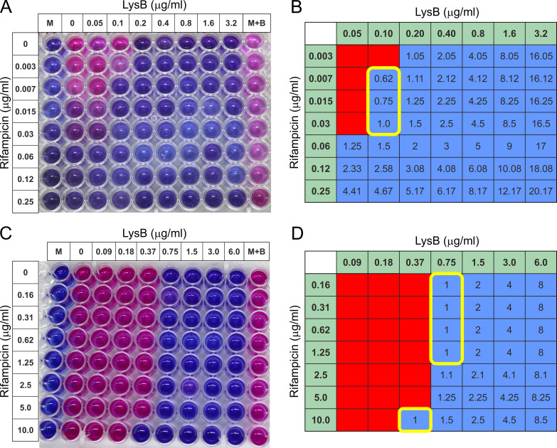
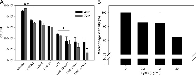
To combat the rapidly emerging drug-resistant M. tuberculosis, it is now essential to look for alternative therapeutics. Mycobacteriophages can be considered as efficient therapeutics due to their natural ability to infect and kill mycobacteria including M. tuberculosis. Here, we have exploited the mycolyl-arabinogalactan esterase property of LysB encoded from mycobacteriophage D29. This study is novel in terms of targeting a multi-drug-resistant pathogenic strain of M. tuberculosis with LysB and also examining the combination of anti-TB drugs and LysB. All the experiments include external administration of LysB. Therefore, the remarkable lytic activity of LysB overcomes the difficulty to enter the complex cell envelope of mycobacteria. Targeting the intracellularly located M. tuberculosis by LysB and non-toxicity to macrophages take the process of the development of LysB as a drug one step ahead, and also, the interaction studies with rifampicin and isoniazid will help to form a new treatment regimen against tuberculosis.

Keywords: D29 mycobacteriophage; additive effect; antibiotic resistance; mycolyl-arabinogalactan esterase; phage therapy; tuberculosis.


Conflict of interest statement

The authors declare no conflict of interest.

Figures

FIG 1
FIG 1
Purified LysB of D29 is an active esterase. Panel A shows a Coomassie Brilliant Blue-stained SDS-polyacrylamide gel with purified D29 LysB (marked with an arrowhead). Panel B shows the Western blot of this protein using anti-LysB antibodies. In both panels, ‘L’ is the protein ladder with molecular weights marked in kilodalton. Panel C shows the activity assay of LysB. The assay was carried out by incubating the substrate nitrophenyl butyrate with purified LysB or BSA, and the reaction was monitored by measuring the absorbance of released 4-nitrophenol at 415 nm with time. Different concentrations of LysB (1.0-, 2.5-, 5.0-, and 10.0-µg/mL reaction volumes as indicated) were taken for the assay. BSA was used as negative control. In this panel, the data represent an average of at least three experiments with error bars representing standard deviation with 99% significant score. BSA, bovine serum albumin.
FIG 2
FIG 2
The in vitro anti-mycobacterial activity of D29 mycobacteriophage-derived LysB against M. tuberculosis was measured using the REMA assay. Mycobacteria were incubated with different concentrations (0.03–12.0 µg/mL) of the LysB against M. tuberculosis (A and C, respectively). (A) The 1 × 10⁴ CFU/well of M. tuberculosis in 100 µL of medium was treated with 100 µL of different concentration of LysB for 6 days. The viability of mycobacteria was determined by a change in color of the resazurin dye; the blue color shows the anti-M. tuberculosis activity/effect, and the pink color indicates bacterial growth. (B) Culture (3 µL) from selected wells from panel A was spotted on MB 7H11 agar plate to confirm the REMA results. (C) M. tuberculosis (2 × 10⁵ CFU/well) in 100 µL of medium was treated with 100 µL of different concentrations of LysB for 48 h. The viability of mycobacteria was determined by a change in color of the resazurin dye added on the third day. (D) Plate layout plan for the experiment. B, M. tuberculosis H37Rv bacteria; LysB, Lysin B; M, 7H9 medium; M + B, M. tuberculosis H37Rv growth controls (without any treatment); PB, buffer used for suspending lyophilized protein; R, rifampicin; REMA, resazurin microtiter assay plate.
FIG 3
FIG 3
The anti-mycobacterial activity of LysB was determined by plate lysis assay on MB 7H11 medium plate in the (A) presence or (B) absence of Tween-80. Anti-mycobacterial activity is indicated by the formation of a lytic zone. In the presence of Tween-80, the clear spot zone was observed at 3 µg/mL. In the absence of Tween-80, the concentration up to which a clear spot zone was observed increased and was visible at 6 µg/mL.
FIG 4
FIG 4
Effect of LysB against M. tuberculosis MDR isolate using the REMA assay. (A) Mycobacteria were incubated with different concentrations (0.0625–20.0 µg/mL) of the anti-TB drugs rifampicin or moxifloxacin or LysB (concentration ranging from 0.05 to 20.0 µg/mL) for 6 days. The viability of mycobacteria was determined by a change in color of the REMA; the blue color shows the anti-M. tuberculosis activity/effect, and the pink color indicates bacterial growth. (B) Plate layout of the experiment shown in panel A. B, M. tuberculosis bacteria; LysB, well containing only Lysin B protein; M, blank media control without inoculation; M + B, M. tuberculosis growth control (without any treatment); Mox, mycobacteria treated with moxifloxacin; Rif, mycobacteria treated with rifampicin.
FIG 5
FIG 5
Checkerboard microtiter plate assay testing the combination of rifampicin and LysB against drug-sensitive M. tuberculosis H37Rv and multi-drug-resistant M. tuberculosis isolate. (A) M. tuberculosis H37Rv strain: twofold serial dilutions were performed ranging from 0.05 to 3.2 µg/mL for LysB and from 0.003 to 0.25 µg/mL for rifampicin. The addition of resazurin dye revealed the viability of the bacteria. (C) M. tuberculosis MDR isolate: twofold serial dilutions were performed ranging from 0.1 to 6.0 µg/mL for LysB and from 0.16 to 10.0 µg/mL for rifampicin. The addition of resazurin dye revealed the viability of the bacteria. (B and D) FICs: wells corresponding to no growth by color development (blue color) during REMA assay were shaded blue, while wells corresponding to growth by visual inspection of color (pink color) developed during REMA assay were shaded red. Values for LysB and rifampicin along x- and y-axes, respectively, represent the concentration of the drug in that column or row alone. The value in each cell is the FICI, or the sum of the FICs, i.e., the ratio of the concentration of the drug in that column or row to the minimum inhibitory concentration of that drug alone of the two drugs in that well. The yellow line-bordered box encloses wells with a FICI of ≤1. The combination is considered additive because the minimum FICI is 0.62 and 1.0 for M. tuberculosis drug-susceptible and MDR isolates, respectively. FIC, fractional inhibitory concentration.
FIG 6
FIG 6
Killing of intracellular M. tuberculosis by D29 LysB. (A) RAW 264.7 macrophages infected with M. tuberculosis (multiplicity of infection [MOI], 10) for 3 h were treated with 0.2, 2.0, or 20.0 µg/mL of LysB either alone or in combination with ATTs, i.e., isoniazid (3 µg/mL) and rifampicin (3 µg/mL) for 48 or 72 h, respectively. The differences in the intracellular survival of M. tuberculosis between different treatments were analyzed by the Student’s t-test. Each bar represents the average value of three independent experiments, and the error bars represent the standard deviations. Significant differences are apparent between groups in pairwise comparisons using a two-tailed t-test at a confidence interval of P < 0.1 (*) or P < 0.05 (**). (B) In vitro cytotoxicity study of RAW 264.7 macrophage cells after 24 h of exposure to LysB. Assays were carried out in triplicate; error bars represent standard deviations. ATT, anti-TB drug.

References

    1. WHO . 2021. Fact sheet details tuberculosis.
    1. Chakaya J, Khan M, Ntoumi F, Aklillu E, Fatima R, Mwaba P, Kapata N, Mfinanga S, Hasnain SE, Katoto P, Bulabula ANH, Sam-Agudu NA, Nachega JB, Tiberi S, McHugh TD, Abubakar I, Zumla A. 2021. Global tuberculosis report 2020 - reflections on the global TB burden, treatment and prevention efforts. Int J Infect Dis 113:S7–S12. doi:10.1016/j.ijid.2021.02.107 - DOI - PMC - PubMed
    1. Grange JM, Zumla A. 2002. The global emergency of tuberculosis: what is the cause J R Soc Promot Health 122:78–81. doi:10.1177/146642400212200206 - DOI - PubMed
    1. (WHO) WHO. 2017. Guidelines for treatment of drug-susceptible tuberculosis and patient care. Available from: https://appswhoint/iris/bitstream/handle/10665/255052/9789241550000-engpdf
    1. Steele MA, Burk RF, DesPrez RM. 1991. Toxic hepatitis with isoniazid and rifampin. A meta-analysis. Chest 99:465–471. doi:10.1378/chest.99.2.465 - DOI - PubMed

LinkOut - more resources