. 2023 Oct 6;19(10):e1011711.
doi: 10.1371/journal.ppat.1011711. eCollection 2023 Oct.

Machine learning-based phenotypic imaging to characterise the targetable biology of Plasmodium falciparum male gametocytes for the development of transmission-blocking antimalarials

Affiliations

Machine learning-based phenotypic imaging to characterise the targetable biology of Plasmodium falciparum male gametocytes for the development of transmission-blocking antimalarials

Oleksiy Tsebriy et al. PLoS Pathog. .

Abstract

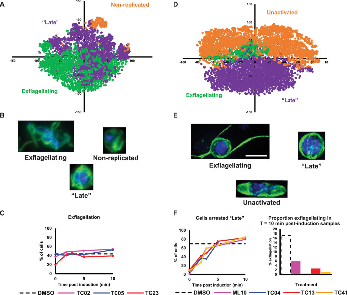
Preventing parasite transmission from humans to mosquitoes is recognised to be critical for achieving elimination and eradication of malaria. Consequently developing new antimalarial drugs with transmission-blocking properties is a priority. Large screening campaigns have identified many new transmission-blocking molecules, however little is known about how they target the mosquito-transmissible Plasmodium falciparum stage V gametocytes, or how they affect their underlying cell biology. To respond to this knowledge gap, we have developed a machine learning image analysis pipeline to characterise and compare the cellular phenotypes generated by transmission-blocking molecules during male gametogenesis. Using this approach, we studied 40 molecules, categorising their activity based upon timing of action and visual effects on the organisation of tubulin and DNA within the cell. Our data both proposes new modes of action and corroborates existing modes of action of identified transmission-blocking molecules. Furthermore, the characterised molecules provide a new armoury of tool compounds to probe gametocyte cell biology and the generated imaging dataset provides a new reference for researchers to correlate molecular target or gene deletion to specific cellular phenotype. Our analysis pipeline is not optimised for a specific organism and could be applied to any fluorescence microscopy dataset containing cells delineated by bounding boxes, and so is potentially extendible to any disease model.


Conflict of interest statement

The authors have declared that no competing interests exist.

Figures

Fig 1
Fig 1. The activity of TCAMS compound identified in the Pf DGFA screen.
(A) 85 molecules were confirmed active in the Pf DGFA. 58 were previously identified in one or more transmission-blocking screens. (B) The majority of molecules displayed similar activity against male and female gametocytes, or biased/specific activity against male gametocytes. No molecule was identified with >4.7-fold greater activity against female gametocytes. Abbreviations (IC50 = concentration giving 50% inhibition, PbODA = P. berghei Ookinete Development Assay [5], PfDGFA = P. falciparum Dual Gamete Formation Assay, PfFGAA = P. falciparum Female Gametocyte Activation Assay [7], PfGCT = P. falciparum Gametocyte Viability ATP Assay [18]).
Fig 2
Fig 2. The molecules active against P. falciparum gametocytes identified in this study clustered based upon chemical structure similarity using the FragFp algorithm.
Terminating nodes at the ends of branches represent compounds and their colour indicates putative function based upon literature and database annotation of similar compounds. The greyscale colour of nodes at branch points indicates similarity of compounds. Dark branchpoints = highly similar; Light branchpoints = divergent chemical structures. Compounds labelled with codes were studied in more detail. The colour of compound name indicates whether it was found to specifically target male gametocytes or target both gametocyte sexes in the initial Pf DFGA screen.
Fig 3
Fig 3. Using phenotypic imaging to cluster drug-treated cells with similar phenotypes.
(A) Raw fluorescence microscopy images were processed in ICY Bioimage Analysis to identify cells and then passed to the PhIDDLI pipeline for machine learning based clustering. (B) PhIDDLI interactive clustering output showing the distribution of cell phenotypes and navigable to view individual cells within each cluster.
Fig 4
Fig 4. Phenotypic distribution of male gametocytes treated with 45 transmission-blocking compounds.
2446 cells (augmented in eight different orientations) from 45 different treatments/controls were analysed by PhIDDLI and visualised by t-SNE and k-means clustering into nine clusters. (A) Localisation of the unactivated control samples (blue shades) and untreated control samples (red shades) within the dataset. Different shades indicate control cells from the six independent experiments comprising the entire screen. (B) The analysed dataset coloured by computed cluster identity. Images show representative cells from each cluster. The colour of the border surrounding the cell matches the cluster it represents. Scale bars = 3μm.
Fig 5
Fig 5. Assignment of drugs to phenotypic clusters based upon principle component analysis and k-means clustering.
The cluster assignment of cells treated with each drug from Fig 4 was analysed by principle component analysis (PCA). Clusters were assigned by k-means clustering and 5 clusters (A-E) was found to be optimal using the Elbow method. Images indicate exemplar cellular phenotypes for each drug treatment. Scale bar = 3μm.
Fig 6
Fig 6. Comparing how male gametogenesis changes phenotypically when cells are treated with compounds at different times post-induction.
(A-C) Cells were treated at 0, 2, 5 or 10 min post-induction of gametogenesis with early-acting TC11, TC14 and TC16. Resultant phenotypes were then assessed at 20 min post-induction when a parallel DMSO treated control showed maximal levels of exflagellation. (D-F) The phenotype of late-acting TC39 and microtubule depolymeriser vinblastine were evaluated if cells were similarly treated 0, 2, 3, 5 or 10 min post-induction of gametogenesis. (A + D) PhIDDLI plots of clusters of identified cellular phenotypes that were manually assigned after visual inspection. (B + E) Representative cells from each identified cluster. (C + F) Quantification of how cells from each compound treatment change phenotypic cluster depending on timing of compound treatment. Scale bar = 4μm.
Fig 7
Fig 7. Compounds targeting early male gametogenesis rapidly lose activity if administered later and compounds preventing induction of gametogenesis with a similar core structure show polypharmacology.
Cells were treated at 0, 2, 3, 5 or 10 min post-induction of gametogenesis and their resultant phenotypes studied at 20 min post-induction when a DMSO-treated control showed maximal levels of exflagellation. (A-C) TC02, TC05 and TC23 all showed early activity in the initial screen and so were compared together in the timecourse assay. (D-F) TC04, TC13 and TC41 which completely prevented induction of gametogenesis in the initial screen were evaluated with a known PKG inihibitor, ML-10. (A + D) PhIDDLI plots of clusters of identified cellular phenotypes that were manually assigned after visual inspection. (B + E) Representative cells from each identified cluster. (C + F) Quantification of how cells from each compound treatment change phenotypic cluster depending on timing of compound treatment. Scale bar = 4μm.
Fig 8
Fig 8. A model summarising the observed activity of studied compounds in the timecourse assays.
Normal male gametogenesis involves the cell rounding up, replicating its genome three times, assembling up to eight microtubule-rich flagellae, and emergence of male gametes. Integrating data from this study, transmission-blocking molecules were either observed to halt male gametogenesis or generate cells with an entirely different phenotype.

References

    1. Sinden RE. Gametocytogenesis of Plasmodium falciparum in vitro: an electron microscopic study. Parasitology. 1982. Feb;84(1):1–11. doi: 10.1017/s003118200005160x - DOI - PubMed
    1. Plouffe DM, Wree M, Du AY, Meister S, Li F, Patra K, et al.. High-Throughput Assay and Discovery of Small Molecules that Interrupt Malaria Transmission. Cell Host & Microbe. 2016. Jan;19(1):114–26. doi: 10.1016/j.chom.2015.12.001 - DOI - PMC - PubMed
    1. Burrows JN, Duparc S, Gutteridge WE, Hooft van Huijsduijnen R, Kaszubska W, Macintyre F, et al.. New developments in anti-malarial target candidate and product profiles. Malaria Journal. 2017;16:26. doi: 10.1186/s12936-016-1675-x - DOI - PMC - PubMed
    1. D’Alessandro S, Camarda G, Corbett Y, Siciliano G, Parapini S, Cevenini L, et al.. A chemical susceptibility profile of the Plasmodium falciparum transmission stages by complementary cell-based gametocyte assays. J Antimicrob Chemother. 2016. May;71(5):1148–58. doi: 10.1093/jac/dkv493 - DOI - PubMed
    1. Delves M, Lafuente-Monasterio MJ, Upton L, Ruecker A, Leroy D, Gamo FJ, et al.. Fueling Open Innovation for Malaria Transmission-Blocking Drugs: Hundreds of Molecules Targeting Early Parasite Mosquito Stages. Front Microbiol. 2019. Sep;10:1–10. - PMC - PubMed

Publication types