

doi: 10.1055/a-2164-0850.
Epub 2023 Oct 6.
Color overlay of contrast-enhanced endoscopic ultrasound for pancreaticobiliary disease
Affiliations
- PMID: 37802115
- PMCID: PMC10558281
- DOI: 10.1055/a-2164-0850
Item in Clipboard
Color overlay of contrast-enhanced endoscopic ultrasound for pancreaticobiliary disease
Endoscopy.
2023 Dec.
No abstract available
Conflict of interest statement
A. Katanuma has received honoraria as a lecture fee from Olympus Co., Tokyo, Japan. H. Toyonaga, T. Hayashi, M. Motoya, T. Kin, and K. Takahashi declare that they have no conflict of interest.
Figures

Case 1: Magnetic resonance cholangiopancreatography image of a branch duct intraductal papillary mucinous neoplasm in the pancreatic head.

Case 1: Contrast-enhanced endoscopic ultrasound indicated enhanced mural nodules in the branch duct intraductal papillary mucinous neoplasm.
a
Left: B-mode; right: normal contrast-enhanced mode.
b
Left: B-mode; right: color overlay mode.
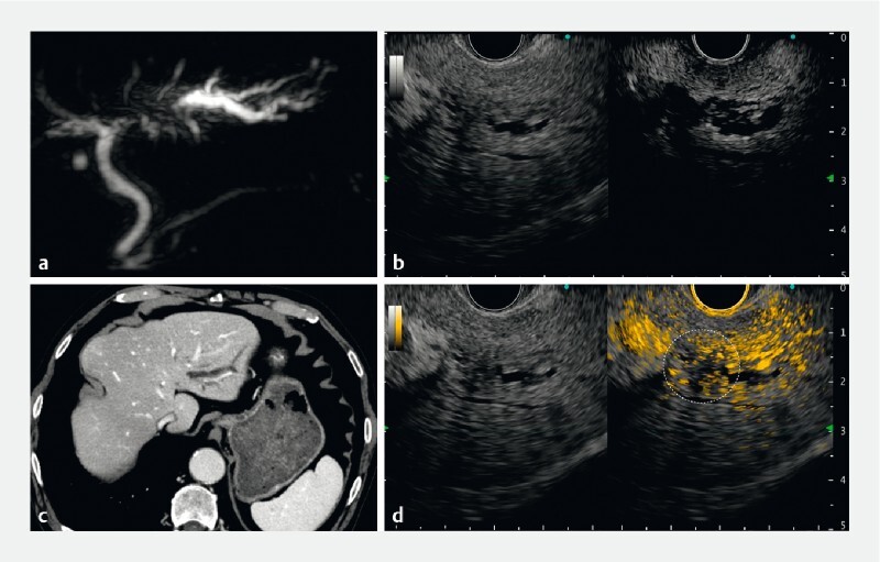
Case 2:
a
Magnetic resonance cholangiopancreatography indicated left hepatic duct obstruction.
b
Contrast-enhanced computed tomography indicated obstruction and upstream dilation of the left hepatic duct; however, no obvious mass could be noted in the obstructed area.
c
Left: B-mode; right: normal contrast-enhanced mode.
d
Left: B-mode; right: color overlay mode. The numerous adjacent vessels and dilated bile ducts made it difficult to recognize the mass lesion in the conventional black and white contrast-enhanced endoscopic ultrasound mode. On switching to color overlay mode, hypovascular areas without orange contrast particles appeared and the lesions causing biliary obstruction could be identified (dashed circle).

Case 3: Contrast-enhanced computed tomography image indicated a hypovascular pancreatic head tumor, 50 mm in diameter, with multiple liver metastases. It was suspected that the inside of the tumor was necrotic.

Case 3:
a
There was a large mass lesion in the pancreatic head, which was hypovascular, and no viable location could be recognized by conventional contrast-enhanced endoscopic ultrasound (EUS).
b
In the color overlay mode, the contrast color map was overlaid onto the B-mode, so the lesion and blood flow could be well recognized even after switching to single view. EUS-guided tissue acquisition from the viable area was performed.
References
-
- Gincul R, Palazzo M, Pujol B et al.Contrast-harmonic endoscopic ultrasound for the diagnosis of pancreatic adenocarcinoma: a prospective multicenter trial. Endoscopy. 2014;46:373–379. - PubMed
-
- Kamata K, Kitano M, Omoto S et al.Contrast-enhanced harmonic endoscopic ultrasonography for differential diagnosis of pancreatic cysts. Endoscopy. 2016;48:35–41. - PubMed
-
- Yamamoto N, Kato H, Tomoda T et al.Contrast-enhanced harmonic endoscopic ultrasonography with time-intensity curve analysis for intraductal papillary mucinous neoplasms of the pancreas. Endoscopy. 2016;48:26–34. - PubMed
-
- Krishna S G, Rao B B, Ugbarugba E et al.Diagnostic performance of endoscopic ultrasound for detection of pancreatic malignancy following an indeterminate multidetector CT scan: a systemic review and meta-analysis. Surg Endosc. 2017;31:4558–4567. - PubMed
MeSH terms
LinkOut - more resources
Full Text Sources
Medical