. 2023 Nov:365:109085.
doi: 10.1016/j.mbs.2023.109085. Epub 2023 Oct 4.

Deconstructing the integrated oscillator model for pancreatic β-cells

Affiliations

Deconstructing the integrated oscillator model for pancreatic β-cells

Richard Bertram et al. Math Biosci. 2023 Nov.

Abstract

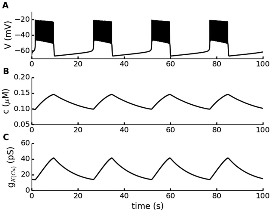
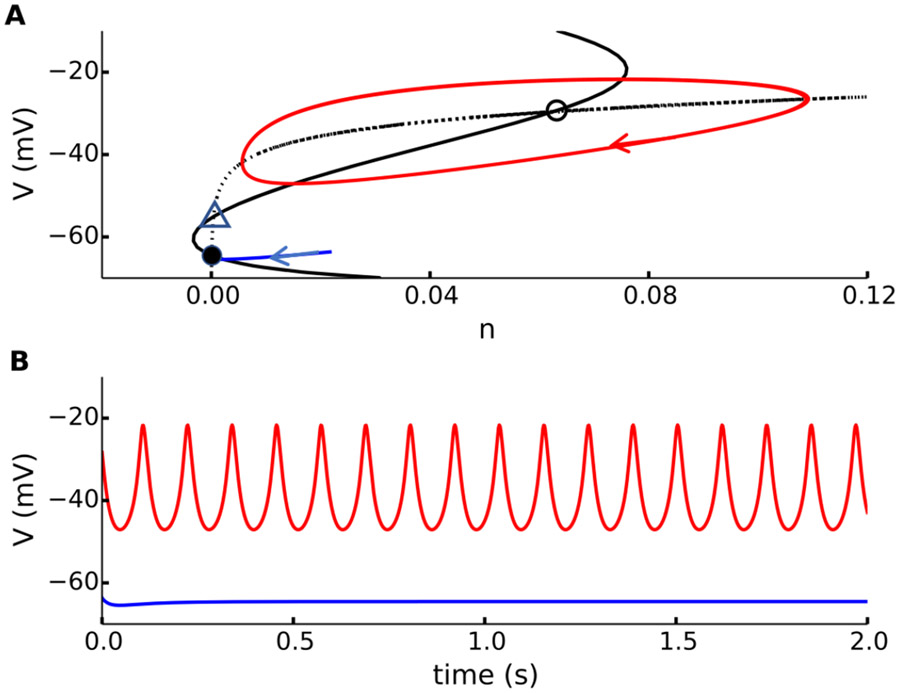
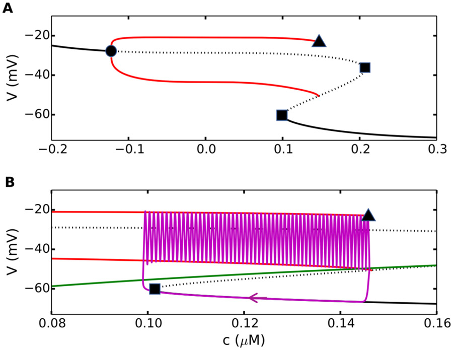
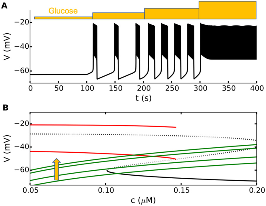
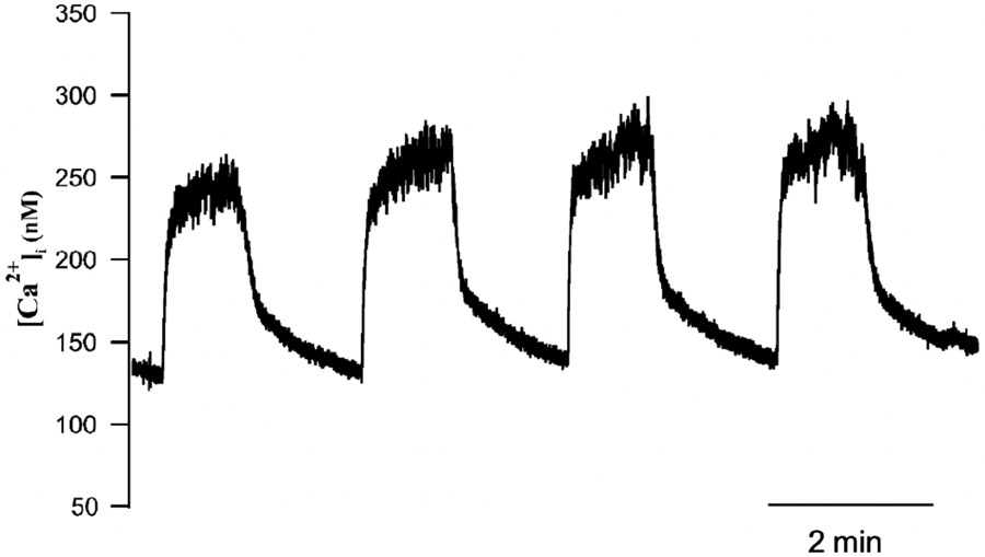
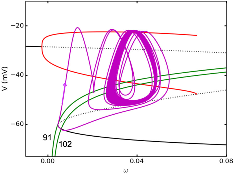
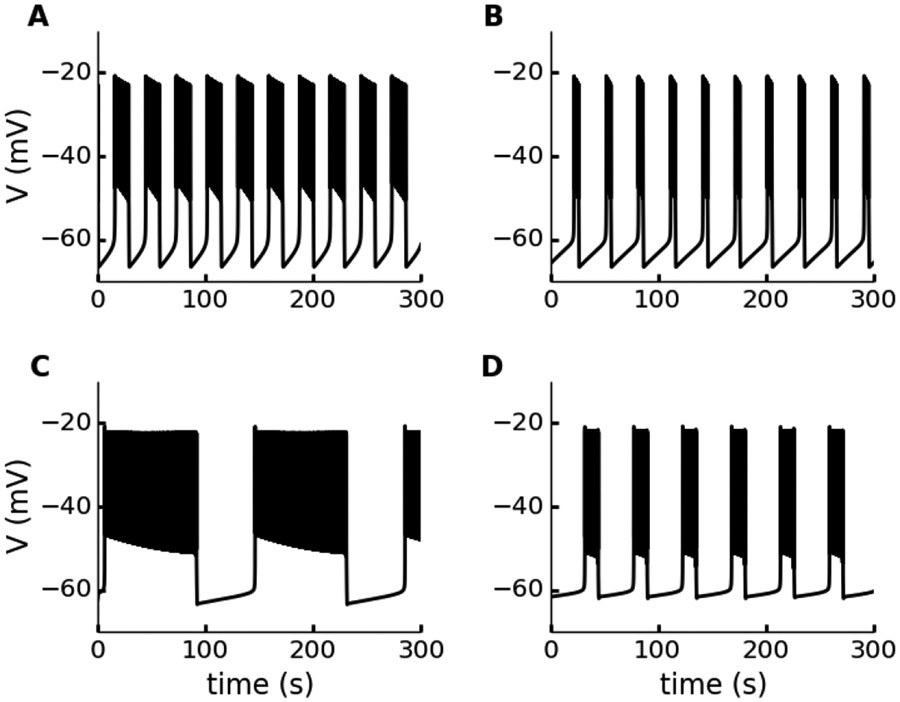
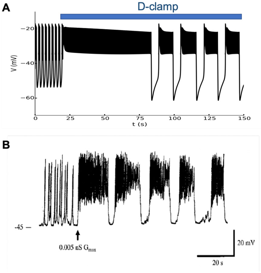
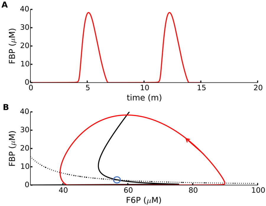
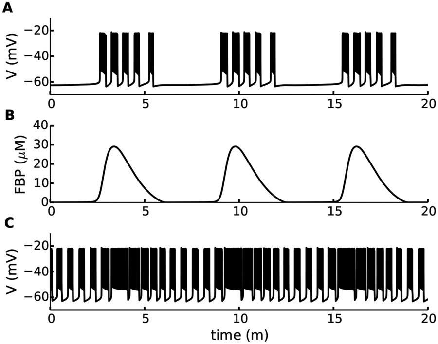
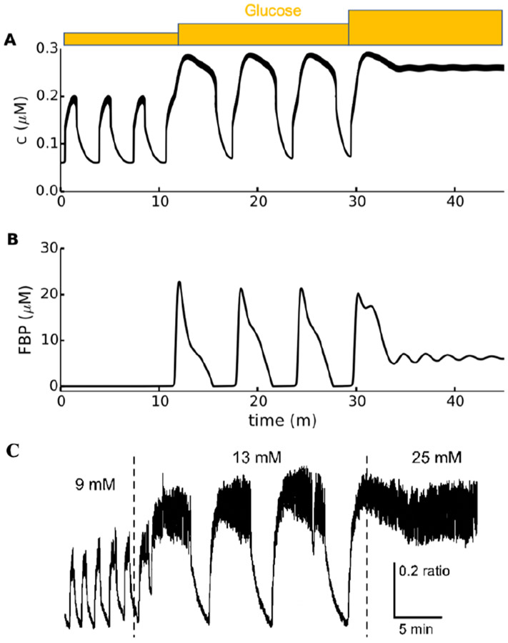
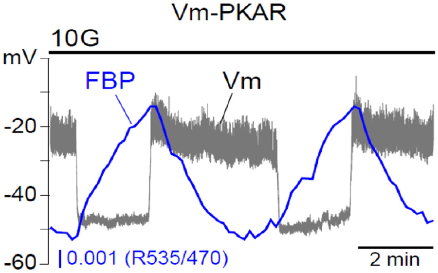
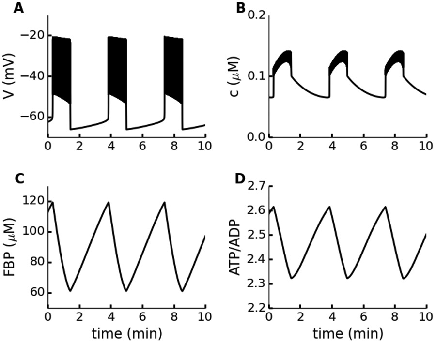
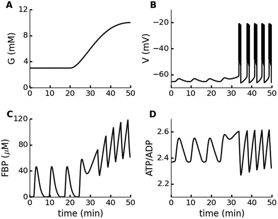
Electrical bursting oscillations in the β-cells of pancreatic islets have been a focus of investigation for more than fifty years. This has been aided by mathematical models, which are descendants of the pioneering Chay-Keizer model. This article describes the key biophysical and mathematical elements of this model, and then describes the path forward from there to the Integrated Oscillator Model (IOM). It is both a history and a deconstruction of the IOM that describes the various elements that have been added to the model over time, and the motivation for adding them. Finally, the article is a celebration of the 40th anniversary of the publication of the Chay-Keizer model.

Keywords: Beta-cells; Electrical bursting; Fast/slow analysis; Islets.


Conflict of interest statement

Declaration of competing interest None.

Figures

Figure 1:
Figure 1:
Bursting produced by a 3-dimensional Chay-Keizer model. (A) Fast bursting, consisting of active phases where the model cell is spiking and silent phases where it is hyperpolarized. (B) The free intracellular Ca2+ concentration has a slow sawtooth pattern, rising during each burst active phase and declining during each silent phase. (C) The K(Ca) conductance responds to the Ca2+ concentration, exhibiting a similar sawtooth pattern. Parameter values are given in Table 1.
Figure 2:
Figure 2:
Bistable dynamics of the Chay-Keizer fast subsystem, with c = 0.12 μM. (A) Phase portrait showing the V nullcline (black, solid), n nullcline (black, dotted), a continuous spiking limit cycle (red), and a trajectory leading to the stable node (blue). The equilibrium points are a stable node (black, filled circle), an unstable spiral (black, unfilled circle), and a saddle point (black, unfilled triangle). (B) Time courses of the continuous spiking limit cycle (red) and trajectory (blue) leading to the low-V stable equilibrium. Parameter values are given in Table 1.
Figure 3:
Figure 3:
Fast-slow analysis of the 3-dimensional Chay-Keizer model. (A) Bifurcation diagram of the fast subsystem. Stable (solid) and unstable (dotted) equilibria are in black. The minimum and maximum V of the stable periodic spiking solutions are in red. There is a subcritical Hopf bifurcation (filled circle), two saddle-node bifurcations (squares), and a homoclinic bifurcation (triangle). (B) The c-nullcline (green) is superimposed on the bifurcation diagram. Also superimposed is the burst trajectory (magenta). Parameter values are given in Table 1.
Figure 4:
Figure 4:
Explanation for the response to glucose in the Chay-Keizer model. (A) Increases in the extracellular glucose concentration are simulated by increasing the parameter for the activity of plasma membrane Ca2+ pumps, kpmca. In this simulation, kpmca (units of ms−1) is increased from 0.025, to 0.035, to 0.055, and finally to 0.065 as indicated. (B) The fast-subsystem bifurcation diagram is unaffected by the change in kpmca, which only acts by shifting the c-nullcline upward. Parameter values are given in Table 1.
Figure 5:
Figure 5:
Recording of the intracellular Ca2+ concentration from a mouse pancreatic islet using the fluorescent dye fura-2. The Ca2+ profile has a sharp rise at the beginning of a burst, followed by a plateau, and then an initial sharp decline followed by a slow falloff during the silent phase. This is in contrast with the sawtooth shape predicted by the Chay-Keizer model (Fig. 1). Reproduced from [110].
Figure 6:
Figure 6:
Bursting produced by a Chay-Keizer model with an added compartment for the Ca2+ concentration in the ER. (A) Bursting electrical activity. (B) The cytosolic Ca2+ concentration no longer has a sawtooth pattern, but instead looks more like a square wave. (C) The ER Ca2+ concentration exhibits a sawtooth pattern; it is the slowest variable in the system. Parameter values are given in Table 2.
Figure 7:
Figure 7:
Fast-slow decomposition of bursting produced by the Chay-Keizer model with an ER compartment. Two ω-nullclines (green) are shown, for cER = 91 μM and cER = 102 μM. The latter intersects the bottom branch of the critical manifold, while the former does not. The burst trajectory is superimposed (magenta). Parameter values are given in Table 2.
Figure 8:
Figure 8:
Burst period varies over a much wider range with a phantom burster than with a standard burst mechanism. (A, B) There is a substantial change in the plateau fraction, but not period, when the K(Ca) channel conductance is increased from gK(Ca) = 250 pS (left) to 900 pS (right) in the Chay-Keizer model. (C, D) There is a substantial change in both plateau fraction and period with the same conductance change in the Chay-Keizer model with an ER. Parameter values for the top and bottom panels are given in Tables 1 and 2, respectively.
Figure 9:
Figure 9:
Adding an appropriate current to a fast bursting cell converts it to a medium burster. (A) Model prediction. The model cell is initially bursting at a high frequency, but when the dynamic-clamp current is added (“D-clamp”) it switches to medium bursting after a transient. Simulation performed using the original phantom bursting model [15]. The dynamic clamp parameter values are gcmp = 12 pS, Vcmp = 100 mV, τz = 50 ms. (B) Experimental recording of electrical activity from a β-cell in which a dynamic clamp current with conductance of 5 pS is added at the arrow. This panel reproduced from [15].
Figure 10:
Figure 10:
Oscillations in the concentration of the glycolytic metabolite FBP, produced by the glycolytic enzyme PFK. (A) The FBP time course exhibits pulses, with period of 7 min. (B) The F6P-nullcline (black dotted curve) and FBP-nullcline (black solid curve) shown in the phase plane. They intersect once at an unstable node (blue circle). The glycolytic oscillation is a limit cycle (red).
Figure 11:
Figure 11:
Compound and accordion bursting produced by the DOM. (A) During compound bursting there are episodes of bursts followed by long periods of quiescence. (B) Intrinsic metabolic oscillations package the bursts into episodes. Each pulse of FBP produces an episode of bursts. (C) A reduction in the K(ATP) conductance converts compound bursting into accordion bursting, in which there is a slow rhythm in the burst plateau fraction.
Figure 12:
Figure 12:
(A) Illustration of the DOM. The top bar represents the glycolytic oscillator, while the bottom represents the electrical oscillator. Open sections indicate a low equilibrium state, hatched sections indicate an oscillatory state, and filled sections indicate a high equilibrium state. The small left arrow indicates a glucose level that produces fast bursting. The larger right arrow is a higher glucose level that produces slow bursting. Movement from one to the other, across the GO threshold, is a regime change as shown in Fig. 13. (B) The GO bar is left-shifted relative to the EO bar to illustrate the transitions that occur in Fig. 16 as the glucose level is slowly increased. The small leftmost arrow indicates a low glucose level, producing the subthreshold metabolic oscillations that occur during the first 30 min of Fig. 16. The large rightmost large arrow indicates a glucose level past the top threshold for the GO and producing the slow bursting with passive metabolic oscillations that occur during the last 20 min of Fig. 16.
Figure 13:
Figure 13:
Regime changes occur when the glucose concentration is increased. (A, B) At the first glucose level the electrical oscillator in the DOM is on and the glycolytic oscillator is off, so c oscillations are due to fast bursting. When glucose is increased past the upper threshold for the EO and lower threshold for the GO, much slower and larger c oscillations are produced, driven by the GO. When glucose is increased past the upper GO threshold, this slow oscillator turns off and the c concentration is tonically elevated while the membrane potential spikes continuously. (C) Experimental Ca2+ recording using the fluorescence dye fura-2. Regime changes occur when the glucose concentration is increased from 9 to 13 mM and later to 25 mM. This panel reproduced from [69].
Figure 14:
Figure 14:
Simultaneous recording of the membrane potential from a β-cell within an islet (Vm ) in gray and fluorescence from the pyruvate kinase activity reporter (PKAR) in blue. PKAR is a Förster resonance energy transfer (FRET) biosensor that reports the FBP concentration in islet cells. Reproduced from [67].
Figure 15:
Figure 15:
Slow bursting with passive metabolic oscillations produced by the IOM. (A, B) Voltage and Ca2+ concentration profiles exhibit 4-min oscillations. These are the variables most readily measured in experiments. (C) The FBP level falls during a burst active phase and rises during a silent phase, as has been observed experimentally using the PKAR sensor [67]. (D) The ATP/ADP ratio has a sawtooth appearance, as has been observed experimentally using the fluorescent biosensor Perceval-HR [67, 57, 61].
Figure 16:
Figure 16:
Ramping from basal to stimulatory glucose levels converts AMOs to PMOs. (A) The glucose concentration begins at a steady basal level and is then ramped up to stimulatory values. (B) The voltage initially exhibits small subthreshold oscillations, and later switches to slow bursting when the K(ATP) conductance is sufficiently small. (C) The FBP time course is pulsatile when glucose is at a substimulatory level and later has a sawtooth shape at stimulatory glucose levels. (D) The ATP/ADP ratio has a pulsatile appearance when the metabolic oscillations are active, and a sawtooth appearance when they are passive.

Similar articles

Cited by

References

    1. Adablah JE, Vinson R, Roper MG, and Bertram R, Synchronization of pancreatic islets by periodic or non-periodic muscarinic agonist pulses, PLoS One, 14 (2019), p. e0211832. - PMC - PubMed
    1. Antunes CM, Salgado AP, Rosário LM, and Santos RM, Differential patterns of glucose-induced electrical activity and intracellular calcium responses in single mouse and rat pancreatic islets, Diabetes, 49 (2000), pp. 2028–2038. - PubMed
    1. Ashcroft FM, Harrison DE, and Ashcroft SJH, Glucose induces closure of single potassium channels in isolated rat pancreatic β-cells, Nature, 312 (1984), pp. 446–448. - PubMed
    1. Ashcroft FM and Rorsman P, KATP channels and islet hormone secretion: new insights and controversies, Nature Rev., 9 (2013), pp. 660–669. - PMC - PubMed
    1. Atwater I, Dawson CM, Scott A, Eddlestone G, and Rojas E, The nature of the oscillatory behavior in electrical activity for pancreatic β-cell, in Biochemistry and Biophysics of the Pancreatic β-cell, Thieme G, ed., Verlag, New York, 1980, pp. 100–107. - PubMed

LinkOut - more resources