. 2023 Oct;11(10):e007230.
doi: 10.1136/jitc-2023-007230.

Bladder cancer intrinsic LRFN2 drives anticancer immunotherapy resistance by attenuating CD8+ T cell infiltration and functional transition

Affiliations

Bladder cancer intrinsic LRFN2 drives anticancer immunotherapy resistance by attenuating CD8+ T cell infiltration and functional transition

Anze Yu et al. J Immunother Cancer. 2023 Oct.

Abstract

Background: Immune checkpoint inhibitor (ICI) therapy improves the survival of patients with advanced bladder cancer (BLCA); however, its overall effectiveness is limited, and many patients still develop immunotherapy resistance. The leucine-rich repeat and fibronectin type-III domain-containing protein (LRFN) family has previously been implicated in regulating brain dysfunction; however, the mechanisms underlying the effect of LRFN2 on the tumor microenvironment (TME) and immunotherapy remain unclear.

Methods: Here we combined bulk RNA sequencing, single-cell RNA sequencing, ProcartaPlex multiple immunoassays, functional experiments, and TissueFAXS panoramic tissue quantification assays to demonstrate that LRFN2 shapes a non-inflammatory TME in BLCA.

Results: First, comprehensive multiomics analysis identified LRFN2 as a novel immunosuppressive target specific to BLCA. We found that tumor-intrinsic LRFN2 inhibited the recruitment and functional transition of CD8+ T cells by reducing the secretion of pro-inflammatory cytokines and chemokines, and this mechanism was verified in vitro and in vivo. LRFN2 restrained antitumor immunity by inhibiting the infiltration, proliferation, and differentiation of CD8+ T cells in vitro. Furthermore, a spatial exclusivity relationship was observed between LRFN2+ tumor cells and CD8+ T cells and cell markers programmed cell death-1 (PD-1) and T cell factor 1 (TCF-1). Preclinically, LRFN2 knockdown significantly enhanced the efficacy of ICI therapy. Clinically, LRFN2 can predict immunotherapy responses in real-world and public immunotherapy cohorts. Our results reveal a new role for LRFN2 in tumor immune evasion by regulating chemokine secretion and inhibiting CD8+ T-cell recruitment and functional transition.

Conclusions: Thus, LRFN2 represents a new target that can be combined with ICIs to provide a potential treatment option for BLCA.

Keywords: Immunotherapy; Tumor Microenvironment; Urinary Bladder Neoplasms.


Conflict of interest statement

Competing interests: None declared.

Figures
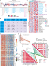
Figure 1
Figure 1
LRFN2 correlated with a non-inflammatory tumor microenvironment in BLCA. (A) LRFN2 expression and cancer immunity cycles in BLCA. The colors represent seven different steps. (B) LRFN2 expression and CD4+ T cell, CD8+ T cell, M1 macrophage, and NK cell infiltration levels in six independent algorithms. (C) LRFN2 expression, infiltrated immune cells and cancer immunity cycles in the Xiangya cohort. (D) Effector genes expression of CD8+ T cells, dendritic cells, NK cells, macrophages, and Th1 cells in high-LRFN2 and low-LRFN2 groups in TCGA-BLCA. (E) LRFN2 expression and T cell-inflamed scores in TCGA-BLCA. (F) LRFN2 expression and T cell-inflamed related genes (bottom left) and immune checkpoint genes (upper right) in the Xiangya cohort. *p<0.05, **p<0.01, ***p<0.001. BLCA, bladder cancer; LRFN2, leucine-rich repeat and fibronectin type-III domain-containing protein; mRNA, messenger RNA; NK, natural killer; TCGA, The Cancer Genome Atlas.
Figure 2
Figure 2
LRFN2 is specifically expressed in BLCA cells and inhibits cytokine and chemokine secretion. (A–B) UMAP plot of all the single cells and LRFN2 expression pattern in the Xiangya scRNA-seq. (C–D) GO enrichment between different LRFN2 expression groups in BLCA cells in the Xiangya scRNA-seq. (E) KEGG enrichment between different LRFN2 expression groups in BLCA cells in the Xiangya scRNA-seq. (F–G) KO and KEGG enrichment between different LRFN2 expression groups in TCGA-BLCA. (H) Venn diagram showed different expressed genes among high LRFN2 expression group, high immune-related genes group and high stromal score group. (I–J) GO enrichment between different LRFN2 expression groups in BLCA cells in GSE135337. (K) qRT-PCR and western blot of LRFN2 knock down in human bladder cancer cell line T24. (L) Heatmap of the chemokines/cytokines secretion in supernatants of T24-shNC and T24-shLRFN2 cells detected by ProcartaPlex multiple immunoassays. (M) qRT-PCR detected CCL2, CCL3, CCL4, CCL5, CXCL9, and CXCL10 RNA express levels in T24-shNC and T24-shLRFN2 cells. *p<0.05, **p<0.01, ***p<0.001, ****p<0.0001. UMAP, Uniform Manifold Approximation and Projection; BLCA, bladder cancer; GO, Gene Ontology; KEGG, Kyoto Encyclopedia of Genes and Genomes; LRFN2, leucine-rich repeat and fibronectin type-III domain-containing protein; qRT-PCR, quantitative reverse transcription PCR; scRNA-seq, single cell RNA sequencing; TCGA, The Cancer Genome Atlas.
Figure 3
Figure 3
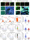
LRFN2+ tumor cells were negatively correlated with CD8+ T-cell infiltration and differentiation in human BLCA. (A–B) Representative multicolor staining of inflamed (A) and non-inflamed (B) phenotypes of patients with BLCA in the Xiangya BLCA tissue microarray (TMA): LRFN2(yellow), CK19 (azure), CD8 (green), TCF-1 (purple), PD-1 (red), and DAPI (blue). (C) Representative flow cytometry-like plots of CD8+PD-1+ (left), LRFN2+PD-1+ (middle) and LRFN2+ CK-19+ cells (right) in TMA, respectively. (D–E) Gradient analysis for multidimensional distances (0–25 µm, 25–50 µm, 50–100 µm, 100–150 µm) showed the spatial distribution of LRFN2+ tumor cells (D) and LRFN2 tumor cells (E) between CD8+ cells, PD-1+ cells and CD8+TCF-1+ cells. (F–H) Percentage of CD8+ cells (F), PD-1+ cells (G), and CD8+TCF-1+ cells (H) in total cells of high-LRFN2 and low-LRFN2 groups in TMA. **p<0.01, ****p<0.0001. MFI, mean fluorescence intensity; BLCA, bladder cancer; LRFN2, leucine-rich repeat and fibronectin type-III domain-containing protein; PD-1, Programmed cell death-1; DAPI, 4',6-diamidino-2-phenylindole; TCF-1, T cell factor-1.
Figure 4
Figure 4
LRFN2 inhibited T-cell differentiation and antitumor ability in mouse bladder cancer in vitro. (A) Flow chart of in vitro studies. (B) Heatmap of the chemokines/cytokines secretion in supernatants of MB49-shNC and MB49-shLRFN2 cells detected by ProcartaPlex multiple immunoassays. (C) Quantitative reverse transcription PCR detected CCL2, CCL3, CCL4, CCL5, CXCL9 and CXCL10 RNA express levels in MB49-shNC and MB49-shLRFN2 cells. (D) Schematic diagram (created with BioRender.com) and histogram of relative migration index of activated CD8+ T cells between MB49-shLRFN2 and MB49-shNC. (E–F) The percentage of CD44CD62L+ naive T cells (TN), CD44+CD62L+ central memory T cells (TCM), and CD44+CD62L effect memory T cells (TEM) in live CD8+ T cells (E) and CD4+ T cells (F) after co-culture with MB49-shNC and MB49-shLRFN2 cells for 12 hours. (G–H) Line chart showed the T cells cytotoxicity (G) and remained tumor cell numbers (H) after co-culture with MB49-shNC and MB49-shLRFN2 cells for 12 hours. ns, p>0.05, *p<0.05, **p<0.01, ***p<0.001, ****p<0.0001. IFN, interferon; IL, interleukin; LRFN2, leucine-rich repeat and fibronectin type-III domain-containing protein; TNF, tumor necrosis factor; LDH, lactate dehydrogenase.
Figure 5
Figure 5
LRFN2 deficiency promote the infiltration and differentiation of CD8+ T cells in vivo. (A) Flow chart of in vivo studies. (B) MB49-shLRFN2 and MB49-shNC growth curve in C57B/L6 mice. (C–D) Tumor images (C) and weight (D) of individual groups. (E) The infiltration of CD8+ TILs within the live CD45+ TILs between MB49-shLRFN2 and MB49-shNC groups. (F) The percentage of TCF-1+PD-1 naive T cells, TCF-1+PD-1+ precursor exhausted T cells, and TCF-1PD-1+ terminal exhausted T cells within the live CD45+CD8+ TILs between MB49-shLRFN2 and MB49-shNC groups. (G) TIGIT expression of live CD8+ TILs in different groups. (H) IFN-γ and TNF-α expression of live CD8+ TILs in different groups. (I) TIM-3 expression of live CD8+ TILs in different groups. ns, p>0.05, *p<0.05, **p<0.01, ***p<0.001, ****p<0.0001. IFN, interferon; LRFN2, leucine-rich repeat and fibronectin type-III domain-containing protein; TNF, tumor necrosis factor; PD-1, Programmed cell death-1;TIM-3,T cell immunoglobulin domain and mucin domain-3; MFI, Mean fluorescence intensity; TCF-1, T cell factor-1; TILs, Tumor infiltrating lymphocytes; TIGIT, T cell immunoglobulin and ITIM domain.
Figure 6
Figure 6
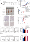
LRFN2 deficiency enhanced the efficacy of anti-PD-1 therapy of bladder cancer in mice. (A) Flow chart of the study. (B) Tumor growth curve among different groups. (C–D) Tumor images (C) and weight (D) of individual groups. (E) Representative IHC images of CD8 staining in individual groups. (F) Mice survival curve of different groups. (G) Percentage of CD62L+ cells within the live CD45+CD8+ TILs between different groups. (H) Ki-67+ cells proportion within the live CD45+CD8+ TILs between different groups. ns, p>0.05, *p<0.05, **p<0.01, ***p<0.001, ****p<0.0001. LRFN2, leucine-rich repeat and fibronectin type-III domain-containing protein; TILs, Tumor Infiltrating Lymphocytes; IHC, Immunohistochemistry; PD-1, Programmed cell death-1.
Figure 7
Figure 7
Potential of LRFN2 in predicting immunotherapy response in real-world immunotherapy cohorts. (A–B) Representative image of multicolor staining of staining of LRFN2 (yellow), CD8 (green), TCF-1 (purple), and PD-1 (red) in ICI response (A) and resistance (B) patient with BLCA. (C) Representative CT image for ICI response (upper) and resistance (bottom) patient with BLCA. (D) Relative percentage of resistance and response patients between different LRFN2 expression in the Xiangya immune cohort. (E) Disease-free survival of patients with BLCA between different LRFN2 expression in the Xiangya immune cohort. (F) Immunohistochemical staining images of LRFN2 in the two patients underwent single cell RNA sequencing. (G) tSNE plot of all the single T cells in the Xiangya scRNA-seq. (H) tSNE plot of T cells in resistance group and response group. (I) tSNE plot of differential gene expression on T cells. (J) Heatmap of differential gene expression on T cells between different resistance (upper) and response (bottom) group. (K) Percentage of exhaustion-like and transition-like cells between resistance and response group. (L) Relative percentage of resistance and response patients between different LRFN2 expression in GSE165252. BLCA, bladder cancer; PD-1, Programmed cell death-1; DAPI, 4’,6-diamidino-2-phenylindole; tSNE, t-Distributed Stochastic Neighbor Embedding; TCF-1, T cell factor-1; ICI, immune checkpoint inhibitor; LRFN2, leucine-rich repeat and fibronectin type-III domain-containing protein; scRNA-seq, single cell RNA sequencing.

Similar articles

Cited by

References

    1. Sung H, Ferlay J, Siegel RL, et al. Global cancer Statistics 2020: GLOBOCAN estimates of incidence and mortality worldwide for 36 cancers in 185 countries. CA Cancer J Clin 2021;71:209–49. 10.3322/caac.21660 - DOI - PubMed
    1. Hu J, Chen J, Ou Z, et al. Neoadjuvant Immunotherapy, chemotherapy, and combination therapy in muscle-invasive bladder cancer: a multi-center real-world retrospective study. Cell Rep Med 2022;3:100785. 10.1016/j.xcrm.2022.100785 - DOI - PMC - PubMed
    1. Cathomas R, Lorch A, Bruins HM, et al. The 2021 updated European Association of Urology guidelines on metastatic urothelial carcinoma. Eur Urol 2022;81:95–103. 10.1016/j.eururo.2021.09.026 - DOI - PubMed
    1. Zeng S, Zhang Z, Xu C. Dose-dense methotrexate, vinblastine, doxorubicin, and cisplatin as the gold standard neoadjuvant chemotherapy regimen for patients with nonmetastatic muscle-invasive bladder cancer remains controversial. J Clin Oncol 2022;40:3093–4. 10.1200/JCO.22.00669 - DOI - PubMed
    1. Zhang S. Neoadjuvant programmed cell death protein 1 and chemotherapy in combination in muscle-invasive urothelial cancer: promising, but not deserving to try further. J Clin Oncol 2022;40:2656. 10.1200/JCO.22.00420 - DOI - PubMed

Publication types