. 2023 Oct 6;13(1):312.
doi: 10.1038/s41398-023-02609-w.

Chronic treatment with D2-antagonist haloperidol leads to inhibitory/excitatory imbalance in striatal D1-neurons

Affiliations

Chronic treatment with D2-antagonist haloperidol leads to inhibitory/excitatory imbalance in striatal D1-neurons

Cátia Santa et al. Transl Psychiatry. .

Abstract

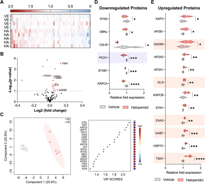
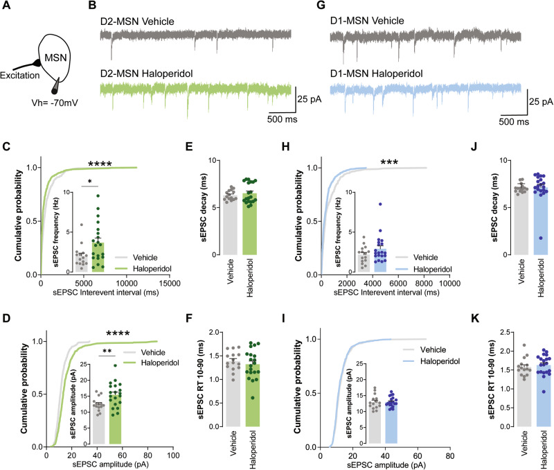
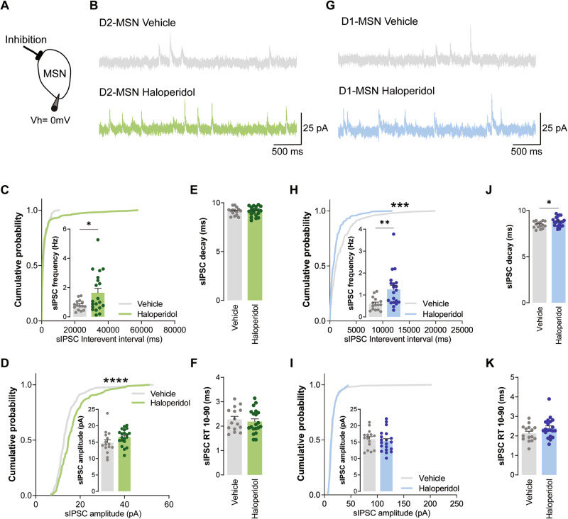
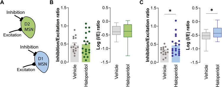
Striatal dysfunction has been implicated in the pathophysiology of schizophrenia, a disorder characterized by positive symptoms such as hallucinations and delusions. Haloperidol is a typical antipsychotic medication used in the treatment of schizophrenia that is known to antagonize dopamine D2 receptors, which are abundantly expressed in the striatum. However, haloperidol's delayed therapeutic effect also suggests a mechanism of action that may go beyond the acute blocking of D2 receptors. Here, we performed proteomic analysis of striatum brain tissue and found more than 400 proteins significantly altered after 30 days of chronic haloperidol treatment in mice, namely proteins involved in glutamatergic and GABAergic synaptic transmission. Cell-type specific electrophysiological recordings further revealed that haloperidol not only reduces the excitability of striatal medium spiny neurons expressing dopamine D2 receptors (D2-MSNs) but also affects D1-MSNs by increasing the ratio of inhibitory/excitatory synaptic transmission (I/E ratio) specifically onto D1-MSNs but not D2-MSNs. Therefore, we propose the slow remodeling of D1-MSNs as a mechanism mediating the delayed therapeutic effect of haloperidol over striatum circuits. Understanding how haloperidol exactly contributes to treating schizophrenia symptoms may help to improve therapeutic outcomes and elucidate the molecular underpinnings of this disorder.


Conflict of interest statement

The authors declare no competing interests.

Figures

Fig. 1
Fig. 1. Proteomic analysis revealed synaptic modulation upon chronic exposure to haloperidol.
A Heatmap view of 93 proteins (x axis) from the “neuronal system” family of the Reactome database. Protein fold enrichment is color-coded relative to the control average (blue: decreased expression; red: increased expression). The Y-axis represents biological replicates (Vehicles (VE): 1–4; Haloperidol (HA): 1–5). B Volcano plot of the 93 proteins with neuronal-related functions according to the Reactome database. Proteins statistically altered (p < 0.1) upon chronic exposure to haloperidol are color-coded (blue: decreased expression; red: increased expression). Proteins involved in glutamatergic and GABAergic synaptic transmission are highlighted. C Supervised multivariate analysis (PLS-DA) with the representation of the first two components accounting for 58.7% of the variability in the dataset (left panel). Variable importance in projection (VIP) score plot for the top 20 most important proteins identified by PLS-DA analysis (right panel). Fold enrichment of each protein in the haloperidol group is color-coded relative to the control average (blue: decreased expression; red: increased expression). D Violin plots of the significantly downregulated neuronal proteins in haloperidol-treated mice. Proteins involved in glutamatergic and GABAergic synaptic transmission are highlighted. E Violin plots of the significantly upregulated neuronal proteins in haloperidol-treated mice. Proteins involved in glutamatergic and GABAergic synaptic transmission are highlighted. Welch’s unpaired t test for D, E; *p < 0.1, **p < 0.05, ***p < 0.01, ****p < 0.001. Statistical details are shown in Supplementary Table S2.
Fig. 2
Fig. 2. Chronic administration of haloperidol increases excitatory synaptic transmission onto D2-MSNs, but not D1-MSNs.
A Illustration of the protocol used to record sEPSC. B, G Representative traces of sEPSC obtained from D2-MSNs and D1-MSNs, respectively. C, H Cumulative distribution of sEPSC inter-event intervals in vehicle (n = 15 cells) or haloperidol-treated (n = 20 cells) D2- and D1-MSNs, respectively. Inset shows average sEPSC frequency. D, I Cumulative distribution of sEPSC amplitudes in vehicle (n = 15 cells) or haloperidol-treated (n = 20 cells) D2- and D1-MSNs, respectively. Inset shows average sEPSC amplitude. E, J Average decay time of sEPSC recorded from D2- and D1-MSNs of vehicle (n = 15 cells) or haloperidol-treated (n = 20 cells) mice. F, K Average 10–90% rise time (RT) of sEPSC recorded from D2- and D1-MSNs of vehicle (n = 15 cells) or haloperidol-treated (n = 20 cells) mice. Welch’s unpaired t test for all panels except for cumulative distributions (Kolmogorov–Smirnov test for cumulatives). *p < 0.05, **p < 0.01, ***p < 0.001, ****p < 0.0001. Statistical details are shown in Supplementary Table S2.
Fig. 3
Fig. 3. Chronic administration of haloperidol increases inhibitory synaptic transmission onto both D2- and D1-MSNs.
A Illustration of the protocol used to record sIPSC. B, G Representative traces of sIPSC obtained from D2-MSNs and D1-MSNs, respectively. C, H Cumulative distribution of sIPSC inter-event intervals in vehicle (n = 15 cells) or haloperidol-treated (n = 20 cells) D2- and D1-MSNs, respectively. Inset shows average sIPSC frequency. D, I Cumulative distribution of sIPSC amplitudes in vehicle (n = 15 cells) or haloperidol-treated (n = 20 cells) D2- and D1-MSNs, respectively. Inset shows average sIPSC amplitude. E, J Average decay time of sIPSC recorded from D2- and D1-MSNs of vehicle (n = 15 cells) or haloperidol-treated (n = 20 cells) mice. F, K Average 10–90% rise time (RT) of sIPSC recorded from D2- and D1-MSNs of vehicle (n = 15 cells) or haloperidol-treated (n = 20 cells) mice. Welch’s unpaired t test for all panels except for cumulative distributions (Kolmogorov–Smirnov test for cumulatives). *p < 0.05, **p < 0.01, ****p < 0.0001. Statistical details are shown in Supplementary Table S2.
Fig. 4
Fig. 4. Chronic administration of haloperidol leads to increased I/E ratio in D1- but not D2-MSNs.
A Illustration of I/E ratio in D1- (blue) and D2-MSNs (green). B, C Average IPSC/EPSC (I/E) frequency ratio (left) and the logarithm of the ratio between IPSC/EPSC frequencies (right) in vehicle and haloperidol-treated D2- and D1-MSNs, respectively. Welch’s unpaired t test for all panels. *p < 0.05. Statistical details are shown in Supplementary Table S2.

References

    1. Global burden of 369 diseases and injuries in 204 countries and territories. 1990-2019: a systematic analysis for the Global Burden of Disease Study 2019. Lancet. 2020;396:1204–22. doi: 10.1016/S0140-6736(20)30925-9. - DOI - PMC - PubMed
    1. Gardner DM, Baldessarini RJ, Waraich P. Modern antipsychotic drugs: a critical overview. CMAJ. 2005;172:1703–11. doi: 10.1503/cmaj.1041064. - DOI - PMC - PubMed
    1. Kapur S, Mamo D. Half a century of antipsychotics and still a central role for dopamine D2 receptors. Prog Neuropsychopharmacol Biol Psychiatry. 2003;27:1081–90. doi: 10.1016/j.pnpbp.2003.09.004. - DOI - PubMed
    1. Schoots O, Seeman P, Guan HC, Paterson AD, Van Tol HH. Long-term haloperidol elevates dopamine D4 receptors by 2-fold in rats. Eur J Pharm. 1995;289:67–72. doi: 10.1016/0922-4106(95)90169-8. - DOI - PubMed
    1. Zanatta G, Nunes G, Bezerra EM, da Costa RF, Martins A, Caetano EWS, et al. Antipsychotic haloperidol binding to the human dopamine D3 receptor: beyond docking through QM/MM refinement toward the design of improved schizophrenia medicines. ACS Chem Neurosci. 2014;5:1041–54. doi: 10.1021/cn500111e. - DOI - PubMed

Publication types