. 2023 Oct 6;14(1):6226.
doi: 10.1038/s41467-023-42008-9.

Chronological adhesive cardiac patch for synchronous mechanophysiological monitoring and electrocoupling therapy

Affiliations

Chronological adhesive cardiac patch for synchronous mechanophysiological monitoring and electrocoupling therapy

Chaojie Yu et al. Nat Commun. .

Abstract

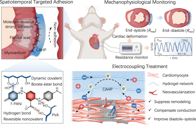
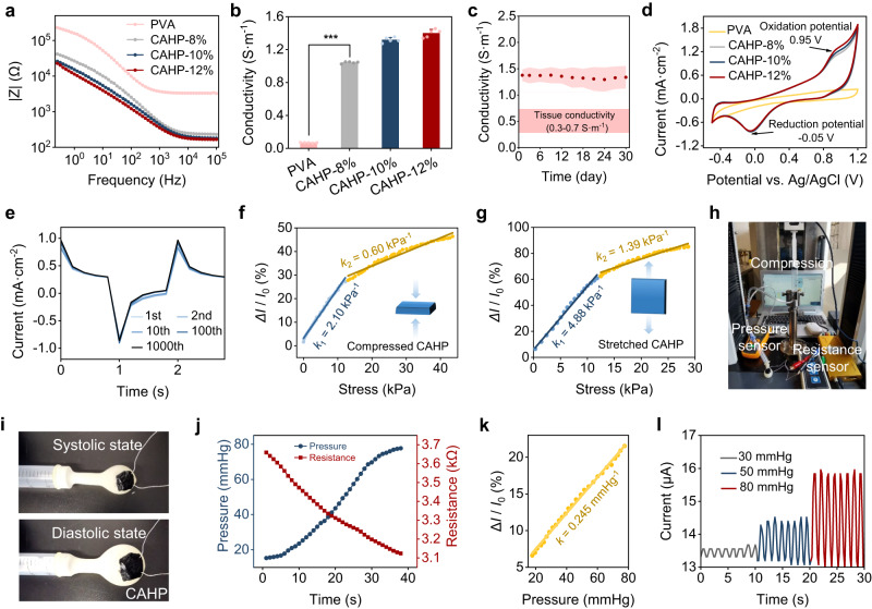
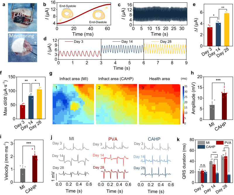
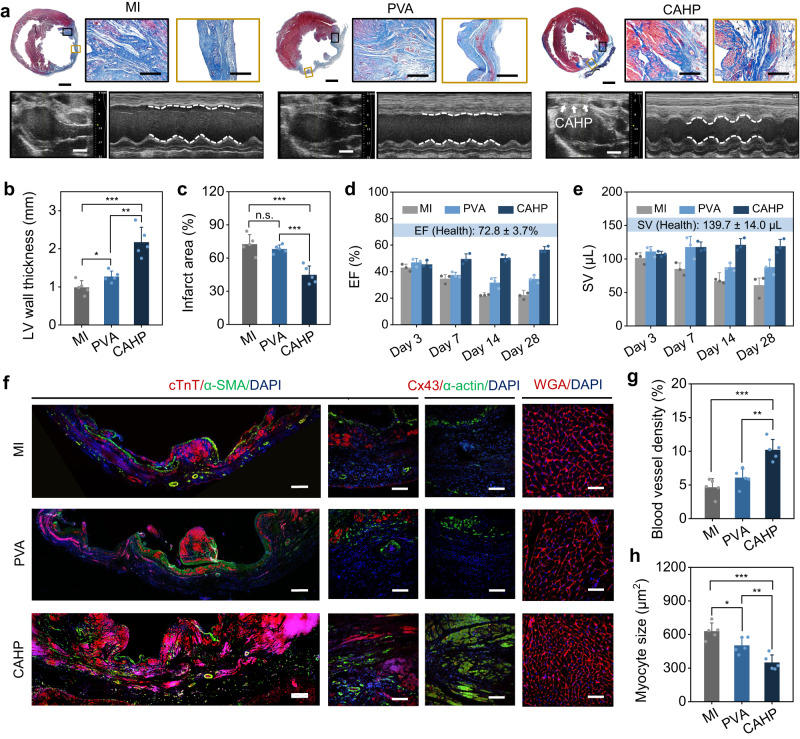
With advances in tissue engineering and bioelectronics, flexible electronic hydrogels that allow conformal tissue integration, online precision diagnosis, and simultaneous tissue regeneration are expected to be the next-generation platform for the treatment of myocardial infarction. Here, we report a functionalized polyaniline-based chronological adhesive hydrogel patch (CAHP) that achieves spatiotemporally selective and conformal embedded integration with a moist and dynamic epicardium surface. Significantly, CAHP has high adhesion toughness, rapid self-healing ability, and enhanced electrochemical performance, facilitating sensitive sensing of cardiac mechanophysiology-mediated microdeformations and simultaneous improvement of myocardial fibrosis-induced electrophysiology. As a result, the flexible CAHP platform monitors diastolic-systolic amplitude and rhythm in the infarcted myocardium online while effectively inhibiting ventricular remodeling, promoting vascular regeneration, and improving electrophysiological function through electrocoupling therapy. Therefore, this diagnostic and therapeutic integration provides a promising monitorable treatment protocol for cardiac disease.


Conflict of interest statement

The authors declare no competing interests.

Figures

Fig. 1
Fig. 1. Schematic illustration of a chronological adhesive hydrogel patch (CAHP) for synergistic cardiac mechanophysiological monitoring and electrocoupling therapy.
Chronological adhesion is mediated by interfacial dynamic covalent/noncovalent interactions between functionalized polyaniline (f-PANi) and polyvinyl alcohol (PVA), achieving molecule-invasive strong adhesion to the myocardium while resisting adhesion to nontarget tissues. CAHP can be used for cardiac mechanophysiological monitoring and simultaneous electrocoupling treatment of MI in rats.
Fig. 2
Fig. 2. Structure and properties of f-PANi.
a Chemical structures of the conducting polymers, including PANi, b-PANi, and f-PANi. b FTIR spectra of PANi, b-PANi, and f-PANi. c XPS survey for PANi, b-PANi, and f-PANi. d High-resolution spectra of C1s for different conducting polymers. e Zeta potential of conducting polymer aqueous dispersions (n = 3 independent experiments). f Diagram of water and oil droplets wetting conducting polymer films. g Water and oil contact angles of PANi, b-PANi, and f-PANi (n = 5 independent experiments). h The particle size of different conducting polymers dispersed in water. i Filtration ratio of PANi, b-PANi, and f-PANi. Inset: Photographs of conducting polymer aqueous dispersions passed through a membrane filter (n = 5 independent experiments). j UV–vis absorbance spectra of PANi, b-PANi, and f-PANi aqueous dispersions. k Conductivity of PANi, b-PANi, and f-PANi (n = 5 independent experiments). PANi: polyaniline, b-PANi: borated polyaniline, f-PANi: functionalized polyaniline. Data are presented as the mean ± standard deviation in (e, g, i, k).
Fig. 3
Fig. 3. Chronological adhesion performance and mechanism of CAHP.
a Storage modulus (G′) and loss modulus (G′′) variation of CAHPs during hydrogel gelation. b Chronological adhesion mechanism of the CAHP. c SEM images of the network structure of CAHPs in the initial and complete gel states. Scale bars, 100 μm (left) and 5 μm (right). d Absorbance at 400 nm of f-PANi infiltrated gelatin substrates during the initial and complete gel states (n = 5 independent experiments). e Peeling adhesion photographs when CAHPs in the initial and complete gel states are in contact with porcine myocardium for 10 min. f SEM images of the hydrogel–tissue interface. The white dotted line outlines the cross-section between the epicardium and CAHP. Scale bars, 20 μm. g, h Representative force–displacement curves for hydrogel–tissue hybrids in peeling (g) and lap-shear tests (h). i, j Interfacial toughness (i) and adhesion strength (j) between CAHPs and porcine myocardium (n = 3 independent experiments). k Hematoxylin-eosin-stained images at the contact interface between CAHPs and the epicardium. Scale bars, 20 μm. CAHP: chronological adhesive hydrogel patch. The measurements in (c, f, k) were repeated three times independently with similar results. Data are presented as the mean ± standard deviation in (d, i, j) and were analyzed using one-way ANOVA with Tukey’s post hoc test in (i, j), ***p < 0.001. i p = 7.34 × 10⁻⁴ (Complete gel vs CAHP-8%). j p = 2.04 × 10⁻⁴ (Complete gel vs CAHP-8%).
Fig. 4
Fig. 4. Paintable ability, mechanical properties, and self-healing performance of the CAHPs.
a Photos of the preparation process and paintable performance of the CAHPs. b Representative extrusion force–displacement curve of the CAHPs. c Injection force of the CAHPs (n = 5 independent experiments). d Young’s modulus of the CAHPs (n = 3 independent experiments). e, f Tensile (e) and compressive (f) stress–strain curves of the CAHPs. g Compressive and tensile modulus of the CAHPs (n = 3 independent experiments). h Representative loading–unloading tensile curves of CAHP-12% at a frequency of 1 Hz and deformation of 25% for 50,000 cycles. i The photos show that the CAHPs could rapidly self-heal and remain connected under stretching. Dynamic covalent borate ester bonds predominantly mediated the self-healing properties. j Modulus self-healing properties of the CAHPs when the alternate step strain was switched from 10 to 400%. k Comparison between the CAHP and other conducting polymer hydrogels,, , , in terms of their injection, modulus, anti-fatigue, self-healing, and adhesion properties. PVA: polyvinyl alcohol, f-PANi: functionalized polyaniline, CAHP chronological adhesive hydrogel patch. Data are presented as the mean ± standard deviation in (c, d, g).
Fig. 5
Fig. 5. Electrochemical properties and resistive sensing investigation of the CAHPs.
a Frequency-dependent impedance (Z) curves of the PVA hydrogel and CAHPs. b Conductivity of the PVA hydrogel and CAHPs (n = 5 independent experiments). c The conductivity of CAHP-12% incubated in PBS at 37 °C for 30 days (n = 3 independent experiments). d CV curves of the PVA hydrogel and CAHPs. e Charge injection curves of CAHP-12% with biphasic pulses of 1 s and ±0.5 V for 1000 cycles. f, g Uniaxial compressive (f) and tensile (g) stress-induced current changes (∆I/I0) in the circuit. k represents the sensitivity of stress-dependent current changes, which is the slope of the fitted curve. h Photograph of the pressure-resistance sensing system of the balloon model. i Photograph of the shape change of CAHP on the balloon surface under systolic and diastolic states. j Intraballoon pressure and interfacial CAHP resistance from model systole to diastole. k Sensitivity of CAHP-based resistive sensing in response to pressure changes in the balloon. l Current change in the CAHP when the internal pressure reaches 30 mmHg, 50 mmHg, and 80 mmHg. PVA: polyvinyl alcohol, CAHP chronological adhesive hydrogel patch. Data are presented as the mean ± standard deviation in (b, c) and were analyzed using one-way ANOVA with Tukey’s post hoc test in (b), ***p < 0.001. b p = 1.09 × 10⁻¹⁵ (PVA vs CAHP-8%).
Fig. 6
Fig. 6. Cardiac mechanophysiology monitoring and electrocoupling treatment by CAHP.
a Photographs of CAHP affixed to the left ventricular surface and mechanophysiological monitoring. b Current variation in CAHP from cardiac end-systole to end-diastole. c Current variation curve during CAHP monitoring of healthy hearts. d Current curves in the CAHP were recorded on days 3, 14, and 28 after MI. e The difference (∆I) between the peak and minimum currents (n = 3 animals). f The maximum value (Max dI/dt) of current variation in each stroke (n = 3 animals). g Heatmap of the activation time of calcium transient signals from the apices propagating to different regions of the heart. h, i Physiological potential amplitude (h) and average conduction velocity (i) in the MI and CAHP groups (n = 5 animals). j Representative ECGs for rats in the MI, PVA, and CAHP groups at 3, 14, and 28 days postoperatively. k QRS interval duration (n = 3 animals). MI myocardial infarction, PVA: polyvinyl alcohol, CAHP chronological adhesive hydrogel patch. Data are presented as the mean ± standard deviation and were analyzed using one-way ANOVA with Tukey’s post hoc test in (e, f, h, i, k), n.s.: no significant difference at p > 0.05, *p < 0.05, **p < 0.01, ***p < 0.001. e p = 0.0484 (Day 3 vs Day 14), p = 1.71 × 10⁻³ (Day 14 vs Day 28). f p = 6.04 × 10⁻³ (Day 3 vs Day 14), p = 0.0239 (Day 14 vs Day 28). h p = 9.90 × 10⁻⁴ (MI vs CAHP). i p = 6.12 × 10⁻⁴ (MI vs CAHP). k Day 3: p = 0.726 (MI vs PVA), p = 0.427 (PVA vs CAHP), p = 0.709 (MI vs CAHP). Day 14: p = 9.34 × 10⁻⁴ (MI vs CAHP), p = 1.05 × 10⁻³ (PVA vs CAHP). Day 28: p = 1.63 × 10⁻⁴ (MI vs CAHP), p = 5.03 × 10⁻⁴ (PVA vs CAHP).
Fig. 7
Fig. 7. Myocardial repair effects of CAHP.
a Masson staining for collagen (blue) and muscle (red) and echocardiography imaging in the MI, PVA and CAHP groups. White arrows represent CAHP on the epicardium, and the white dotted line outlines diastolic-systolic ventricular dimension changes. Measurements of Masson staining and echocardiography were repeated independently five and three times, respectively, with similar results. b, c Quantitative analysis of the wall thickness (b) and infarcted area (c) in the MI, PVA, and CAHP groups (n = 5 animals). d, e The ejection fraction (EF) (d) and stroke volume (SV) (e) of the left ventricle after 3, 7, 14, and 28 days postoperatively in the MI, PVA, and CAHP groups (n = 3 animals). f Representative immunofluorescence staining of the infarct region in the MI, PVA, and CAHP groups for cardiac markers (cTnT and α-actin), vascularization marker (α-SMA), intercellular electrical contraction coupling marker (Cx43), cardiomyocyte profiles (WGA), and nuclei (DAPI). g, h Quantitative analysis of blood vessel density (g) and myocyte size (h) (n = 5 animals). Scale bars, 2 mm in overall view, 300 μm in border and infarct area, and 4 mm in echocardiography (a); 300 μm (left) and 100 μm (right) in cTnT/α-SMA staining, 100 μm in Cx43/α-actin staining, and 50 μm in WGA staining (f). MI myocardial infarction, PVA: polyvinyl alcohol, CAHP chronological adhesive hydrogel patch. Data are presented as the mean ± standard deviation in (b, c, d, e, g, h) and were analyzed using one-way ANOVA with Tukey’s post hoc test in (b, c, g, h), n.s.: no significant difference at p > 0.05, *p < 0.05, **p < 0.01, ***p < 0.001. b p = 0.0241 (MI vs PVA), p = 1.29 × 10⁻³ (PVA vs CAHP), p = 2.66 × 10⁻⁴ (MI vs CAHP). c p = 0.332 (MI vs PVA), p = 3.51 × 10⁻⁴ (PVA vs CAHP), p = 7.67 × 10⁻⁴ (MI vs CAHP). g p = 2.74 × 10⁻⁴ (MI vs CAHP), p = 1.79 × 10⁻³ (PVA vs CAHP). h p = 0.0232 (MI vs PVA), p = 2.45 × 10⁻⁴ (MI vs CAHP), p = 8.27 × 10⁻³ (PVA vs CAHP).

Similar articles

Cited by

References

    1. Contessotto P, et al. Elastin-like recombinamers-based hydrogel modulates post-ischemic remodeling in a non-transmural myocardial infarction in sheep. Sci. Transl. Med. 2021;13:eaaz5380. - PubMed
    1. Hu H, et al. A wearable cardiac ultrasound imager. Nature. 2023;613:667–675. - PMC - PubMed
    1. Schiavone G, Lacour SP. Conformable bioelectronic interfaces: mapping the road ahead. Sci. Transl. Med. 2019;11:eaaw5858. - PubMed
    1. Choi YS, et al. Fully implantable and bioresorbable cardiac pacemakers without leads or batteries. Nat. Biotechnol. 2021;39:1228–1238. - PMC - PubMed
    1. Li Y, Li N, De Oliveira N, Wang S. Implantable bioelectronics toward long-term stability and sustainability. Matter. 2021;4:1125–1141.

Publication types