. 2023 Oct 6;13(1):16885.
doi: 10.1038/s41598-023-42743-5.

Quantifying innervation facilitated by deep learning in wound healing

Affiliations

Quantifying innervation facilitated by deep learning in wound healing

Abijeet Singh Mehta et al. Sci Rep. .

Abstract

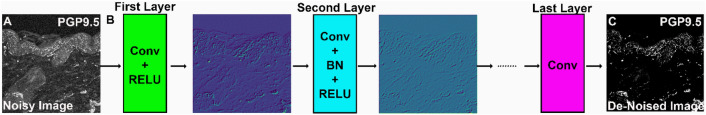
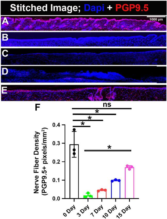
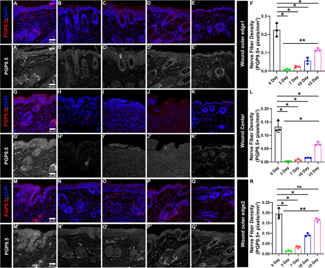
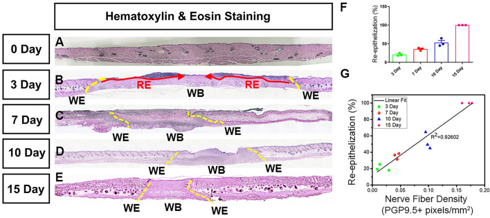
The peripheral nerves (PNs) innervate the dermis and epidermis, and are suggested to play an important role in wound healing. Several methods to quantify skin innervation during wound healing have been reported. Those usually require multiple observers, are complex and labor-intensive, and the noise/background associated with the immunohistochemistry (IHC) images could cause quantification errors/user bias. In this study, we employed the state-of-the-art deep neural network, Denoising Convolutional Neural Network (DnCNN), to perform pre-processing and effectively reduce the noise in the IHC images. Additionally, we utilized an automated image analysis tool, assisted by Matlab, to accurately determine the extent of skin innervation during various stages of wound healing. The 8 mm wound is generated using a circular biopsy punch in the wild-type mouse. Skin samples were collected on days 3, 7, 10 and 15, and sections from paraffin-embedded tissues were stained against pan-neuronal marker- protein-gene-product 9.5 (PGP 9.5) antibody. On day 3 and day 7, negligible nerve fibers were present throughout the wound with few only on the lateral boundaries of the wound. On day 10, a slight increase in nerve fiber density appeared, which significantly increased on day 15. Importantly, we found a positive correlation (R2 = 0.926) between nerve fiber density and re-epithelization, suggesting an association between re-innervation and re-epithelization. These results established a quantitative time course of re-innervation in wound healing, and the automated image analysis method offers a novel and useful tool to facilitate the quantification of innervation in the skin and other tissues.


Conflict of interest statement

The authors declare no competing interests.

Figures

Figure 1
Figure 1
The experimental design and schematic depicting the methodology used to quantify skin innervation. (A) A biopsy punch of 8 mm in diameter is used to create the wound, and skin samples are collected and fixed on days 3, 7, 10 and 15. After fixation, the wounded tissue is paraffin-embedded and sectioned (5 μm thickness) for immunofluorescence analysis against PGP9.5 protein, a neuron-specific marker. (BE) Illustration portraying different stages of wound healing. (B) The homeostatic phase lasts a few hours during which nerve fibers in the wound bed are damaged followed by the (C) inflammatory phase that can last between hours and days. (D) The proliferative phase lasts a few weeks during which re-innervation might be initiated and (E) during the remodeling phase wound matures and can last between weeks to years. In our study, we chose to quantify skin innervation at days 3, 7, 10 and 15 as an attempt to cover all phases of wound healing. (F) The immunohistochemistry (IHC) samples are analyzed using automated Matlab-assisted tools aided by DnCNN-based image denoising. The images were created with BioRender.com.
Figure 2
Figure 2
DnCNN network architecture for image denoising. (A) Noisy image as DnCNN input. (B) The DnCNN network architecture consists of multiple convolutional layers. Each convolutional layer includes batch normalization (BN), convolution (Conv), and rectified linear unit (ReLU) layers. The first layer takes the noisy image as an input, and the subsequent layers process the image to remove noise. (C) Output image after de-noising.
Figure 3
Figure 3
Gradual increase in Re-innervation in the wound bed. PGP9.5 is a pan-neuronal marker and DAPI stains nuclei. (A) Uninjured skin. Skin sample collected on (B) day 3, (C) day 7, (D) day 10 and (E) day 15 of wound healing. (F) Quantification of skin innervation for the whole wound bed represented as mean ± SD, n = 3 wounds from three mice in each group, *P < 0.05, ns is non-significant. The wound bed is recognized by the absence of hair follicles. Scale bar = 1000 μm.
Figure 4
Figure 4
Gradual increase in Re-innervation at lateral wound edges and wound center. PGP9.5 is a pan-neuronal marker (in red) and DAPI stains nuclei of cell (in blue). (A′–E′, G′–K′, M′–Q′) Split images (in grey) show PGP9.5 immunoreactivity. (AE and A′–E′) Immunoreactivity to PGP9.5 at wound outer edge 1 for skin samples. (A,A′) Uninjured, (B,B′) day 3, (C,C′) day 7, (D,D′) day 10, (E,E′) day 15. (F) Quantification of skin innervation at wound outer edge 1. (GK and G′–K′) Immunoreactivity to PGP9.5 at wound center for skin samples. (G,G′) Uninjured, (H,H′) day 3, (I,I′) day 7, (J,J′) day 10, (K,K′) day 15. (L) Quantification of skin innervation at wound center. (MQ and M′–Q′) Immunoreactivity to PGP9.5 at wound outer edge 2 for skin samples. (M,M′) Uninjured, (N,N′) day 3, (O,O′) day 7, (P,P′) day 10, (Q,Q′) day 15. (R) Quantification of skin innervation at wound outer edge 2. All quantification data are represented as mean ± SD, n = 3 wounds from three mice in each group, *P < 0.05, **P < 0.001, ns is non-significant. Scale bar = 50 μm.
Figure 5
Figure 5
Positive correlation between re-innervation and re-epithelization. A representative image of H&E staining of the skin sample collected on (A) day 0, (B) day 3, (C) day 7, (D) day 10, (E) day 15. The original wound edge (yellow dashed lines) on each side is determined by the absence of subdermal adipose tissue. Re-epithelialization (red arrows) is defined by epithelial cell growth. (F) quantification of re-epithelization, (G) correlation between nerve fiber density and re-epithelization at day 3, 7, 10 and 15 of wound healing. R2 = 0.926 show strong positive relation. WE wound edge, WB wound bed, RE re-epithelization.

Update of

References

    1. Ashrafi M, Baguneid M, Bayat A. The role of neuromediators and innervation in cutaneous wound healing. Acta Derm. Venereol. 2016;96(5):587–594. doi: 10.2340/00015555-2321. - DOI - PubMed
    1. Bagood MD, Isseroff RR. TRPV1: Role in skin and skin diseases and potential target for improving wound healing. Int. J. Mol. Sci. 2021;22(11):6135. doi: 10.3390/ijms22116135. - DOI - PMC - PubMed
    1. Falanga V, Isseroff RR, Soulika AM, Romanelli M, Margolis D, Kapp S, Granick M, Harding K. Chronic wounds. Nat. Rev. Dis. Primers. 2022;8(1):50. doi: 10.1038/s41572-022-00377-3. - DOI - PMC - PubMed
    1. Evers LH, Bhavsar D, Mailänder P. The biology of burn injury. Exp. Dermatol. 2010;19(9):777–783. doi: 10.1111/j.1600-0625.2010.01105.x. - DOI - PubMed
    1. Kähler CM, Herold M, Reinisch N, Wiedermann CJ. Interaction of substance P with epidermal growth factor and fibroblast growth factor in cyclooxygenase-dependent proliferation of human skin fibroblasts. J. Cell. Physiol. 1996;166(3):601–608. doi: 10.1002/(SICI)1097-4652(199603)166:3<601::AID-JCP15>3.0.CO;2-9. - DOI - PubMed

Publication types