. 2023 Oct 6;20(1):225.
doi: 10.1186/s12985-023-02189-7.

Unveiling the biology of defective viral genomes in vitro and in vivo: implications for gene expression and pathogenesis of coronavirus

Affiliations

Unveiling the biology of defective viral genomes in vitro and in vivo: implications for gene expression and pathogenesis of coronavirus

Ching-Hung Lin et al. Virol J. .

Abstract

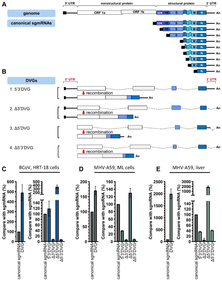
Background: Defective viral genome (DVG) is a truncated version of the full-length virus genome identified in most RNA viruses during infection. The synthesis of DVGs in coronavirus has been suggested; however, the fundamental characteristics of coronavirus DVGs in gene expression and pathogenesis have not been systematically analyzed.

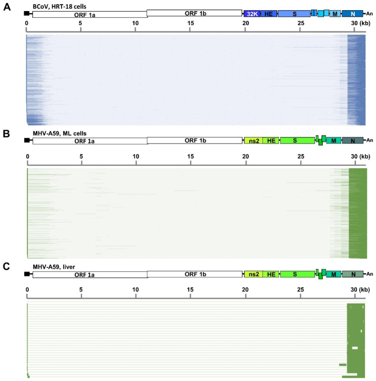
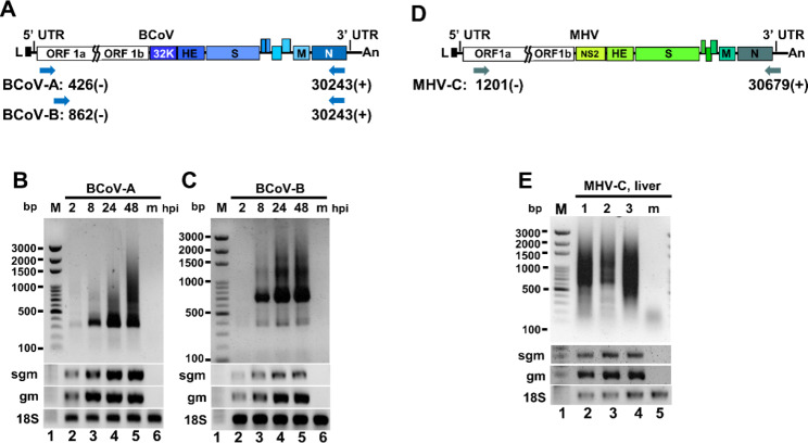
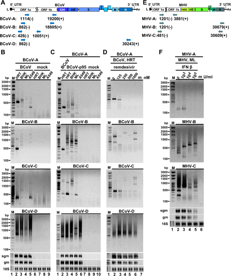
Methods: Nanopore direct RNA sequencing was used to investigate the characteristics of coronavirus DVGs in gene expression including reproducibility, abundance, species and genome structures for bovine coronavirus in cells, and for mouse hepatitis virus (MHV)-A59 (a mouse coronavirus) in cells and in mice. The MHV-A59 full-length genomic cDNAs (~ 31 kilobases) were in vitro constructed to experimentally validate the origin of coronavirus DVG. The synthesis of DVGs was also experimentally identified by RT-PCR followed by sequencing. In addition, the alterations of DVGs in amounts and species under different infection environments and selection pressures including the treatment of antiviral remdesivir and interferon were evaluated based on the banding patterns by RT-PCR.

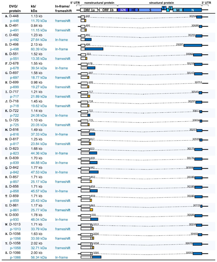
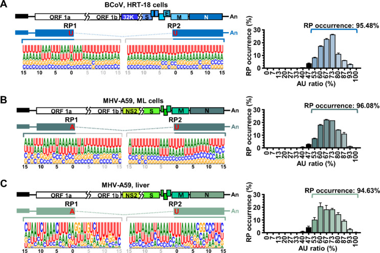
Results: The results are as follows: (i) the structures of DVGs are with diversity, (ii) DVGs are overall synthesized with moderate (MHV-A59 in cells) to high (BCoV in cells and MHV-A59 in mice) reproducibility under regular infection with the same virus inoculum, (iii) DVGs can be synthesized from the full-length coronavirus genome, (iv) the sequences flanking the recombination point of DVGs are AU-rich and thus may contribute to the recombination events during gene expression, (v) the species and amounts of DVG are altered under different infection environments, and (vi) the biological nature of DVGs between in vitro and in vivo is similar.

Conclusions: The identified biological characteristics of coronavirus DVGs in terms of abundance, reproducibility, and variety extend the current model for coronavirus gene expression. In addition, the biological features of alterations in amounts and species of coronavirus DVGs under different infection environments may assist the coronavirus to adapt to the altered environments for virus fitness and may contribute to the coronavirus pathogenesis. Consequently, the unveiled biological features may assist the community to study the gene expression mechanisms of DVGs and their roles in pathogenesis, contributing to the development of antiviral strategy and public health.

Keywords: Coronavirus; Coronavirus genome structure; Defective viral genome; Gene expression; Pathogenesis.


Conflict of interest statement

The authors declare that they have no competing interests.

Figures

Fig. 1
Fig. 1
The classification and abundance of coronavirus DVGs based on nanopore direct RNA sequencing. (A) The structures of the BCoV full-length genome and canonical sgmRNAs. (B) The DVGs can be divided into 4 subgroups Δ5’3’DVG, Δ3’DVG, Δ5’DVG and 5’3’ DVG based on whether DVGs contain 5’ and/or 3’ UTR sequence (partial or complete). Note that DVGs may consist of 1, 2 or more than 2 fragments based on the criteria and definitions for the classes as described in the figure legend of Figure S1. Shown here are DVGs with 4 fragments to emphasize that they are recombination products derived from the different portions of the genome including part of ORF1a, ORF1b, S and N genes. The dashed line indicates the truncated genome in DVGs. (C)-(E) Left panel: The relative amounts of total DVGs and canonical sgmRNAs in BCoV-and MHV-A59-infected cells and MHV-A59 infected mice. Right panel: The relative amounts of each classified DVG subgroup and canonical sgmRNA in BCoV-and MHV-A59-infected cells and MHV-A59 infected mice. Panels (C)-(E) show the mean of two biological replicates; error bars indicate the standard deviation
Fig. 2
Fig. 2
Reproducibility of DVGs. (A)-(C) The overall reproducibility of DVGs for BCoV in BCoV-infected cells, MHV-A59-infected cells and mice. (D)-(F) The reproducibility of 5’3’DVG, Δ3’DVG and Δ5’DVG in BCoV-infected cells. (G)-(I) The reproducibility of 5’3’DVG, Δ3’DVG and Δ5’DVG in MHV-A59-infected cells. (J) The reproducibility of Δ5’DVG in MHV-A59-infected mice. Reproducibility was measured for transcripts with a read count of ≥ 5 based on RNA (BCoV RNA1 and BCoV RNA2) collected from the two biological replicates. The reproducibility is measured in reads per kilobase per million mapped sequence reads (RPKM) and evaluated by Spearman’s correlation coefficient. R = 0.000-0.3999, low reproducibility; R = 0.4000-0.5999, moderate reproducibility; R = 0.6000-1.0000, high reproducibility
Fig. 3
Fig. 3
Schematic diagram showing that the 5’3’DVG species contain different lengths of 5’ and 3’ terminal sequences of the genome for BCoV in HRT-18 cells (A), MHV-A59 in ML cells (B) and MHV-A59 in mice (C). The 5’ and 3’ terminal sequences are highlighted with darker bars on the x-axis. The results are derived from two biological replicates
Fig. 4
Fig. 4
Detection of coronavirus DVGs by RT-PCR. (A) Diagram depicting the primer sets used for determining the synthesis of BCoV DVGs in Figures (B)-(C). (B)-(C) Detection of BCoV DVG synthesis at different time points of BCoV infection by RT-PCR. HRT-18 cells were infected with 0.1 MOI of BCoV followed by total cellular RNA collection at 2, 8, 24 and 48 and RT-PCR with primers shown in Figure (A). (D) Diagram depicting the primer sets used for determining the synthesis of MHV-A-59 DVGs in Figure (E). (E) Detection of DVGs by RT-PCR (lanes 2–4) from 3 individual mice at 3 days postinfection with MHV-A59. m, mock-infected cells or mice. bp, base-pair; M, DNA size marker; hpi, hours postinfection; sgm, sgmRNA N; gm, genome; 18 S, 18 S rRNA
Fig. 5
Fig. 5
The structures of experimentally identified DVGs and the predicted encoded proteins. The numbers shown in each DVG structure are the nucleotide positions at which the recombination occurs. The dashed line indicates the truncated genome in DVG. The encoded fusion protein from D-448, D-491, D-551, D-697, D-717, D-718, D-725, D-817, D-857 and D-859 contains part of nsp1 and protein resulted from frameshift in N protein gene. The encoded fusion protein from D-492, D-678, D-722, D-816, D-823, D-839, D-842, D858 and D930 contains part of nsp1 and N protein. The encoded fusion protein from D-699 contains part of nsp1 and protein resulted from frameshift in gene from 3’UTR. The encoded fusion protein from D-861 contains part of nsp1 and protein resulted from frameshift in N protein gene and gene from 3’UTR. The encoded fusion protein from D-1013, D-1056 and D-1058 contains nsp1, part of nsp2 and protein resulted from frameshift in N protein gene. The encoded fusion protein from D-1066 contains nsp1 and part of nsp2 and N protein. The encoded fusion protein from D-498 contains part of nsp1 and complete N protein
Fig. 6
Fig. 6
Determination of the origin of DVGs using MHV-A59 full-length genomic cDNA. (A) Diagram depicting the primer sets used for determining the synthesis of MHV-A59 DVGs in Figures (B)-(D). (B)-(D) Determination of the origin of DVGs using MHV-A59 full-length genomic cDNA. After assembly and in vitro transcription of MHV-A59 full-length genomic cDNA, the full-length viral RNA was transfected into BHK-MHVR cells. After 48 h of transfection, total cellular RNA was harvested (VP0), and the supernatant was collected to infect fresh BHK-MHVR cells. The virus passage step was repeated until VP2. DVGs were detected by RT-PCR with primers shown in Figure (A). bp, base-pair; M, DNA size marker; hpi, hours postinfection; m, mock-infected cells; sgm, sgmRNA N; gm, genome; VP, virus passage; 18 S, 18 S rRNA
Fig. 7
Fig. 7
Sequences flanking the recombination points of DVGs are AU-rich. (A)-(C) Left panel: Characterization of the sequence flanking the recombination points (RP1 and RP2) of in BCoV-and MHV-A59-infected cells and MHV-A59 infected mice based on databases obtained from nanopore direct RNA sequencing. Right panel: Correspondence between the AU-rich sequence and the occurrence of recombination point. X-axis represents the percentage of AU sequence flanking the recombination points and y-axis represents the percentage of RP occurrence. RP, recombination point. The results are derived from the two biological replicates
Fig. 8
Fig. 8
The species and amounts of DVGs are altered under different infection environments and selection pressures. (A) Diagram depicting the primer sets used for determining the synthesis of BCoV DVGs in Figures (B)-(D). (B) Detection of DVGs synthesized in different cells infected with BCoV by RT-PCR. HRT-18, BHK, ML and A549 cells were infected with BCoV at an MOI of 0.1, followed by total cellular RNA collection at 24 hpi and RT-PCR. The “mock” indicates cells without infection with virus. (C) Detection of DVGs synthesized in different cells infected with BCoV-p95 by RT-PCR. HRT-18, BHK, ML and A549 cells were infected with BCoV-p95 at an MOI of 0.1, followed by total cellular RNA collection at 24 hpi and RT-PCR. (D) Detection of BCoV DVGs synthesized in HRT-18 cells infected with BCoV (0.1 MOI) and treated with antiviral remdesivir at final concentrations of 125, 250, 500 or 1000 nM. Total cellular RNA was collected at 48 hpi and DVGs were detected by by RT-PCR. (E) Diagram depicting the primer sets used for determining the MHV-A59 DVGs in Figure (F). (F) Detection of MHV-A59 DVGs synthesized in ML cells infected with MHV-A59 and treated with IFN β by RT-PCR. ML cells in 2 ml of DMEM were treated with IFN β at final concentrations of 103, 104 or 105 U/ml. After 16 h of treatment, IFN β-treated ML cells were infected with 0.1 MOI of MHV-A59 followed by total cellular RNA collection at 16 hpi. bp, base-pair; M, DNA size marker; m, mock-infected cells; HRT, human rectal tumor cells-18; BHK, baby hamster kidney cells; ML, mouse L cells; A549, adenocarcinomic human alveolar basal epithelial cells; hpi, hours postinfection; sgm, sgmRNA N; gm, genome; 18 S, 18 S rRNA

Similar articles

Cited by

References

    1. Brian DA, Baric RS. Coronavirus genome structure and replication. Curr Top Microbiol Immunol. 2005;287:1–30. - PMC - PubMed
    1. Gorbalenya AE, Enjuanes L, Ziebuhr J, Snijder EJ. Nidovirales: evolving the largest RNA virus genome. Virus Res. 2006;117(1):17–37. doi: 10.1016/j.virusres.2006.01.017. - DOI - PMC - PubMed
    1. Xiao SY, Wu YJ, Liu H. Evolving status of the 2019 novel coronavirus infection: proposal of conventional serologic assays for disease diagnosis and infection monitoring. J Med Virol. 2020;92(5):464–7. doi: 10.1002/jmv.25702. - DOI - PMC - PubMed
    1. Yang WJ, Cao QQ, Qin L, Wang XY, Cheng ZH, Pan AS, et al. Clinical characteristics and imaging manifestations of the 2019 novel coronavirus disease (COVID-19):a multi-center study in Wenzhou city, Zhejiang, China. J Infect. 2020;80(4):388–93. doi: 10.1016/j.jinf.2020.02.016. - DOI - PMC - PubMed
    1. Zaki AM, van Boheemen S, Bestebroer TM, Osterhaus AD, Fouchier RA. Isolation of a novel coronavirus from a man with pneumonia in Saudi Arabia. N Engl J Med. 2012;367(19):1814–20. doi: 10.1056/NEJMoa1211721. - DOI - PubMed

Publication types

LinkOut - more resources