Review
. 2024 Feb;20(7):e2305426.
doi: 10.1002/smll.202305426. Epub 2023 Oct 6.

Nanomaterial-Enabled Photothermal Heating and Its Use for Cancer Therapy via Localized Hyperthermia

Affiliations
Review

Nanomaterial-Enabled Photothermal Heating and Its Use for Cancer Therapy via Localized Hyperthermia

Song Shen et al. Small. 2024 Feb.

Abstract

Photothermal therapy (PTT), which employs nanoscale transducers delivered into a tumor to locally generate heat upon irradiation with near-infrared light, shows great potential in killing cancer cells through hyperthermia. The efficacy of such a treatment is determined by a number of factors, including the amount, distribution, and dissipation of the generated heat, as well as the type of cancer cell involved. The amount of heat generated is largely controlled by the number of transducers accumulated inside the tumor, the absorption coefficient and photothermal conversion efficiency of the transducer, and the irradiance of the light. The efficacy of treatment depends on the distribution of the transducers in the tumor and the penetration depth of the light. The vascularity and tissue thermal conduction both affect the dissipation of heat and thereby the distribution of temperature. The successful implementation of PTT in the clinic setting critically depends on techniques for real-time monitoring and management of temperature.

Keywords: gold nanoparticles; hyperthermia; photothermal therapy; temperature monitoring; thermotolerance.


Figures

Figure 1.
Figure 1.
Schematic illustration showing the parameters that affect the efficacy of PTT in vivo: ⅰ) the attenuation in tissue, penetration depth, and irradiance of the laser; ) the absorption coefficient, photothermal conversion efficiency, photostability, particle size, shape, and surface property of the photothermal transducers; ) the spatial distribution of the transducers; ) the heat-sink effect, thermal conductivity, thermotolerance of the lesion; and ) the actual temperature involved in the therapy.
Figure 2.
Figure 2.
(A) Schematic illustration of the tissue absorption of light with different wavelengths. (B) Penetration depths of UV radiation in human skin. The wavelength dependence of the penetration depth is similar at the volar (formula image) and dorsal (formula image) aspect of the forearm. Spectral characteristics are different at the thenar (formula image). The penetration depth is only very slowly rising in the UVB and increases steeply in the UVA. Reproduced with permission.[40] Copyright 2008, SCImago. (C) Fraction of total reduced scattering attributed to Rayleigh scattering in skin. Reproduced with permission.[41] Copyright 2005, IOP science. (D) Optical absorption spectra of major endogenous chromophores at typical concentrations occurring in living mammalian tissues. The first (NIR-I) and second (NIR-II) windows, where optical absorption is minimized, are indicated. Reproduced with permission.[42] Copyright 2005, Royal Society of Chemistry. (E) Absorption spectrum of liquid water with the wavelength in the range of 10 nm and 10 m. Reproduced with permission.[47] Copyright 1981, University of Missouri.
Figure 3.
Figure 3.
Extinction, absorption, and scattering spectra calculated using the DDA method for a Au nanoparticle dispersed in water (n = 1.33): (A) a Au nanobox with an inner edge length of 50 nm and a wall thickness of 5 nm; (B) a Au nanobox with an inner edge length of 30 nm and a wall thickness of 5 nm; (C) a Au nanobox with an inner edge length of 30 nm and a wall thickness of 3 nm; and (D) an Au nanobox with an inner edge length of 30 nm and a wall thickness of 5 nm, together with holes of 5 nm in edge length in the corners. Reproduced with permission.[49] Copyright 2005, Wiley-VCH.
Figure 4.
Figure 4.
Schematic showing the localized surface plasmon resonances and extinction spectra of (A) Au nanorods with different aspect ratios, (B) Au nanostars with different aspect ratios, (C) SiO2@Au core-shell nanoshells with different shell thicknesses, and (D) Au nanocages with different wall thicknesses. (A) and (C) were reproduced with permission.[48] Copyright 2014, Royal Society of Chemistry. (B) Reproduced with permission.[71] Copyright 2014, American Chemistry Society. (D) Reproduced with permission.[64] Copyright 2011, Springer Nature.
Figure 5.
Figure 5.
(A) Schematic illustration of the aggregate formation and the carboxylesterase-triggered disassembly. (B) TEM images and schematic illustrations of the aggregates before (left) and after (right) the treatment with carboxylesterase. (C) Fluorescence images of the multicellular spheroids showing the penetration depth of carboxylesterase-responsive tetrachloroperylene monoimide-human serum albumins nanoclusters (FHP) and carboxylesterase-irresponsive nanoclusters (HP3) through orthogonal section view of x/z axes (top) and y/z axes (right). scale bar: 200 μm. (A-C) Reproduced with permission.[118] Copyright 2020, Wiley-VCH. (D) Schematic showing the formation of pH-sensitive doxorubicin and IR780 loaded poly(4-formylphenyl methacrylate-co-2-(diethylamino) ethyl methacrylate)-b-polyoligoethyleneglycol methacrylate (PDMs). (E) TEM image of PDMs at pH=7.4 (left) and pH=6.6 (right) showing the assembly and disassembly, respectively. (D-F) Reproduced with permission.[119] Copyright 2020, Wiley-VCH.
Figure 6.
Figure 6.
(A) Cellular uptake of gold nanoparticles as a function of size. Reproduced with permission.[115] Copyright 2006, American Chemistry Society. (B) Herceptin-gold nanoparticles (Her-GNPs) with a size of 40 nm in diameter could down-regulate the membrane ErbB2 receptor (red) and redistribute the receptor from the cell surface to the cytoplasm, while 2 and 70 nm Her-GNPs did not show this effect. Scale bars: 10 μm. Reproduced with permission.[128] Copyright 2008, Springer Nature. (C) Schematic showing that nanoparticles with a size of 40–60 nm are more readily to be uptaken than those smaller than 40 nm or larger than 60 nm. Nanoparticles smaller than 5.5 nm can enter into the nucleus. (D) Uptake of the PEGylated gold nanohexapods, nanocages, and nanorods by MDA-MB-435 cells after incubation for different periods of time. The cells were positioned in an inverted configuration. The initial concentration of the gold nanoparticles in the culture medium were 10 μg/mL for all samples. Reproduced with permission.[57] Copyright 2013, American Chemistry Society.
Figure 7.
Figure 7.
A) Digital images showing the color changes of a suspension of the thermochromic nanoparticles when its temperature was raised from 25 to 45, and then decreased to 25 °C. B) Temperature profiles of ICG and (2′-phenylamino)-6′-diethylamino fluoran/β-naphthol (TFG/Naph) nanoparticles upon the irradiation of NIR light. C). Schematic illustration showing the thermostatic PTT in a tumor. D) Digital images showing the penetration depth of the light. E) The H&E staining sections of the tumor and adjacent tissues after PTT. Reprinted with permission.[150] Copyright 2020, American Chemistry Society.
Figure 8.
Figure 8.
A) Monitoring of temperature in vivo using a nanodiamond (ND) thermometer. B) A schematic illustration of the energy diagram of the nitrogen-vacancy centers. C) Microscopy image of the worm labelled with nanodiamonds. Scale bar: 20 μm. D) The fluorescence from nanodiamond at different continuous wave frequency. E) Time profiles of the total photon counts (Itot) and the estimated temperature of the nanodiamonds. F) Schematic illustration of the setup for the detection of the eigen temperature of csUCNP@C. G) Cells with and without csUCNP@C were irradiated with 730-nm and 980-nm lasers, respectively. Scale bar: 30 mm. (A-E) Reprinted with permission.[168] Copyright 2020, American Association for the Advancement of Science. (F-G) Reprinted with permission.[169] Copyright 2016, Springer Nature.

Similar articles

Cited by

References

    1. Ijff M, Crezee J, Oei AL, Stalpers LJA, Westerveld H, Int. J. Gynecol. Cancer 2022, 32, 288–296. - PMC - PubMed
    1. Liebl CM, Kutschan S, Doerfler J, Kaesmann L, Hubner J, Clin. Exp. Med. 2022, 22, 519–565. - PMC - PubMed
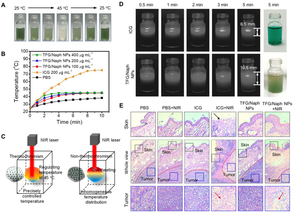
    1. Deng Z, Jiang C, Younis MR, Lei S, He Y, Zheng H, Huang P, Lin J, Chin. Chem. Lett. 2021, 32, 2411–2414.
    1. Sun Y.-d., Zhang H, Liu J.-z., Xu H.-r., Wu H.-y., Zhai H.-z., Lu C.-y., Zhao X, Chen Y.-q., Zhou L.-l., Han J-J, Thoracic Cancer 2019, 10, 543–550. - PMC - PubMed
    1. Jeanjean P, El Hamrani D, Genevois C, Quesson B, Couillaud F, Adv. Mater. Technol. 2022, 7, 2101258.

Publication types

LinkOut - more resources