. 2023 Oct 7;42(1):261.
doi: 10.1186/s13046-023-02835-6.

Anoikis resistance and immune escape mediated by Epstein-Barr virus-encoded latent membrane protein 1-induced stabilization of PGC-1α promotes invasion and metastasis of nasopharyngeal carcinoma

Affiliations

Anoikis resistance and immune escape mediated by Epstein-Barr virus-encoded latent membrane protein 1-induced stabilization of PGC-1α promotes invasion and metastasis of nasopharyngeal carcinoma

Chaoliang Liao et al. J Exp Clin Cancer Res. .

Abstract

Background: Epstein-Barr virus (EBV) is the first discovered human tumor virus that is associated with a variety of malignancies of both lymphoid and epithelial origin including nasopharyngeal carcinoma (NPC). The EBV-encoded latent membrane protein 1 (LMP1) has been well-defined as a potent oncogenic protein, which is intimately correlated with NPC pathogenesis. Anoikis is considered to be a physiological barrier to metastasis, and avoiding anoikis is a major hallmark of metastasis. However, the role of LMP1 in anoikis-resistance and metastasis of NPC has not been fully identified.

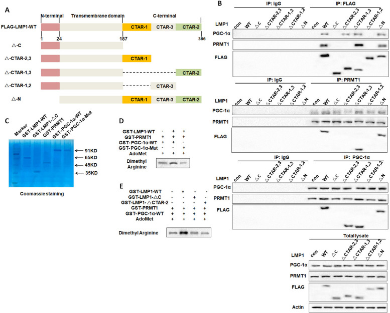
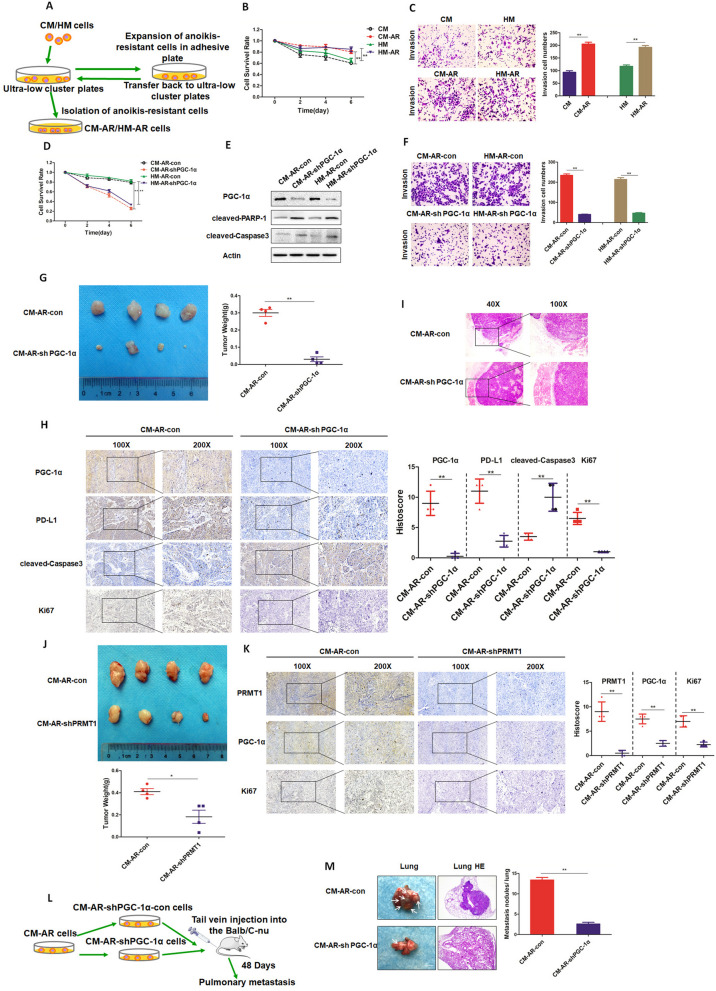
Methods: Trypan blue staining, colony formation assay, flow cytometry, and TUNEL staining, as well as the detection of apoptosis and anoikis resistance-related markers was applied to evaluate the anoikis-resistant capability of NPC cells cultured in ultra-low adhesion condition. Co-immunoprecipitation (Co-IP) experiment was performed to determine the interaction among LMP1, PRMT1 and PGC-1α. Ex vivo ubiquitination assay was used to detect the ubiquitination level of PGC-1α. Anoikis- resistant LMP1-positive NPC cell lines were established and applied for the xenograft and metastatic animal experiments.

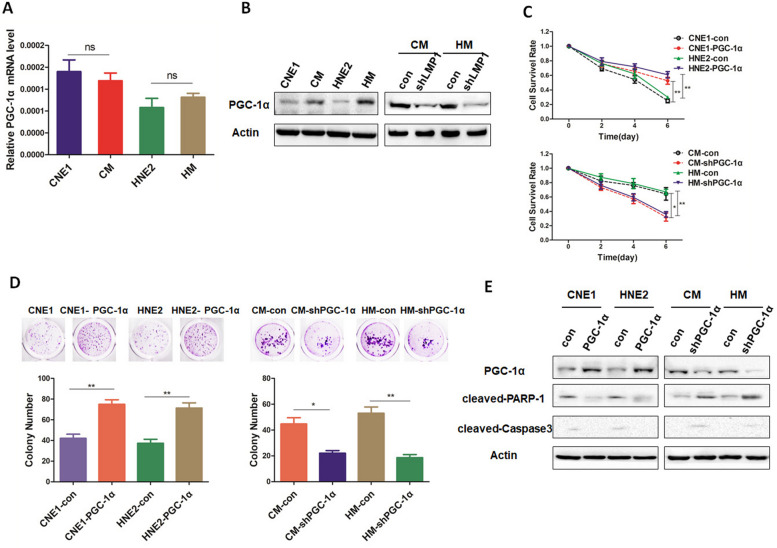
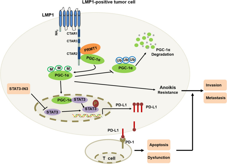
Results: Our current findings reveal the role of LMP1-stabilized peroxisome proliferator activated receptor coactivator-1a (PGC-1α) in anoikis resistance and immune escape to support the invasion and metastasis of NPC. Mechanistically, LMP1 enhances PGC-1α protein stability by promoting the interaction between arginine methyltransferase 1 (PRMT1) and PGC-1α to elevate the methylation modification of PGC-1α, thus endowing NPC cells with anoikis-resistance. Meanwhile, PGC-1α mediates the immune escape induced by LMP1 by coactivating with STAT3 to transcriptionally up-regulate PD-L1 expression.

Conclusion: Our work provides insights into how virus-encoded proteins recruit and interact with host regulatory elements to facilitate the malignant progression of NPC. Therefore, targeting PGC-1α or PRMT1-PGC-1α interaction might be exploited for therapeutic gain for EBV-associated malignancies.

Keywords: Anoikis resistance; Immune escape; LMP1; Nasopharyngeal carcinoma; PGC-1α.


Conflict of interest statement

The authors declare that they have no competing interests.

Figures

Fig. 1
Fig. 1
LMP1 promotes anoikis resistance and invasion of NPC cells. A Cell survival rate of CNE1/CM, HNE2/HM, CM-con/CM-shLMP1, HM-con/HM-shLMP1 cells after 0, 2, 4, or 6 days suspension (**p < 0.01, *p < 0.05). B CNE1/CM, HNE2/HM, CM-con/CM-shLMP1, HM-con/HM-shLMP1 cells were cultured in suspension for 48 h, and the expression of LMP1, cleaved-PARP-1, and cleaved-Caspase 3 was detected by Western blotting. C CNE1/CM, HNE2/HM, CM-con/CM-shLMP1, and HM-con/HM-shLMP1 cells were cultured in suspension for 7 days, and the colony formation rate of the cells reattached was observed (**p < 0.01, *p < 0.05). D CNE1/CM, HNE2/HM, CM-con/CM-shLMP1, and HM-con/HM-shLMP1 cells were cultured in suspension for 48 h, and the apoptotic rate of cells was detected by flow cytometry (**p < 0.01). E CNE1/ CM/CM-shLMP1 and HNE2/HM/HM-shLMP1 cells were cultured in suspension for 48 h, and the apoptotic rate of cells was detected by TUNEL assay, respectively (***p < 0.001,**p < 0.01, *p < 0.05). bar, 200μm. CNE1/CM, HNE2/HM, CM-con/CM-shLMP1, and HM-con/HM-shLMP1 cells were cultured in suspension for 7 days, and (F) the migration and (G) invasion abilities were detected by cell migration assay (**p < 0.01, *p < 0.05)
Fig. 2
Fig. 2
PGC-1α mediates LMP1-induced anoikis resistance in NPC cells. A CNE1/CM and HNE2/HM cells were cultured in suspension for 48 h, and then the transcription level of PGC-1α was detected by real-time fluorescence quantitative PCR. B CNE1/CM, HNE2/HM, CM-con/CM-shLMP1, and HM-con/HM-shLMP1 cells were cultured in suspension for 48 h, and the protein levels of PGC-1α were detected by Western blotting. C Survival rate of CNE1-con/CNE1-PGC-1α, HNE2-con/HNE2-PGC-1α, CM-con/CM-shPGC-1α, and HM-con/HM- shPGC-1α cells was assessed after 0, 2, 4, and 6 days suspension culture (**p < 0.01, *p < 0.05). D CNE1-con/CNE1-PGC-1α, HNE2-con/HNE2-PGC-1α, CM-con/CM-shPGC-1α, and HM-con/HM- shPGC-1α cells were cultured in suspension for 7 days, and the colony formation rate of the cells reattached was measured (**p < 0.01, *p < 0.05). E CNE1-con/CNE1-PGC-1α, HNE2-con/HNE2- PGC-1α, CM-con/CM-shPGC-1α, and HM-con/HM-shPGC-1α cells were cultured in suspension for 48 h and Western blotting was used to detect the expression of PGC-1α, cleaved-PARP-1 and cleaved-Caspase 3
Fig. 3
Fig. 3
LMP1 promotes the PRMT1-PGC-1α interaction and induces arginine methylation of PGC-1α to enhance the stability of PGC-1α. A CNE1/CM and HNE2/HM cells were cultured in suspension and treated with CHX for 0, 1, 2, 4, 8, and 24 h, respectively, and the expression of PGC-1α protein was detected by Western blotting. B CNE1/CM and HNE2/HM cells were cultured in suspension for 48 h, and the arginine methylation level of PGC-1α was detected by Western blotting after purification of PGC-1α. C CNE1/CM and HEN2/HM cells were cultured in suspension, and the interaction between PRMT1-PGC-1α was detected by co- immunoprecipitation. D LMP1 was knocked down in CM and HM cells. Cells were cultured in suspension, and the interaction between PRMT1-PGC-1α was detected by co- immunoprecipitation. E PRMT1 was overexpressed in CNE1 and HNE2 cells, cultured in suspension for 48 h, and the arginine methylation level of PGC-1α was detected by Western blot analysis after purification of PGC-1α. F PRMT1 was overexpressed in CNE1 and HNE2 cells. The cells were cultured in suspension and treated with CHX for 0, 1, 2, 4, 8 and 24 h, respectively, and the expression of PGC-1α protein was detected by Western blotting. G The shRNA targeting PRMT1 was synthesized, and shRNA was transfected to CM and HM cells for 48 h, and then the expression level of PRMT1 was detected by Western blot analysis. H PRMT1 was knocked down in CM and HM cells. Cells were cultured in suspension for 48 h, and the arginine methylation level of PGC-1α was detected by Western blotting after purification of PGC-1α. I PRMT1 was knocked down in CM and HM cells. The cells were cultured in suspension and treated with CHX for 0, 1, 2, 4, 8 and 24 h, respectively, and the expression of PGC-1α protein was detected by Western blot analysis
Fig. 4
Fig. 4
The C-terminal CTAR-2 domain of LMP1 promotes the interaction of PRMT1 with PGC-1α to enhance PGC-1α arginine methylation. A Structure diagram of each truncation of LMP1. B The LMP1 truncated plasmid, PRMT1 overexpression plasmid, and PGC-1α overexpression plasmid were simultaneously overexpressed in 293T cells, and the PRMT1-PGC-1α interaction was detected by co-immunoprecipitation. C GST-LMP1-WT, GST-LMP1-△C, GST-PRMT1, GST-PGC-1α-WT, and GST-PGC-1α-Mut fusion proteins were purified, and then visualized by Coomassie brilliant blue staining. D The GST-LMP1-WT and GST-PRMT1 proteins were incubated with GST-PGC-1α-WT or mutant protein GST-PGC-1α-Mut and AdoMet for in vitro methylation reaction. The level of arginine methylation modification of PGC-1α was detected by Western blotting. E GST-LMP1-WT, GST-LMP1-△C, and GST-LMP1-△CTAR-2 were incubated with GST-PRMT1 protein, GST-PGC-1α-WT protein, and AdoMet, respectively, and the level of arginine methylation modification of PGC-1α was detected by Western blot analysis
Fig. 5
Fig. 5
PRMT1 mediates LMP1-inhibited PGC-1α ubiquitination by enhancing arginine methylation of PGC-1α. A PRMT1 was overexpressed in CNE1 and HNE2 cells. The cells were cultured in suspension and treated with the proteasome inhibitor MG132, and the expression of PGC-1α was detected by Western blotting. B PRMT1 was knocked down in CM and HM cells. The cells were cultured in suspension and treated with the proteasome inhibitor MG132. The expression of PGC-1α was detected by Western blotting. C PRMT1 was overexpressed in CNE1 and HNE2 cells. The cells were cultured in suspension and treated with the proteasome inhibitor MG132, and the ubiquitination level of PGC-1α was detected by in vivo ubiquitination assay. D PRMT1 was knocked down in CM and HM cells. The cells were cultured in suspension and treated with the proteasome inhibitor MG132, and the ubiquitination level of PGC-1α was detected by in vivo ubiquitination assay. E The FLAG-PGC-1α-WT/FLAG-PGC-1α-Mut and PRMT1 plasmids were transfected into 293T cells, and then 293T cells were treated with proteasome inhibitor MG132. The ubiquitination level of PGC-1α was then detected by in vivo ubiquitination assay. F The FLAG-PGC-1α-WT/FLAG-PGC-1α-Mut and shPRMT1 plasmids were transfected into 293T cells. The 293T cells were treated with the proteasome inhibitor MG132, and the ubiquitination level of PGC-1α was detected by in vivo ubiquitination assay
Fig. 6
Fig. 6
PGC-1α mediates LMP1 upregulation of PD-L1 to promote immune escape of NPC cells. A CNE1/CM, HNE2/HM, CM-con/CM-shLMP1, and HM-con/HM-shLMP1 cells were cultured in suspension for 48 h, and the protein levels of PD-L1, TNFα and VEGF were detected by Western blotting. B CNE1/CM, HNE2/HM, CM-con/CM-shLMP1, and HM-con/HM-shLMP1 cells were co-cultured with Jurkat cells or T cells for 24 h, and IFN-γ secreted by T cells was detected by ELISA (**p < 0.01, *p < 0.05). C PGC-1α was overexpressed in CNE1 and HNE2 cells or PGC-1α was knocked down in CM and HM cells, and the transcriptional levels of PGC-1α and PD-L1 were detected by real-time fluorescence quantitative PCR (**p < 0.01, *p < 0.05). D PGC-1α was overexpressed in CNE1 and HNE2 cells or PGC-1α was knocked down in CM and HM cells, and the protein expression level of PD-L1 was detected by Western blotting. E CNE1/HNE2 cells were transfected with PGC-1α overexpression plasmid and cultured for 48 h, and then co-cultured with T cells for 24 h, and the apoptotic rate of T cells was detected by flow cytometry. F PGC-1α was overexpressed in CNE1 and HNE2 cells or PGC-1α was knocked down in CM and HM cells. The secretion of IFN-γ by T cells was detected by ELISA (**p < 0.01, *p < 0.05). G PGC-1α was overexpressed in CNE1 and HNE2 cells, and then these cells were co-cultured with Jurkat cells or T cells. NPC cells were treated with sufficient anti-PD-L1 to block PD-L1 activity, and the secretion of IFN-γ in T cells was detected by ELISA (**p < 0.01, *p < 0.05). H PGC-1α was overexpressed in CNE1 and HNE2 cells or PGC-1α was knocked down in CM and HM cells, and the transcriptional levels of transcription factors STAT1, STAT3, and IRF2 were detected by real-time fluorescence quantitative PCR (**p < 0.01, *p < 0.05). I CNE1/CM and HNE2/HM cells were cultured in suspension, and the interaction of PGC-1α-STAT3 was detected by co-immunoprecipitation. J CNE1/CM cells were cultured in suspension, and the interaction of PGC-1α-STAT1 and PGC-1α-IRF2 was detected by co-immunoprecipitation. K Dual luciferase reporter assay was used to detect the effect of STAT3 on the PD-L1 promoter (**p < 0.01, *p < 0.05). L A dual luciferase reporter gene assay was used to detect the effect of PGC-1α and STAT3 on PD-L1 promoter (**p < 0.01, *p < 0.05). M CNE1/HNE2 cells were transfected with STAT3 overexpression plasmid, and the expression of PGC-1α, STAT3, and PD-L1 was detected by Western blotting. N CNE1/HNE2 cells were transfected with a PGC-1α overexpression plasmid and a STAT3 inhibitor was added; and the expression of PGC-1α, STAT3, and PD-L1 was detected by Western blotting
Fig. 7
Fig. 7
PGC-1α mediates the invasion and metastasis of anoikis-resistant LMP1-positive NPC cells. A Flow chart of anoikis-resistant LMP1-positive NPC cell lines. B The survival rates of CM/CM-AR and HM/ HM-AR cells were detected after 0, 2, 4, and 6 days of suspension culture (**p < 0.01, *p < 0.05). C CM/CM-AR and HM/HM-AR cells were cultured in suspension for 7 days, and the invasive ability of the cells was detected by cell invasion assay (**p < 0.01, *p < 0.05). D CM-AR-con/CM-AR-sh PGC-1α cells and HM-AR-con/HM-AR-sh PGC-1α cells were cultured in suspension, and the survival rate of cells was detected at 0, 2, 4, and 6 days, respectively (**p < 0.01, *p < 0.05). E CM-AR-con/CM-AR-shPGC-1α cells and HM-AR-con/HM-AR-shPGC-1α cells were cultured in suspension for 48 h, and the protein expression levels of cleaved-PARP-1 and cleaved-Caspase 3 were detected by Western blotting. F CM-AR-con/CM-AR-sh PGC-1α cells and HM-AR-con/HM-AR-sh PGC-1α cells were cultured in suspension for 7 days, and the invasive ability of the cells was detected by a cell invasion assay (**p < 0.01, *p < 0.05). G Nude mice were subcutaneously injected with CM-AR-con and CM-AR-shPGC-1α cells, and the tumor volume and mass were detected at 14 days (**p < 0.01, *p < 0.05). H Immunohistochemistry was used to analyze the expression of PGC-1α, PD-L1, cleaved-Caspase 3, and Ki67 in mouse tissue sections (**p < 0.01, *p < 0.05). I Nude mice were subcutaneously injected with CM-AR-con and CM-AR-shPGC-1α cells, and the infiltration of the tumor margins was detected by H&E staining. J Nude mice were subcutaneously injected with CM-AR-con and CM-AR-shPRMT1 cells, and the tumor volume and mass were detected at 14 days (**p < 0.01, *p < 0.05). K Immunohistochemistry was used to analyze the expression of PRMT1, PGC-1α, and Ki67 in mouse tissue sections (**p < 0.01, *p < 0.05). L Flow chart of lung metastasis of anoikis-resistant LMP1-positive NPC cells in nude mice. M CM-AR-con and CM-AR-shPGC-1α cells were injected into the tail vein of nude mice, and 48 days later, the lung metastatic nodules were observed by H&E staining (**p < 0.01, *p < 0.05)
Fig. 8
Fig. 8
Mechanism of LMP1-induced stabilization of PGC-1α promoting invasion and metastasis of nasopharyngeal carcinoma. LMP1 promotes PRMT1-PGC-1α interaction to up-regulate the arginine methylation levels of PGC-1α. This action inhibits ubiquitination and degradation of PGC-1α, thus enhancing its protein stability. Elevated PGC-1α by LMP1 co-activates STAT3 to boost PD-L1 expression, consequently facilitating immune escape of cancer cells. These mechanisms coordinate to enhance the anoikis-resistance, invasion, and metastasis of cancer cells

Similar articles

Cited by

References

    1. Lieberman PM. Virology. Epstein-Barr virus turns 50. Science. 2014;343:1323–1325. doi: 10.1126/science.1252786. - DOI - PMC - PubMed
    1. Matsusaka K, et al. Classification of Epstein-Barr virus-positive gastric cancers by definition of DNA methylation epigenotypes. Cancer Res. 2011;71:7187–7197. doi: 10.1158/0008-5472.CAN-11-1349. - DOI - PubMed
    1. Raab-Traub N. Epstein-Barr virus in the pathogenesis of NPC. Semin Cancer Biol. 2002;12:431–441. doi: 10.1016/s1044579x0200086x. - DOI - PubMed
    1. Luo X, et al. DNMT1 mediates metabolic reprogramming induced by Epstein-Barr virus latent membrane protein 1 and reversed by grifolin in nasopharyngeal carcinoma. Cell Death Dis. 2018;9:619. doi: 10.1038/s41419-018-0662-2. - DOI - PMC - PubMed
    1. Tsao SW, Tramoutanis G, Dawson CW, Lo AK, Huang DP. The significance of LMP1 expression in nasopharyngeal carcinoma. Semin Cancer Biol. 2002;12:473–487. doi: 10.1016/s1044579x02000901. - DOI - PubMed

MeSH terms