Review
. 2023 Oct 6;27(1):99.
doi: 10.1186/s40824-023-00433-3.

Exosome-bearing hydrogels and cardiac tissue regeneration

Affiliations
Review

Exosome-bearing hydrogels and cardiac tissue regeneration

Hassan Amini et al. Biomater Res. .

Abstract

Background: In recent years, cardiovascular disease in particular myocardial infarction (MI) has become the predominant cause of human disability and mortality in the clinical setting. The restricted capacity of adult cardiomyocytes to proliferate and restore the function of infarcted sites is a challenging issue after the occurrence of MI. The application of stem cells and byproducts such as exosomes (Exos) has paved the way for the alleviation of cardiac tissue injury along with conventional medications in clinics. However, the short lifespan and activation of alloreactive immune cells in response to Exos and stem cells are the main issues in patients with MI. Therefore, there is an urgent demand to develop therapeutic approaches with minimum invasion for the restoration of cardiac function.

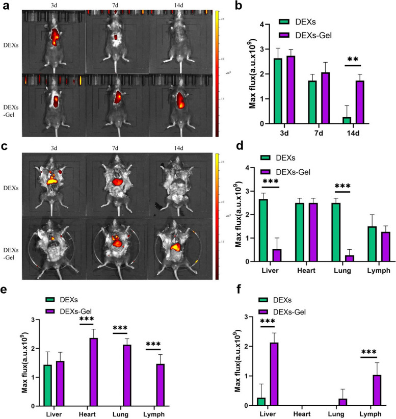
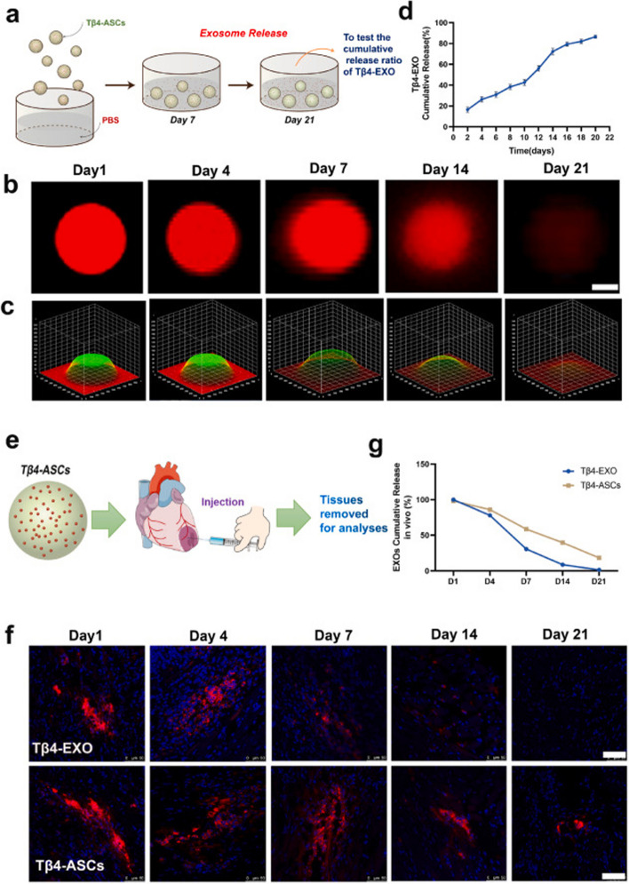
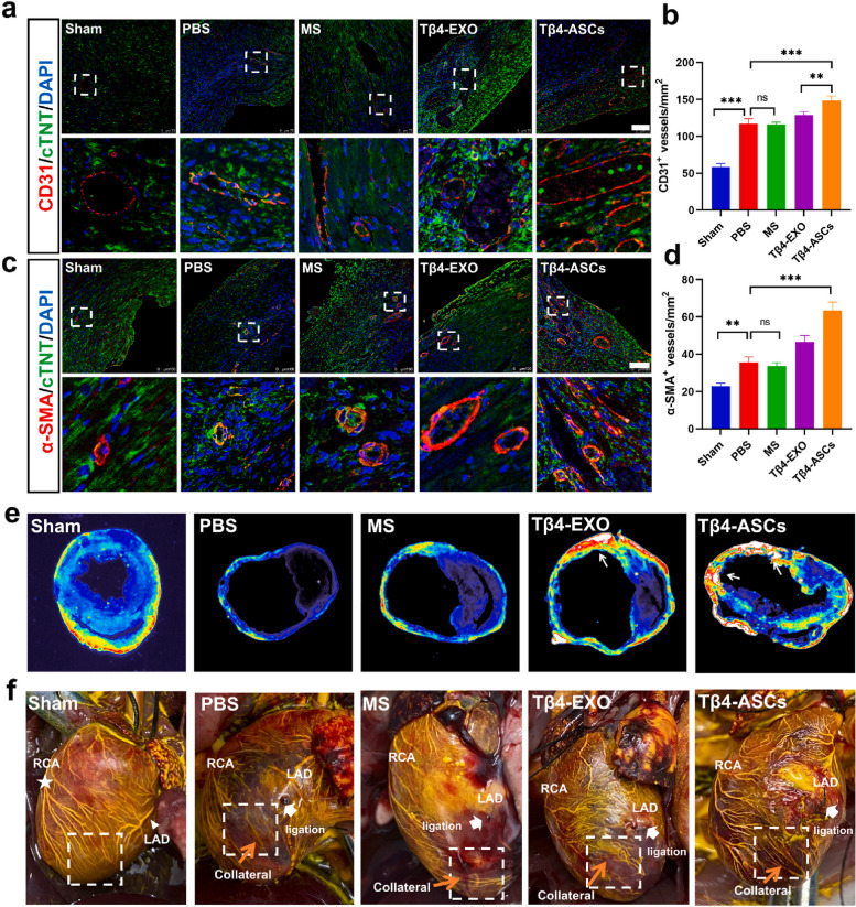
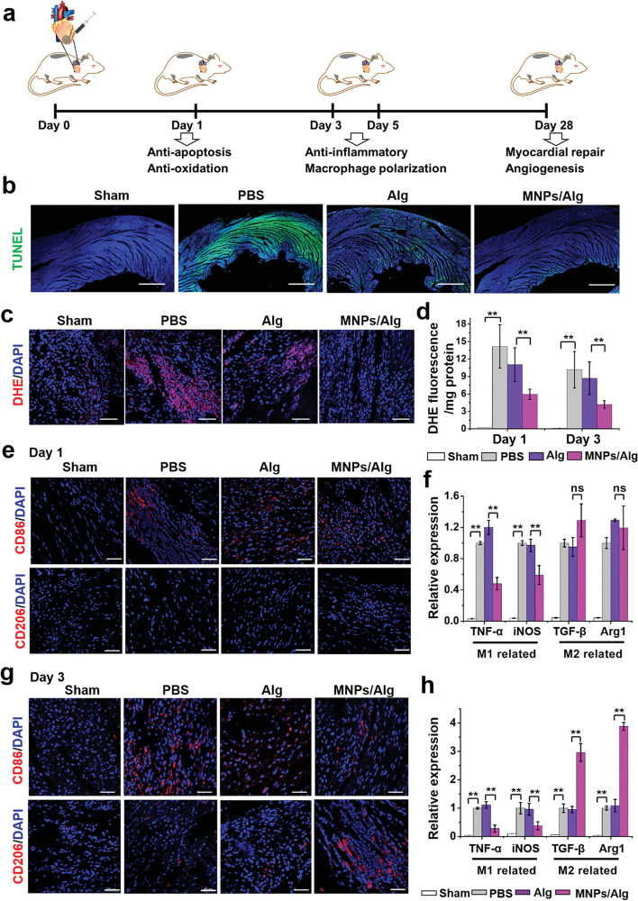
Main body: Here, we focused on recent data associated with the application of Exo-loaded hydrogels in ischemic cardiac tissue. Whether and how the advances in tissue engineering modalities have increased the efficiency of whole-based and byproducts (Exos) therapies under ischemic conditions. The integration of nanotechnology and nanobiology for designing novel smart biomaterials with therapeutic outcomes was highlighted.

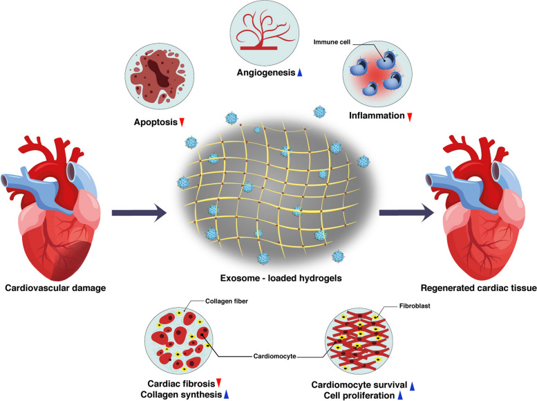
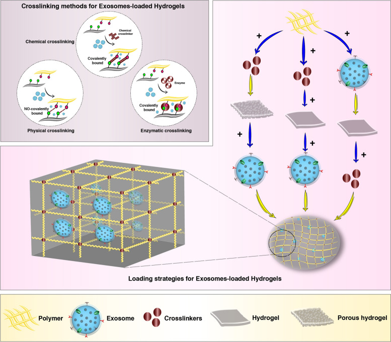
Conclusion: Hydrogels can provide suitable platforms for the transfer of Exos, small molecules, drugs, and other bioactive factors for direct injection into the damaged myocardium. Future studies should focus on the improvement of physicochemical properties of Exo-bearing hydrogel to translate for the standard treatment options.

Keywords: Cardiac tissue engineering; Encapsulation; Exosomes; Hydrogels; Infarction; Regeneration.


Conflict of interest statement

The authors declare that they have no competing interests.

Figures

Fig. 1
Fig. 1
The structure of exosomes (Exos) with the lipid bilayer. Exos can transfer specific cargo (protein and genetic contents) from donor cells to recipient cells in a paracrine manner. The type and levels of specific signaling molecules can induce/inhibit certain molecular pathways inside the target cells
Fig. 2
Fig. 2
Molecular pathways associated with Exo generation and release. An endosomal system composed of early, and late endosomes and multi-vesicular bodies (MVBs) plays a key role in the production of Exos. Inside the endosomes, numerous ILVs are formed by the invagination of the membrane. Upon secretion into the ECM, ILVs are named Exos. Different factors such as ESCRT complex (ESCRT-0, -I, -II, and -III) and tetraspanins sequestrate the signaling molecules into the ILVs. In the latter steps, the SNARE system in collaboration with several GTPases directs the MVBs toward the cell membrane to release the Exos
Fig. 3
Fig. 3
Exo-loaded hydrogels for cardiac tissue regeneration
Fig. 4
Fig. 4
Common strategies used to encapsulate Exos inside the hydrogels. Hydrogel construction is done by the promotion of polymer-polymer interactions with chemical, physical, and enzymatic crosslinking approaches. Exos are integrated into the hydrogel before, during, or after the crosslinking process
Fig. 5
Fig. 5
Alginate-based hydrogel is an appropriate platform for the sustained release of dendritic cell Exos (DEXs) into the targeted tissue over 14 days. Near-IR fluorescence indicated that the maximum flux values (a.u.) were high in mice that received DEX-loaded hydrogel compared to the DEX group after 14 days (a-c). Data indicated different a.u. values in several organs on days 3, 7, and 14 (d-f) (n = 3). Unpaired t-test. *p < 0.05, **p < 0.01, ***p < 0.001 [132]. Copyright 2021. Journal of Nanobiotechnology
Fig. 6
Fig. 6
Hydrogels can release functional Exos for therapeutic purposes for long periods. The cumulative release of PKH-26-labeled Thymosin β4 (Tβ4)-Exos bearing GelMA/PEGDA microspheres has been documented for 21 days in in vitro conditions (a). Using confocal microscopy, fluorescence images at a 3D surface plot were prepared (b - c). Continuous delivery led to the reduction of fluorescence intensity inside the microspheres by increasing the release time. A fluorescence spectrophotometer indicates the cumulative release of PKH26-labeled Tβ4-Exos over 20 days after being injected into infarcted myocardium (e). Immunofluorescence images revealed an appropriate release of loaded Tβ4-Exos in in vivo conditions (f-g). Scale bar: 15 μm [135]. Copyright 2022. Bioactive Materials
Fig. 7
Fig. 7
Exo-bearing hydrogels can be used for the induction of vascularization in ischemic changes. The angiogenic potential of Tβ4-Exos bearing GelMA/PEGDA microspheres was examined in a mouse model of MI (a-f). Using immunofluorescence staining, the capillary intensity (CD31+ vessels) was calculated in different groups including sham, infarct + PBS (PBS), GelMA/PEGDA microsphere alone (MS), Tβ4-Exos + GelMA/PEGDA microsphere (Tβ4-Exos), and Tβ4-Exo producing stem cells + GelMA/PEGDA microsphere (Tβ4-Exos + ASC) after 28 days (Scale bar: 75 μm) (a and b).The number of α-SMA+ arterioles (c and d). Cross-section of cardiac tissue and monitoring entire CD31 expression (e). The gross appurtenance of hearts after microfilling (f). White star: RCA (right coronary artery); White triangle: LCA (left anterior descending); White arrow: (the ligation of LAD). Microspheres (MS); Phosphate-buffered saline (PBS). One-way analysis of variance (ANOVA). **p < 0.01; ***p < 0.001 [135]. Copyright 2022. Bioactive Materials
Fig. 8
Fig. 8
Measuring the cardiogenic potential of Exo-bearing conductive thiolated hyaluronic acid hydrogel in an ischemic/reperfusion model of rat after 28 days. Masson’s-trichrome, Hematoxylin-Eosin, and Sirius red staining (a). Left ventricle thickness (b), Fibrosis size (c), and Sirius Red positive area (d). (n = 3; **p < 0.01 versus Sham group; #p < 0.05 and ## p < 0.01 vs. ischemic reperfusion model). (I: Sham; II: ischemic/reperfusion model; III: Free-Exo group; IV: EHBPE/HA-SH; V: AT-EHBPE/HA-SH; VI: AT-EHBPE/HA-SH@Exo; and VII: AT-EHBPE/HA-SH/CP05@. [136]. Copyright 2021. ACS Applied Materials & Interfaces
Fig. 9
Fig. 9
Monitoring regenerative potential of melanin nanoparticle-loaded alginate hydrogel (MNPs/Alg hydrogel) on reactive oxygen species (ROS) and immune response in a rat model of MI. A time-dependent healing process occurred within the ischemic area after MNPs/Alg hydrogel transplantation (a). Detection of apoptotic cells in the infarcted zone using TUNEL assay 1-day post-hydrogel administration (b). Immunofluorescence images related to O2•– levels using DHE staining 1 day after hydrogel injection (c). Detection of O2•– levels using ROS detection kit (DHE) on days 1 and 3 (d). The levels of CD86 and CD206 positive macrophages within the infarct zone were visualized using immunofluorescence images (e). Expression of pro-inflammatory (TNF-α and iNOS), and anti-inflammatory (TGF-β and Arg1) cytokines 1 day after injection (f). CD86 and CD206 positive macrophages after 3 days (g). Expression of pro-inflammatory, and anti-inflammatory cytokines on day 3 (n = 3). Scale bar: 50 μm; Student’s t-test. *p < 0.05, **p < 0.01; and nsp > 0.05 [153]. Copyright 2021. Advanced Science. 2021
Fig. 10
Fig. 10
Regenerative potential of Exo-loaded hyaluronic acid hydrogel in a rat model of transverse aortic constriction (TAC). H & E staining was performed 28 days after hydrogel administration (a; Scale bar: 200 μm). Different functional parameters, including were LVIDd, LVIDs, LVFS, LVEDV, LVESV, and LVEF were measured using echocardiography before TAC (b) and 28 days after TAC induction (c). The levels of fibrotic changes were evaluated using Masson’s Trichrome staining in rats that received PBS, hydrogel alone (HA), and Exo-loaded hydrogel (ExoGel) 28 days after transplantation (d; Scale bar: 400 μm). LV chamber area (e; n = 5); LV wall thickness (f; n = 5); Interstitial fibrosis rate (g). One-Way ANOVA analysis with post hoc Bonferroni test. *p < 0.05; **p < 0.01; ****p < 0.0001. HA: heart failure; LV: left ventricle; LV internal diameter end diastole: LVIDd; End-systole: LVIDs; LV fractional shortening: LVFS; LV end-diastolic volume: LVEDV; LV end-systolic volume: LVESV; and LV ejection fraction: LVEF. Copyright 2022 [165]. Journal of Molecular and Cellular Cardiology

References

    1. Lodrini AM, Goumans M-J. Cardiomyocytes cellular phenotypes after myocardial infarction. Front Cardiovasc Med. 2021;8:750510. 10.3389/fcvm.2021.750510. - PMC - PubMed
    1. Amini H, et al. Cardiac progenitor cells application in cardiovascular disease. J Cardiovasc Thorac Res. 2017;9(3):127–32. - PMC - PubMed
    1. Madonna R, et al. Position paper of the European Society of Cardiology Working Group Cellular Biology of the heart: cell-based therapies for myocardial repair and regeneration in ischemic heart disease and heart failure. Eur Heart J. 2016;37(23):1789–98. - PMC - PubMed
    1. Britten M, et al. Infarct remodeling after intracoronary progenitor cell treatment in patients with acute myocardial infarction (TOPCARE-AMI) mechanistic insights from serial contrast-enhanced magnetic resonance imaging. Circulation. 2003;108(18):2212–8. - PubMed
    1. Schächinger V, et al. Transplantation of progenitor cells and regeneration enhancement in acute myocardial infarction: final one-year results of the TOPCARE-AMI Trial. J Am Coll Cardiol. 2004;44(8):1690–9. - PubMed