. 2023 Dec 21;36(1):194-212.
doi: 10.1093/plcell/koad259.

The LIKE SEX FOUR 1-malate dehydrogenase complex functions as a scaffold to recruit β-amylase to promote starch degradation

Affiliations

The LIKE SEX FOUR 1-malate dehydrogenase complex functions as a scaffold to recruit β-amylase to promote starch degradation

Jian Liu et al. Plant Cell. .

Abstract

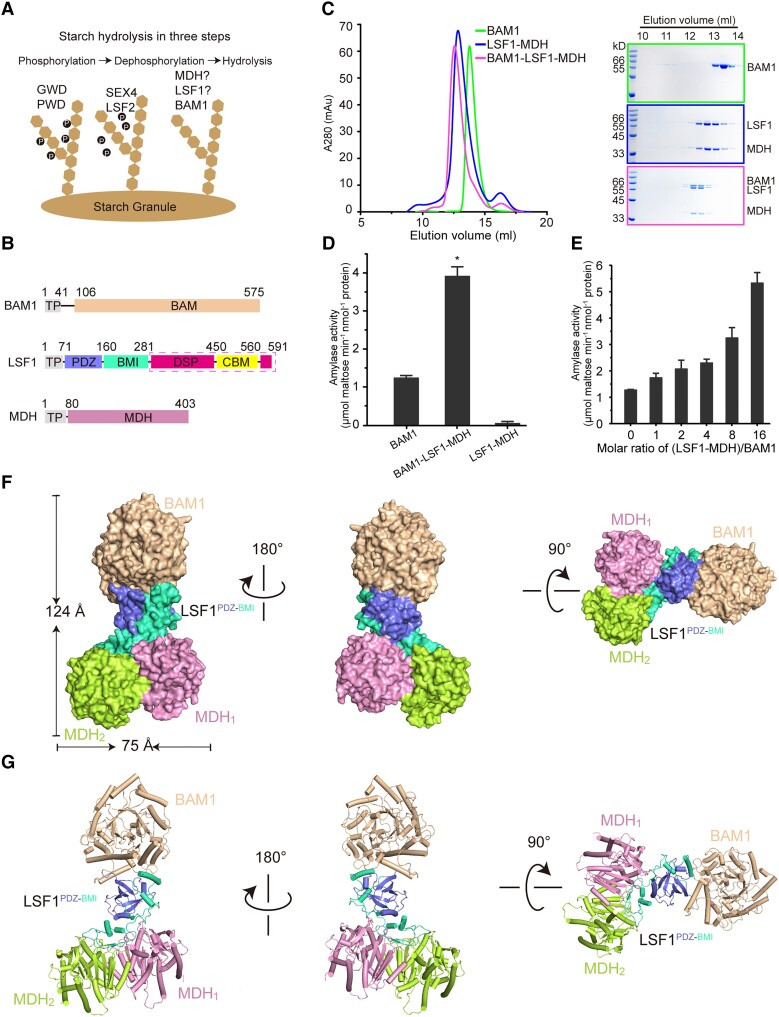
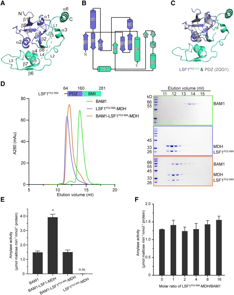
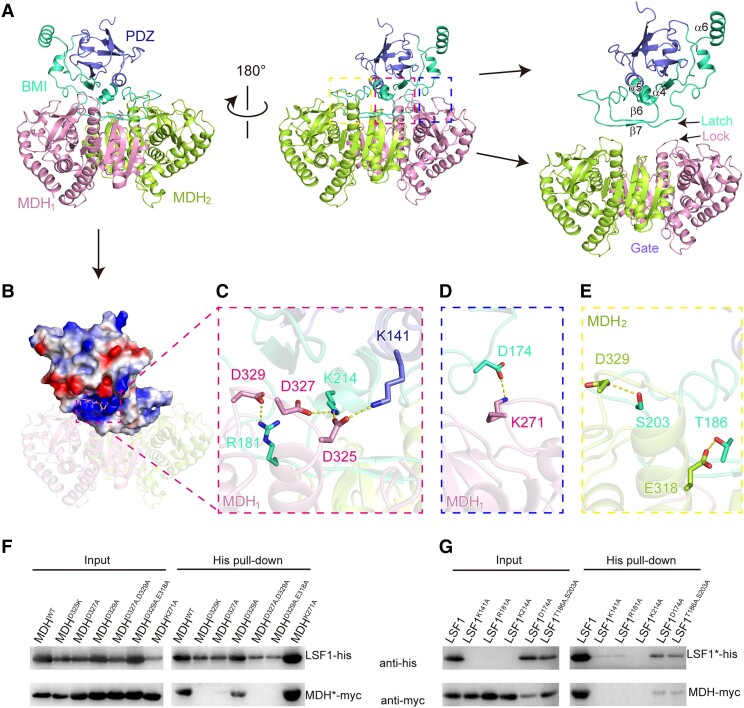
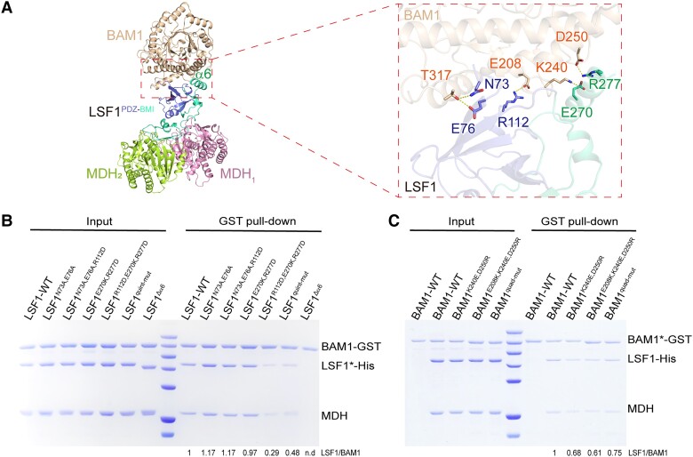
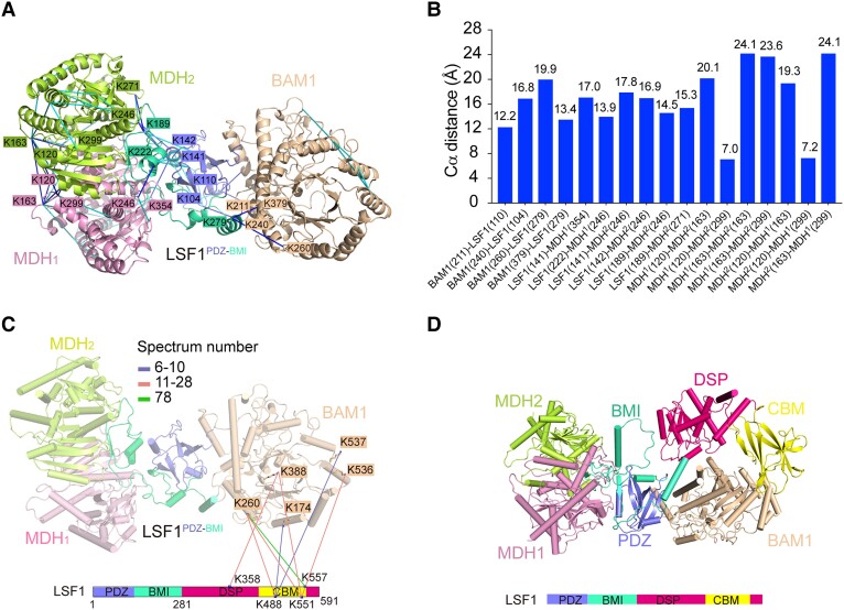
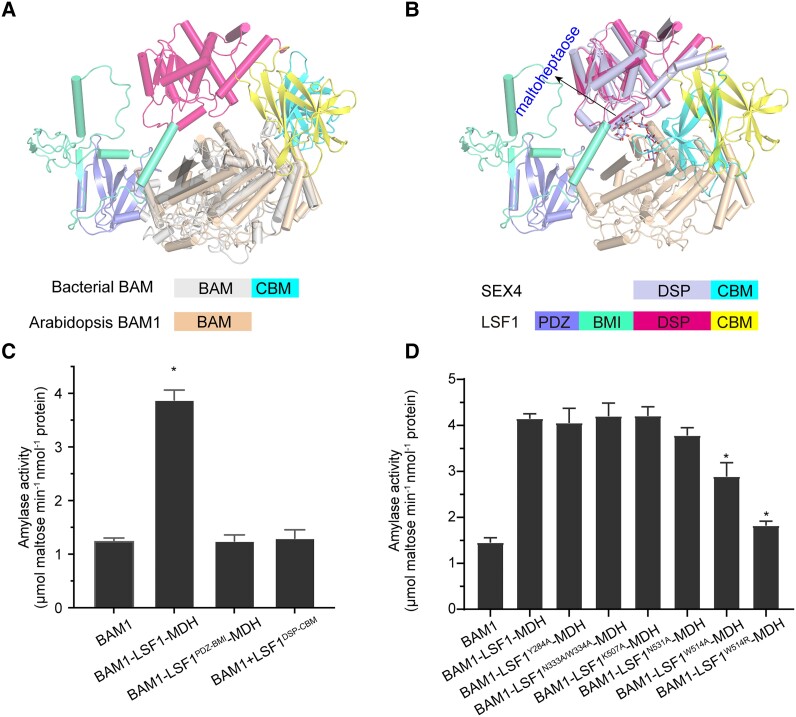
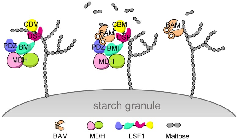
In plant leaves, starch is composed of glucan polymers that accumulate in chloroplasts as the products of photosynthesis during the day; starch is mobilized at night to continuously provide sugars to sustain plant growth and development. Efficient starch degradation requires the involvement of several enzymes, including β-amylase and glucan phosphatase. However, how these enzymes cooperate remains largely unclear. Here, we show that the glucan phosphatase LIKE SEX FOUR 1 (LSF1) interacts with plastid NAD-dependent malate dehydrogenase (MDH) to recruit β-amylase (BAM1), thus reconstituting the BAM1-LSF1-MDH complex. The starch hydrolysis activity of BAM1 drastically increased in the presence of LSF1-MDH in vitro. We determined the structure of the BAM1-LSF1-MDH complex by a combination of cryo-electron microscopy, crosslinking mass spectrometry, and molecular docking. The starch-binding domain of the dual-specificity phosphatase and carbohydrate-binding module of LSF1 was docked in proximity to BAM1, thus facilitating BAM1 access to and hydrolysis of the polyglucans of starch, thus revealing the molecular mechanism by which the LSF1-MDH complex improves the starch degradation activity of BAM1. Moreover, LSF1 is phosphatase inactive, and the enzymatic activity of MDH was dispensable for starch degradation, suggesting nonenzymatic scaffold functions for LSF1-MDH in starch degradation. These findings provide important insights into the precise regulation of starch degradation.


Conflict of interest statement

Conflict of interest statement. None declared.

Figures

Figure 1.
Figure 1.
Structure of the BAM1–LSF1–MDH complex. A) Diagram of starch degradation. Proteins involved in starch phosphorylation, dephosphorylation, and hydrolysis are indicated. B) Diagrams of the domain architecture of BAM1, LSF1, and MDH. TP, chloroplast TP, were omitted for protein production. BAM1 and MDH are colored in beige and pink. For LSF1, the individual domain of PDZ, BMI, DSP, and CBM are colored in slate, green-cyan, hot pink, and yellow, respectively. The domains of DSP-CBM absent from the cryo-EM structure are indicated by the dashed magenta box. C) Reconstitution of the ternary complex by gel filtration. Left, gel filtration chromatography. Right, SDS-PAGE corresponding to the chromatography. The incubation of BAM1 with LSF1–MDH led to the formation and coelution of the ternary BAM1–LSF1–MDH complex. D) Starch hydrolytic activity of the enzymes by detecting the maltose product. n.m., no measured activity. E) Titration assay of the starch-degradation activity of BAM1 upon the addition of the binary LSF1–MDH complex with increased molar ratios. All data reported here were obtained from 3 independent experiments and presented as mean ± Sd of triplicate experiments. Significant differences were determined using 2-tailed Student's t-test (*P < 0.05). F and G) Cryo-EM density of the BAM1–LSF1–MDH complex F) and its cartoon representation G). BAM1 is colored in beige, MDH1 and MDH2 are in pink and lemon, and the PDZ and BMI of LSF1 are in slate and green-cyan, respectively.
Figure 2.
Figure 2.
The LSF1 N terminus is sufficient for ternary complex assembly. A) Structure of the LSF1 N terminus. Secondary structural elements are labeled. The PDZ and BMI domains are colored in slate and green-cyan, respectively. B) Topological model of the LSF1 N terminus. The number of secondary structural elements are labeled. C) Structural superposition of the LSF1 N terminus with the canonical PDZ domain. The structure of 2QG1 is shown in gray. D) The N-terminal PDZ-BMI domain of LSF1 is sufficient to mediate ternary complex formation. Left, a representative gel filtration chromatography; right, SDS-PAGE corresponding to the chromatography. E) Starch hydrolytic activity examined for the BAM1–LSF1PDZ-BMI–MDH complex. n.m., no measured activity. Two-tailed Student's t-tests are used for statistical analysis (*P < 0.05). Error bar, Sd. F) Titration assay of the starch-degradation activity of BAM1 upon the addition of the binary LSF1PDZ-BMI–MDH complex with increased molar ratios. LSF1PDZ-BMI–MDH is unable to promote the starch hydrolytic activity of BAM1. Two-tailed Student's t-tests are used for statistical analysis (*P < 0.05). Error bar, Sd.
Figure 3.
Figure 3.
A gate-latch-lock recognition mode between LSF1 and MDH. A) The interaction surface between LSF1 and dimeric MDH. The PDZ and BMI of LSF1, and the protomers of MDH are shown in slate, green-cyan, pink and lemon, respectively. In the right panel, the location of the gate (dimeric MDH), latch (the sheets β6β7 and its preceding and following loops in LSF1), and lock (a loop) are individually indicated. B) Electrostatic potential analysis of the interaction surface. The positively charged pocket of LSF1 is labeled by a dashed rectangle. The negatively charged residues from MDH are shown in sticks. C) Residues involved in the gate-latch-lock recognition. The interaction residue pairs are shown in sticks and indicated by dashed lines. D) A salt bridge interaction between LSF1 D174 and MDH1 K271. E) Hydrogen-bonded interactions between LSF1 and MDH2. The interaction residue pairs are shown in sticks and indicated by dashed lines. F and G) Validation of critical residues involved in the LSF1–MDH interaction by coexpression and immunoblot analysis. LSF1 and MDH were constructed with His and myc tag, respectively. For each lane, the relative wild-type or mutant constructs are shown above the gel. The star symbol on LSF1 or MDH indicates the relative mutants corresponding to each lane labeled above the gel. Mutations in MDH F) or LSF1 G).
Figure 4.
Figure 4.
Interaction between BAM1 and LSF1. A) The interaction surface between BAM1 and LSF1. BAM1 is shown in beige, PDZ and BMI are colored in slate and green-cyan respectively. The unique α6 is labeled. Key residues involved in the interaction are shown in sticks on the right panel. B and C) Critical residues involved in the BAM1–LSF1 interaction validated by in vitro GST pull-down assay by mutations in either LSF1 B) or BAM1 C). Relative mutants are shown on top of the gel, and the star symbols on LSF* or BAM1* represent relative mutants corresponding to each gel lane. The wild-type binary His-LSF1–MDH or mutant binary His-LSF1*–MDH complexes were obtained by coproduction, and incubated with GST-BAM1. LSF1 quint-mut indicates the quintuple mutant combination N73A, E76A, R112D, E270K, R277D. BAM1 quad-mut indicates the quadruple mutant combination E208K, K240E, D250R, T317A. The band intensities were estimated by ImageJ bundled with 64-bit Java 8. The lane of the wild-type BAM1 and LSF1 was set to 1; the intensities for other lanes of BAM1* and LSF1 were calculated relative to the wild type. The ratio of LSF1/BAM1* for each is calculated and provided below the gel.
Figure 5.
Figure 5.
Structural modeling of the complete BAM1–LSF1–MDH complex on the basis of CXMS data and AlphaFold2 prediction. A) Crosslinks mapped onto the structure of BAM1–LSF1–MDH complex. The blue and cyan lines indicate intersubunit and intrasubunit crosslinked residue pairs, respectively. For clarity, residues involved in the intersubunit interaction are shown, whereas those involved in the intrasubunit interaction are omitted. Residues are colored consistent with the corresponding subunits. B) The distance distribution of all crosslinked residue pairs mapped onto the structure of BAM1–LSF1–MDH complex. The 26 Å cutoff value was used to filter the crosslinks, well below the 35 Å upper limit. C) The intersubunit crosslinks between LSF1DSP-CBM and BAM1. The DSP and CBM domains of LSF1 are shown in hot pink, and yellow, respectively. The identified crosslink pairs are indicated by colored lines based on the spectrum number. For clarity, the LSF1PDZ-BMI-mediated crosslinked residue pairs are omitted. D) Structure model of the complete BAM1–LSF1–MDH complex based on CXMS data and AlphaFold2 predictions. BAM1, MDH1, and MDH2 are colored in beige, pink, and lemon, respectively. The LSF1 domains PDZ, BMI, DSP, and CBM are shown in slate, green-cyan, hot pink, and yellow, respectively. The DSP-CBM of LSF1 was docked in close proximity to BAM1 based on CXMS data.
Figure 6.
Figure 6.
The presence of the C-terminal DSP-CBM of LSF1 is essential for the increased starch degradation of the BAM1–LSF1–MDH complex. A) Structure alignment of bacterial BAM with MDH–LSF1-bound BAM1. For clarity, the structures of MDH are omitted. The bacterial BAM core and CBM are shown in gray and cyan, respectively. Arabidopsis BAM1 is shown in beige. The PDZ, BMI, DSP, and CBM domains of LSF1 are shown in slate, green-cyan, hot pink, and yellow, respectively. B) Structure alignment of maltoheptaose-bound SEX4 with BAM1- and MDH-bound LSF1. For clarity, the structures of MDH are omitted. Maltoheptaose is shown in sticks and indicated. The DSP and CBM segments of SEX4 are shown in light blue and cyan, respectively. The PDZ, BMI, DSP, and CBM segments of LSF1 are shown in slate, green-cyan, hot pink, and yellow, respectively. C) The essential role for both PDZ-BMI and DSP-CBM of LSF1 on the improved starch-hydrolysis activity of BAM1. D) The effect of mutations of the C-terminal DSP and CBM of LSF1 on the starch degradation activity of the BAM1–LSF1–MDH mutant complex. All data reported here were obtained from 3 independent experiments and presented as mean ± Sd of triplicate experiments. Significant differences were determined using 2-tailed Student's t-test (*P < 0.05).
Figure 7.
Figure 7.
A proposed working model for the ternary BAM–LSF1–MDH complex in starch degradation. Left, LSF1 in complex with MDH functions as a scaffold anchored to the starch granule. Right, BAM1 (or BAM3) alone possesses weak starch hydrolysis activity. Middle panel, BAM1 (or BAM3)–LSF1–MDH exhibits strikingly increased starch degradation activity, generating more maltose products. Specifically, the N-terminal PDZ-BMI domain of LSF1 interacts with MDH and recruits BAM, whereas the C-terminal DSP-CBM segment of LSF1 binds to the polyglucan chain, anchors to the starch granule, and presents the polyglucan to the catalytic pocket of BAM, thus promoting starch degradation.

Similar articles

Cited by

References

    1. Adams PD, Afonine PV, Bunkoczi G, Chen VB, Davis IW, Echols N, Headd JJ, Hung LW, Kapral GJ, Grosse-Kunstleve RW, et al. . PHENIX: a comprehensive Python-based system for macromolecular structure solution. Acta Crystallogr D Biol Crystallogr. 2010:66(2):213–221. 10.1107/S0907444909052925 - DOI - PMC - PubMed
    1. Baunsgaard L, Lutken H, Mikkelsen R, Glaring MA, Pham TT, Blennow A. A novel isoform of glucan, water dikinase phosphorylates pre-phosphorylated alpha-glucans and is involved in starch degradation in Arabidopsis. Plant J. 2005:41(4):595–605. 10.1111/j.1365-313X.2004.02322.x - DOI - PubMed
    1. Beeler S, Liu HC, Stadler M, Schreier T, Eicke S, Lue WL, Truernit E, Zeeman SC, Chen J, Kotting O. Plastidial NAD-dependent malate dehydrogenase is critical for embryo development and heterotrophic metabolism in Arabidopsis. Plant Physiol. 2014:164(3):1175–1190. 10.1104/pp.113.233866 - DOI - PMC - PubMed
    1. Chen S, McMullan G, Faruqi AR, Murshudov GN, Short JM, Scheres SH, Henderson R. High-resolution noise substitution to measure overfitting and validate resolution in 3D structure determination by single particle electron cryomicroscopy. Ultramicroscopy. 2013:135:24–35. 10.1016/j.ultramic.2013.06.004 - DOI - PMC - PubMed
    1. Chen T, Ma J, Liu Y, Chen Z, Xiao N, Lu Y, Fu Y, Yang C, Li M, Wu S, et al. . iProX in 2021: connecting proteomics data sharing with big data. Nucleic Acids Res. 2022:50(D1):D1522–D1527. 10.1093/nar/gkab1081 - DOI - PMC - PubMed

MeSH terms

LinkOut - more resources