Review
. 2023 Nov 21;44(44):4622-4633.
doi: 10.1093/eurheartj/ehad637.

Mavacamten: a first-in-class myosin inhibitor for obstructive hypertrophic cardiomyopathy

Affiliations
Review

Mavacamten: a first-in-class myosin inhibitor for obstructive hypertrophic cardiomyopathy

Eugene Braunwald et al. Eur Heart J. .

Erratum in

Abstract

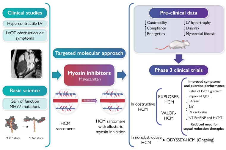
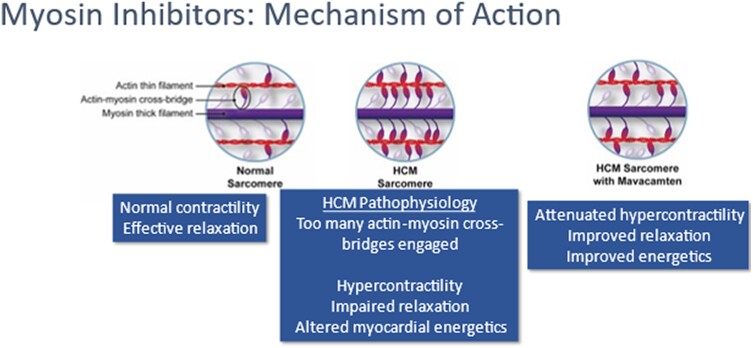
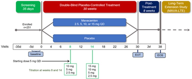
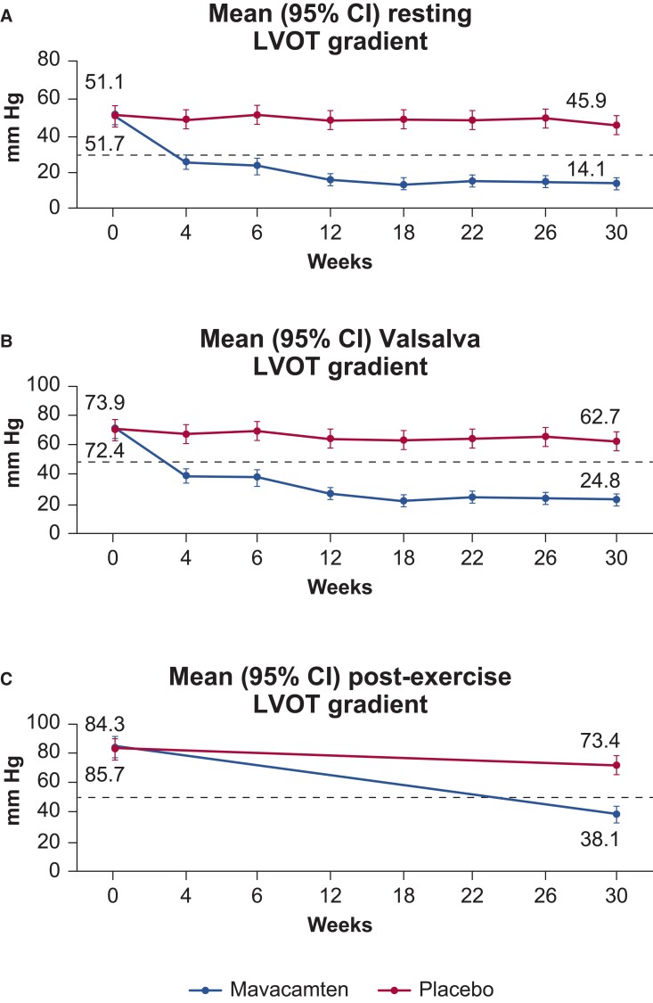
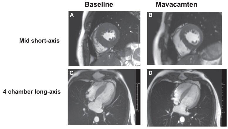
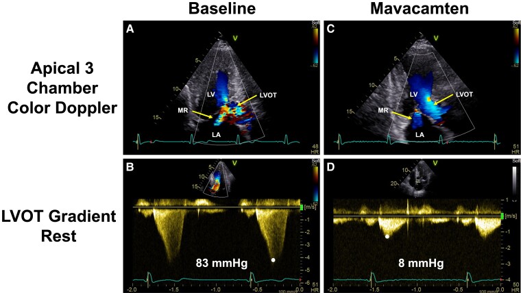
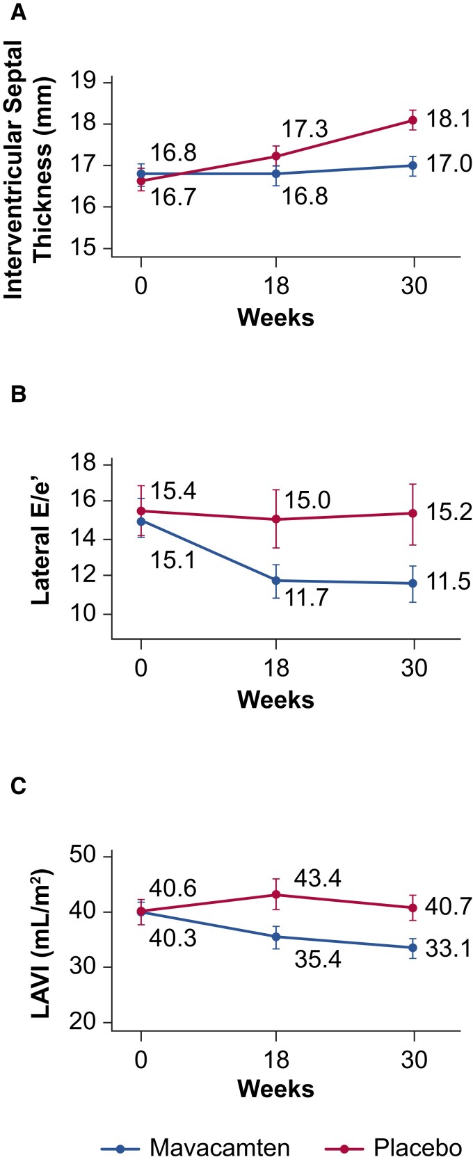
Mavacamten is a first-in-class, targeted, cardiac-specific myosin inhibitor approved by the US Food and Drug Administration for the treatment of adults with symptomatic New York Heart Association Classes II and III obstructive hypertrophic cardiomyopathy (oHCM). Mavacamten was developed to target the hyper-contractile phenotype, which plays a critical role in the pathophysiology of the disease. In Phase 2 and 3 clinical trials, mavacamten was well tolerated, reduced left ventricular outflow tract gradients, improved exercise capacity and symptoms, and was associated with improvements in other clinically relevant parameters, such as patient-reported outcomes and circulating biomarkers. In addition, treatment with mavacamten was associated with evidence of favourable cardiac remodelling in multi-modality imaging studies. Mavacamten substantially reduced guideline eligibility for septal reduction therapy candidates with oHCM and drug-refractory symptoms. In this article, the available efficacy and safety data from completed and ongoing clinical studies of mavacamten in patients with symptomatic oHCM are reviewed. Longer term extension studies may help address questions related to the positioning of mavacamten in current oHCM management algorithms, interactions with background therapy, as well as the potential for disease modification beyond symptomatic relief of left ventricular outflow tract obstruction.

Keywords: Ejection fraction; Myectomy; Myosin; Pressure gradient.


Figures

Graphical Abstract
Graphical Abstract
The path to treatment of obstructive hypertrophic cardiomyopathy. (Top left) Haemodynamic observations demonstrated. Left ventricular (LV) obstruction and symptoms related to LV hypertrophy. (Bottom left) Discovery of genetic variants in ∼40% of patients. (Centre) Sarcomeres in obstructive hypertrophic cardiomyopathy (oHCM) show excess of myosin–actin cross-bridges that are normalized by mavacamten. (Top right) Pre-clinical observations in mouse and pig models of oHCM. (Bottom right) The two placebo-controlled clinical trials of mavacamten in oHCM. HCM, hypertrophic cardiomyopathy; hsTnT, high-sensitivity troponin T; LA, left atrial; LV, left ventricular; LVOT, left ventricular outflow tract; NT-proBNP, N-terminal pro-B-type natriuretic peptide; QOL, quality of life.
Figure 1
Figure 1
Rationale for the use of mavacamten in hypertrophic cardiomyopathy. Molecular basis of hyper-contractility in hypertrophic cardiomyopathy and the effect of mavacamten. Hypertrophic cardiomyopathy-causing mutations may lead to a gain-of-function effect, increasing the proportion of myosin heads in the active state and leading to adverse energetic, structural, and clinical consequences. Mavacamten binds to the myosin molecules and reduces their likelihood of being in the active state, thus attenuating hyper-contractility and its adverse metabolic effects., Reprinted from Ho et al. (https://www.ahajournals.org/doi/10.1161/CIRCHEARTFAILURE.120.006853) with permission from Wolters Kluwer Health, Inc.
Figure 2
Figure 2
EXPLORER-HCM study design. Patients with baseline left ventricular outflow tract pressure gradient >50 mmHg and New York Heart Association Classes II and III symptoms were randomized 1:1 to receive once-daily oral mavacamten (starting dose of 5 mg with a two-step dose titration) or placebo for 30 weeks. Adapted from Ho et al. with permission from Wolters Kluwer Health, Inc.
Figure 3
Figure 3
Left ventricular outflow tract gradients at baseline and after 30 weeks of mavacamten. (A) Gradients at rest. (B) Valsalva gradient. (C) Post-exercise gradient. Adapted from Olivotto et al. with permission from Elsevier.
Figure 4
Figure 4
Cine end-diastolic cardiac magnetic resonance images of a patient with obstructive hypertrophic cardiomyopathy: effects of mavacamten. Baseline before treatment (A and C) and after 30 weeks of mavacamten (B and D). Mid short axis (A and B). Four-chamber long axis (C and D). Compared with baseline, at Week 30, left ventricular mass and maximal wall thickness were reduced from 149 g/m2 and 26 mm to 117 g/m2 and 20 mm, respectively; maximal left atrial size fell from 77 to 59 mL/m2. Left atrial total emptying fraction increased from 27% to 50%. Data are from the EXPLORER-HCM trial., Images courtesy of Dr Raymond Kwong, Brigham and Women’s Hospital.
Figure 5
Figure 5
Echocardiographic images of a patient with obstructive hypertrophic cardiomyopathy: effects of mavacamten. (A) At baseline, colour Doppler shows flow acceleration in the left ventricular outflow tract and significant mitral regurgitation. (B) At baseline, continuous wave Doppler through the left ventricular outflow tract shows a late peak consistent with dynamic left ventricular outflow tract obstruction and a peak gradient of 83 mmHg. (C) Continuous wave Doppler flow through the left ventricular outflow tract demonstrates an early peak and reduction of the peak gradient to 8 mmHg. (D) Following 30 weeks of mavacamten, the colour Doppler flow in the left ventricular outflow tract and in the mitral regurgitation jet are consistent with resolution of the left ventricular outflow tract obstruction and reduction in mitral regurgitation, respectively. Data are from the EXPLORER HCM trial., Images courtesy of Dr Sheila Hegde, Brigham and Women’s Hospital.
Figure 6
Figure 6
Echocardiographic findings in EXPLORER-HCM. Line graphs show mean (95% confidence interval) echocardiographic parameters over time. (A) Inter-ventricular septal thickness. (B) Lateral E/eʹ. (C) Left atrial volume index. E/eʹ, ratio between early mitral inflow velocity and mitral annular early diastolic velocity; LAVI, left atrial volume index. Adapted from Hegde et al. with permission from Elsevier.

References

    1. Ommen SR, Mital S, Burke MA, Day SM, Deswal A, Elliott P et al. 2020 AHA/ACC guideline for the diagnosis and treatment of patients with hypertrophic cardiomyopathy: a report of the American College of Cardiology/American Heart Association joint committee on clinical practice guidelines. Circulation 2020;142:e558–631. 10.1161/CIR.0000000000000937 - DOI - PubMed
    1. Marian AJ, Braunwald E. Hypertrophic cardiomyopathy: genetics, pathogenesis, clinical manifestations, diagnosis, and therapy. Circ Res 2017;121:749–70. 10.1161/CIRCRESAHA.117.311059 - DOI - PMC - PubMed
    1. Lehman SJ, Crocini C, Leinwand LA. Targeting the sarcomere in inherited cardiomyopathies. Nat Rev Cardiol 2022;19:353–63. 10.1038/s41569-022-00682-0 - DOI - PMC - PubMed
    1. Morrow AG, Braunwald E. Functional aortic stenosis; a malformation characterized by resistance to left ventricular outflow without anatomic obstruction. Circulation 1959;20:181–9. 10.1161/01.cir.20.2.181 - DOI - PubMed
    1. Braunwald E, Lambrew CT, Rockoff SD, Ross J Jr, Morrow AG. Idiopathic hypertrophic subaortic stenosis. I. A description of the disease based upon an analysis of 64 patients. Circulation 1964;30 Suppl 4:3–119. 10.1161/01.cir.29.5s4.iv-3 - DOI - PubMed